Résumé : université de Tours
←
→
Transcription du contenu de la page
Si votre navigateur ne rend pas la page correctement, lisez s'il vous plaît le contenu de la page ci-dessous

Année 2017-2018 - Demande d’allocation doctorale
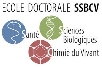
ED Santé, Sciences Biologiques et Chimie du Vivant (SSBCV) n°549
1. Informations administratives :
Nom de l’encadrant responsable de la thèse : C Destrieux
Unité : Imagerie et Cerveau, Inserm U930 Université de Tours
Equipe (si unité multi-équipes): 3, « Imagerie, Biomarqueurs et Thérapeutique »
Email de l’encadrant : christophe.destrieux@univ-tours.fr
Co-encadrant éventuel : Frédéric Andersson, IGR Inserm
(frederic.andersson@univ-tours.fr)
2. Titre de la thèse : établissement d’un atlas IRM multi échelles du tronc cérébral
humain
3. Résumé :
Contexte. Le tronc cérébral contient de multiples structures grises (noyaux des nerfs crâniens, noyaux
propres, substance réticulée) et blanches (faisceaux d’association et de projection). Son exploration en
IRM (Imagerie par Résonance Magnétique) in vivo clinique 1,5 ou 3T est limitée par la résolution
spatiale et le contraste qui ne permettent qu’une étude globale, sans distinction de ses sous-structures.
Celles-ci sont en revanche accessibles à des méthodes d’IRM précliniques sur pièces anatomiques ;
elles améliorent la résolution spatiale et le contraste grâce à des valeurs élevées de champ magnétique
(11,7T versus 1,5 ou 3T en clinique) et de pondération en diffusion (b=4500mT/m versus 40mT/m),
mais aussi grâce à des temps d’acquisition potentiellement illimités et à l’absence de bruit
physiologique.
Objectif. Réaliser un atlas multimodal du tronc cérébral humain à partir d’acquisitions IRM ex vivo à
très haut champ (11,7T), puis l’appliquer à des images in vivo afin d’obtenir, en condition de routine
clinique (3T), une segmentation automatique de structures qui y sont peu ou pas visibles directement.
Contributions.
1) Etablissement d’un atlas anatomique probabiliste haute résolution du tronc cérébral humain. 8
pièces anatomiques de tronc cérébral humain sont actuellement en cours d’acquisition sur l’imageur
préclinique 11,7T de Neurospin : images pondérées en T2 (résolution spatiale : 100-300μm isotrope)
et en diffusion (350μm isotrope). Le candidat y segmentera manuellement les structures grises du
tronc cérébral à l’aide d’un protocole qui a été développé dans notre laboratoire (71 structures sur
environ 800 coupes par pièce anatomique). Un atlas probabiliste sera alors calculé depuis ces
segmentations manuelles qui segmentera automatiquement des pièces anatomiques « neuves ». Une
validation de type jack-knife sera proposée afin de s’assurer de la concordance entre segmentations
manuelle et automatique.
2) application à l’IRM in vivo. L’atlas ex vivo haute résolution obtenu en 1) sera alors appliqué à des
images IRM in vivo afin d’y segmenter automatiquement les structures grises du tronc cérébral. Le
problème consistera à limiter la segmentation à une sous-partie des images in vivo (tronc cérébral) et à
utiliser des images de résolution spatiale et de contrastes très différents entre l’atlas et le jeu de
données à segmenter.
3) validation de la segmentation anatomique automatique in vivo. La validation de la méthode
développée en (2) ne sera pas triviale puisqu'une grande partie des structures segmentées sera invisible
sur les images in vivo acquises sur un imageur clinique 3T avec une résolution spatiale limitée au
millimètre; il ne sera donc pas possible de comparer directement la segmentation automatisée de ces
images à une vérité terrain in vivo. Nous utiliserons des données issues du projet ANR Fibratlas qui
constitue une cohorte de sujets âgés pour lesquels des images IRM in vivo (résolution clinique à 3T) et
ex vivo (résolution submillimétrique à 11,7T) seront disponibles. Pour 3 de ces sujets, les structures du
tronc cérébral seront segmentées automatiquement sur les images in vivo à 3T (segmentation à valider)
et manuellement (vérité terrain) sur les images ex vivo à 11,7T. Ces deux segmentations seront alors
comparées afin de valider la méthode de segmentation automatique.
Retombées. L’atlas développé pourra être utilisé dans des buts académique (segmentation automatisée
de cohortes, par exemple pour des études morphométriques ou pour l’établissement de régions
d’intérêt reproductibles) ou clinique (localisation de cibles de stimulation profonde sur des données
d’IRM 3T où elles sont invisibles). Dans ce cadre, il fera l’objet d’une valorisation industrielle.
Faisabilité. Les 8 pièces anatomiques qui vont être utilisées pour la constitution de l’atlas sont en
cours d’acquisition IRM dans le cadre d’une collaboration avec un des leaders de l’imagerie très haut
champ ex vivo, Neurospin ; les IRM in vivo et ex vivo utilisées dans les étapes 2 et 3 seront financées
par l’ANR Fibratlas ; les règles anatomiques de segmentation du tronc cérébral ont été établies lors
d’un travail de master 2. Le laboratoire d’accueil dispose de compétences en traitement d’images et
d’une expertise internationalement reconnue en anatomie cérébrale ; il a notamment été à l’origine de
la publication d’un atlas probabiliste du cortex cérébral humain largement distribué à la communauté
(C Destrieux, Neuroimage, 2010). Le doctorant disposera des ressources des projets ANR Fibratlas
puis régional Fibravasc.

4. Résumé en anglais : Multiscale MRI atlas of the human brainstem

Context. The brainstem contains multiple grey (cranial nerves, proper and reticular nuclei) and white
structures (association and projection tracts). Its in vivo exploration, using clinical 1.5 or 3T MRI
(Magnetic Resonance Imaging) scanners, is limited by the spatial resolution and contrast of this
method. It indeed only permits a global exploration of the brainstem, but cannot distinguish its finer
components. However, some of the latter become visible using preclinical scanner and anatomical
specimens; it increases the resulting spatial resolution and contrast, thanks to the high strength of the
magnetic field (B0=11,7T versus 1.5 to 3T for clinical practice) and gradients used to explore diffusion
(b=4500mT/m versus 40mT/m), but also to potentially unlimited scanning time and absence of
physiological noise.
Long-term aim. Build a multimodal atlas of the human brainstem from ex vivo ultra high filed MRI,
and use it to automatically segment in vivo images obtained at a lower resolution.
Contributions.
1) High resolution anatomical probabilistic atlas of the human brainstem. 8 anatomical specimens are
being scanned on the preclinical 11.7T scanner in Neurospin: T2 (spatial resolution 100-300μm
isotropic) and diffusion weighted (350μm isotropic) images. The PhD student will use a protocol we
previously develop to manually segment 71 grey matter structures of the brainstem (about 800 slices
per specimen). The segmented data will be used to compute a probabilistic atlas, which will be able to
automatically segment new anatomical specimens obtained at the same resolution. For validation, a
jack-knifing leave one out procedure will be used to check the similarity between automated and
manual segmentations.
2) Application of the high-resolution atlas to in vivo MRI. The high resolution ex vivo atlas will be
used to automatically segment grey structures of the brainstem in in vivo images. This will imply to
limit the segmentation to a subpart of in vivo images (namely the brainstem) and to use data having
very different contrast and spatial resolution (in vivo images versus ex vivo atlas).
3) Validation of the automated segmentation of in vivo images. No direct manual segmentation of in
vivo images will be possible since most of the studied structures won’t be visible on this dataset
obtained at a millimetric resolution on a 3T scanner. In other words, no ground truth will be available
in vivo to validate the developed automated atlas. For this purpose, we will use subjects from the ANR
Fibratlas project, which collects in and ex vivo MRI data for the same subjects. The ultra-high field
MRI-scanner used for ex vivo acquissitons will provide high contrast and resolution, allowing a
manual segmentation. At least 3 in/ex vivo datasets should be available until the beginning of this
PhD: brainstem structures will be automatically segmented in in vivo images (segmentation to be
validated) and manually in ex vivo images (ground truth). Both segmentations will then be compared
to validate the automated method.
Spin-off. The proposed atlas will have academic (automated segmentation of cohorts, for instance for
morphometric analysis or the definition of reproducible regions of interest) or clinical applications
(definition of targets for deep brain stimulation using clinical images where these targets are not
visible). In this last context, this atlas will be proposed for an industrial partnership.
Feasibility. The 8 anatomical specimens which will be used for building the atlas are being scanned in
the frame of a partnership with Neurospin, one of the leader laboratories in the field of ultra high field
ex vivo imaging. MRI data used for steps #2 and #3 will be obtained from the Fibratlas ANR. The
anatomical rules and method for segmentation of the brainstem were defined during a master thesis.
The host laboratory includes experts in the field of Neuroanatomy and imaging process; among other
things this laboratory developed an automated atlas of the human cortex widely distributed in the
community (C Destrieux, Neuroimage, 2010).
Vous pouvez aussi lire