Canadian Ophthalmological Society evidence-based clinical practice guidelines for the management of glaucoma in the adult eye Guide factuel de ...
←
→
Transcription du contenu de la page
Si votre navigateur ne rend pas la page correctement, lisez s'il vous plaît le contenu de la page ci-dessous
VOL. 44, SUPPL. 1   JUNE/JUIN 2009

Canadian Ophthalmological Society
evidence-based clinical practice
guidelines for the management of
glaucoma in the adult eye

Guide factuel de pratique clinique de la
Société canadienne d’ophtalmologie pour la
gestion du glaucome chez l’adulte
                                                                       The Canadian Journal of Ophthalmology is the official journal of the Canadian
                                                                         Ophthalmological Society and is committed to timely publication of
                                                                           original, peer-reviewed ophthalmology and vision science articles.

                                                                        Le Journal canadien d’ophtalmologie, organe officiel de la Société canadienne
                                                                         d’ophtalmologie, s’engage à publier des articles originaux évalués par
                                                                                  les pairs en ophtalmologie et en science de la vision.

                                        Editorial Board / Comité de rédaction
                                                                   EDITOR-IN-CHIEF / RÉDACTEUR EN CHEF
                                                                            Phil Hooper, MD, FRCSC

610-1525 Carling Ave.
                                        SECTION EDITORS                                           INTERNATIONAL ADVISORY BOARD
Ottawa ON K1Z 8R9                       RÉDACTEURS DE SECTION                                     CONSEIL CONSULTATIF INTERNATIONAL
613-729-6779
800-267-5763                                                    Cataract & optics / Cataracte & optique
Fax 613-729-7209                        Susan K. Lindley, MD, FRCSC                      Randall Olson, MD
cjo@eyesite.ca                          Suren Sanmugasunderam, MD, FRCSC                 Salt Lake City, Utah, USA

eyesite.ca                                                  Cornea & external disease & refractive surgery
                                                        Maladies externes & de la cornée & chirurgie réfractive
PRESIDENT                               Simon P. Holland, MB, FRCSC                     Prashant Garg, MD
PRÉSIDENT                               Alexander C. Tokarewicz, MD, FRCSC              Banjara Hills, Hyderabad, India
Lorne Bellan, MD, FRCSC                                                     Genetics / Génétique
EXECUTIVE DIRECTOR
                                        Michael Walter, PhD                                Mansoor Sarfarazi, PhD
                                                                                           Farmington, Connecticut, USA
DIRECTEUR GÉNÉRAL
                                                                           Glaucoma / Glaucome
Hubert Drouin, CAE
                                        Graham E. Trope, MB, PhD, FRCSC                  Tarek Shaarawy, MD
                                                                                         Geneva, Switzerland
MANAGING EDITOR (acting)
DIRECTRICE DE LA RÉDACTION                                           Low vision / Basse vision
(intérimaire)                           Samuel N. Markowitz, MD, FRCSC                August Colenbrander, MD
                                                                                      Novato, California, USA
Victoria Bell, MA Translation
                                                            Neuro-ophthalmology / Neuro-ophtalmologie
CONTRIBUTING EDITORS                    Agnes Wong, MD, PhD, FRCSC                   Andrew Lee, MD
RÉDACTEURS INVITÉS                                                                   Iowa City, Iowa, USA

Ian McIlraith, MD, FRCSC                                          Ocular oncology / Oncologie oculaire
Martin J. Steinbach, PhD                Christine Corriveau, MD, FRCSC                  Miguel Materin, MD
                                                                                        New Haven, Connecticut, USA
TRANSLATION
                                                           Oculoplastic surgery / Chirurgie plastique oculaire
TRADUCTION
                                        Bryan P. Arthurs, MD, FRCSC                      John Woog, MD
Claude Gendron,                                                                          Rochester, Minnesota, USA
Les Rédactions Oléron limitée
                                                 Ophthalmic & general pathology / Pathologie ophtalmique & générale
TRANSLATION EDITOR                      J. Godfrey Heathcote, MB, PhD, FRCPC          Paul Hiscott, PhD
                                                                                      Liverpool, UK
DIRECTEUR DE LA TRADUCTION

Caroline Lajoie, MD, MSc, FRCSC, DABO                   Pediatric ophthalmology / Ophtalmologie pédiatrique
                                        Robert K. Koenekoop, MD, PhD, FRCSC           Elias Traboulsi, MD
EDITORES EMERITI                                                                      Cleveland, Ohio, USA

 Dr. David J. Addison                                             Retina & vitreous / Rétine & vitré
 Dr. Miguel N. Burnier, Jr.             David A.L. Maberley, MD, FRCSC, MSc Epid        Alex P. Hunyor, MB, BS, FRANZCO, FRACS
†Dr. Cecil C. Ewing (2006)              Tom G. Sheidow, MD, FRCSC, M Math               Chatswood, Australia

 Dr. Ian M. MacDonald                                                           Uveitis / Uvéite
 Dr. Brent J. MacInnis                  William G. Hodge, MD, PhD, FRCSC                     Marc D. de Smet, MD
†Dr. J. Clement McCulloch (2007)                                                             Amsterdam, Netherlands
 Dr. Donald W. Mills                                             Vision science / Science de la vision
 Dr. Howard N. Reed                     Neeru Gupta, MD, PhD, FRCSC, DABO                Robert N. Weinreb, MD
 Dr. Graham E. Trope                                                                     La Jolla, California, USA
SERVICE INFORMATION                                                                           RENSEIGNEMENTS AUX LECTEURS
Subscriptions                                                                                 Abonnements
Requests for subscriptions, renewals, and single or back numbers, and all                     Les demandes d’abonnement, de renouvellement, de numéros uniques
remittances should be addressed to: Subscription Office, NRC Research                         ou anciens ainsi que les paiements doivent être adressées à Bureau
Press, 1200 Montreal Rd., Ottawa ON K1A 0R6; 613-993-9084 or                                  des abonnements, Presses scientifiques du CNRC, 1200, chemin de
800-668-1222, fax 613-952-7656, pubs@nrc-cnrc.gc.ca. Remittances                              Montréal, Ottawa ON K1A 0R6; 613-993-9084 ou 800-668-1222,
should be made payable to the Receiver General for Canada, credit                             télécopieur 613-952-7656, pubs@nrc-cnrc.gc.ca. Les paiements doivent
National Research Council Canada.                                                             être faits à l’ordre du Receveur général du Canada, au crédit du Conseil
                                                                                              national de recherches Canada.
Replacing missing issues
Claims for missing issues must be made within 3 months of the issue                           Remplacement de numéros manquants
date (subject to availability).                                                               Pour recevoir gratuitement (sous réserve de sa disponibilité) un numéro
                                                                                              manquant, vous devez présenter votre demande dans les 3 mois de la date
Offprints/Reprints                                                                            du numéro en question.
Offprints of papers in the forthcoming issue and bulk reprints of previ-
ously published papers may be ordered in minimum quantities of 50.                            Tirés à part et réimpressions
Contact NRC Research Press at 613-990-2254, fax 613-952-7656,                                 On peut commander des tirés à part d’articles du prochain numéro ou des
rp.business@nrc-cnrc.gc.ca. Reprint orders depend on availability of                          réimpressions en gros d’articles déjà publiés, en lots d’au moins
plates.                                                                                       50 exemplaires. Joindre les presses scientifiques du CNRC par téléphone au
                                                                                              613-990-2254, télécopie au 613-992-7656 ou courriel à
Microform                                                                                     rp.business@nrc-cnrc.gc.ca. Le suivi des commandes de réimpression
CJO is available in microform or via electronic databases from                                dépendra de la disponibilité des plaques.
University Microfilms, Inc., PO Box 1346, Ann Arbor MI
48106-1346, USA; 800-521-0600, http://www.umi.com.                                            Microfiches
                                                                                              Le JCO est disponible sur microfiches ou sur base de données électronique
Permissions                                                                                   chez University Microfilms, Inc., PO Box 1346, Ann Arbor MI
All material in CJO is copyrighted and may not be reproduced by any                           48106-1346, USA; 800-521-0600, http://www.umi.com.
means without prior permission from the Managing Editor, Canadian
Journal of Ophthalmology, 610-1525 Carling Ave., Ottawa ON                                    Permissions
K1Z 8R9; 613-729-6779 or 800-267-5763, fax 613-729-7209,                                      Tous les articles publiés dans le JCO sont assujettis au droit d’auteur; il est
cjo@eyesite.ca.                                                                               interdit de les reproduire sous quelque forme que ce soit sans la permission
                                                                                              de la directrice de la rédaction, Journal canadien d’ophtalmologie, 610-1525,
Change of address                                                                             avenue Carling, Ottawa ON K1Z 8R9; 613-729-6779 ou 800-267-5763,
Please send your new address and the effective date to:                                       télécopieur 613-729-7209, cjo@eyesite.ca.
Canadian Journal of Ophthalmology, 610-1525 Carling Ave., Ottawa
                                                                                              Changement d’adresse
ON K1Z 8R9; 613-729-6779 or 800-267-5763, fax 613-729-7209,
                                                                                              Veuillez envoyer votre nouvelle adresse et la date d’entrée en vigueur au
cos@eyesite.ca.
                                                                                              Journal canadien d’ophtalmologie, 610-1525, avenue Carling, Ottawa ON
Advertising sales                                                                             K1Z 8R9; 613-729-6779, 800-267-5763, télécopieur
Canadian Ophthalmological Society, 610-1525 Carling Ave., Ottawa                              613-729-7209, cos@eyesite.ca.
ON K1Z 8R9; 613-729-6779 or 800-267-5763, fax 613-729-7209,                                   Annonces publicitaires
jdavis@eyesite.ca.                                                                            Société canadienne d’ophtalmologie, 610-1525, avenue Carling, Ottawa
Printing                                                                                      ON K1Z 8R9; 613-729-6779 ou 800-267-5763, télécopieur
Reprographic and Publication Distribution Services                                            613-729-7209, jdavis@eyesite.ca.
National Research Council Canada                                                              Impression
1200 Montreal Rd., Ottawa ON K1A 0R6                                                          Services de reprographie et de diffusion des publications
              Publishing services                                                             Conseil national de recherches Canada
Publishing services provided by NRC Research Press, National Research                         1200, chemin de Montréal, Ottawa ON K1A 0R6
Council Canada, Ottawa ON K1A 0R6, http://pubs.nrc-cnrc.gc.ca.
                                                                                                             Services d’édition
Manager, Client Journals, Carol McKinley; Publication Officer,                                Les services d’édition sont fournis par les Presses scientifiques du CNRC,
Lianne Johnsen; JournalsCJO.CISTI@nrc-cnrc.gc.ca.                                             Conseil national de recherches Canada, Ottawa, ON K1A 0R6;
                                                                                              http://pubs.nrc-cnrc.gc.ca.
                                                                                              Gestionnaire, Revues affiliées, Carol McKinley; Agente de publication,
                                                                                              Lianne Johnsen; JournalsCJO.CISTI@nrc-cnrc.gc.ca.

The CJO is the official journal of the Canadian Ophthalmological Society and is committed     Le JCO, organe officiel de la société canadienne d’ophtalmologie, s’engage à publier des
to timely publication of original, peer-reviewed ophthalmology and vision science articles.   articles originaux évalués par les pairs en ophtalmologie et en science de la vision.
The CJO is owned by the Canadian Ophthalmological Society, and editorial material             Le JCO est la propriété de la Société canadienne d’ophtalmologie, qui détient un droit de
appearing in it is copyrighted. Information for authors: http://eyesite.ca/english/program-   publication exclusif sur tous les articles qui y sont publiés. L’information aux auteurs :
and-services/cjo/cjo-info.html. Scientific issues are published in February, April, June,     http://eyesite.ca/francais/programmes-et-services/cjo/cjo-info.html. Des numéros
August, October, and December. CJO is supplied to members of COS as a benefit of              scientifiques paraissent en février, avril, juin, août, octobre, et décembre. Les membres en
membership; others may subscribe yearly. Rates for 2009: Canada $85 plus GST, United          règle de la SCO reçoivent gratuitement le JCO; toute autre personne peut s’abonner. Tarifs
States US$100, elsewhere US$110; for first-class postage add $25, US$35, and US$85,           annuels pour 2009 : Canada, 85 $ TPS en sus; États-Unis, 100 $ US; ailleurs, 110 $ US.
respectively. Single copies (subject to availability): Canada $25 plus GST, US or foreign     Pour courrier première classe ajouter 25 $, 35 $ US, et 85 $ US, respectivement. Au numéro
destinations US$18 (plus shipping & handling). All subscriptions are payable in advance.      (selon les disponibilités) : Canada 25 $ TPS en sus, 18 $ US l’exemplaire (manutention et
Visa, MasterCard, and American Express are accepted. Remittances should be made               transport en sus). Les abonnements sont payables d’avance. Les cartes Visa, MasterCard,
payable to the Receiver General for Canada, credit National Research Council Canada,          et American Express sont acceptées. Prière d’envoyer le paiement au Receveur général
and sent to the Subscription Office, NRC Research Press, 1200 Montreal Rd., Ottawa            du Canada au crédit du Conseil national de recherches Canada, et envoyez à Bureau des
ON K1A 0R6; 613-993-9084 or 800-668-1222; fax 613-952-7656;                                   abonnements, Presses scientifiques du CNRC, 1200, chemin de Montréal, Ottawa ON
pubs@nrc-cnrc.gc.ca.                                                                          K1A 0R6; 613-993-9084 ou 800-668-1222; télécopieur 613-952-7656;
ISSN 0008-4182 (print) ISSN 1715-3360 (online)                                                pubs@nrc-cnrc.gc.ca.

All editorial matter in the Canadian Journal of Ophthalmology represents                      ISSN 0008-4182 (version imprimée) ISSN 1715-3360 (version en ligne)
the opinions of the authors and not necessarily those of the Canadian                         Tous les articles à caractère éditorial dans le Journal canadien d’ophtalmologie représentent
Ophthalmological Society.                                                                     les opinions de leurs auteurs et n’engagent pas la Société canadienne d’ophtalmologie.
Publications mail agreement no. 40036504.                                                     Poste-publications – convention no 40036504.

    International mailing: CJO – Canadian Journal of Ophthalmology, ISSN 0008-4182, is published bimonthly, 6 times per year (Feb., Apr., June, Aug., Oct., Dec.) by
    National Research Council Canada at 26 Power Dam Way, Ste. S1-S3, Plattsburgh NY 12901. Periodicals postage paid at Plattsburgh, NY. POSTMASTER send address
    changes to CJO c/o USACAN Media. PO Box 2888, Plattsburgh NY 12901.

S2       CAN J OPHTHALMOL—VOL. 44, SUPPL. 1, 2009
The Canadian Journal of Ophthalmology is the official journal of the Canadian

                                                                              Ophthalmological Society and is committed to timely publication of
                                                                                original, peer-reviewed ophthalmology and vision science articles.

                                                                             Le Journal canadien d’ophtalmologie, organe officiel de la Société canadienne
                                                                              d’ophtalmologie, s’engage à publier des articles originaux évalués par
                                                                                       les pairs en ophtalmologie et en science de la vision.

CONTENTS                                                                                   Volume 44, Supplement 1, June/juin 2009

Canadian Ophthalmological Society evidence-                              Diagnostic tests in glaucoma                       S15
based clinical practice guidelines for the                                 S15 Role of imaging devices in diagnosing
management of glaucoma in the adult eye                                         glaucoma
                                                                                S15 ONH and RNFL analyzers
Introduction                                                      S7            S15 Confocal scanning laser tomography
                                                                                S15 Optical coherence tomography
Methods                                                           S7            S15 Scanning laser polarimetry
                                                                           S16 Performance of ONH/RNFL analyzers in the
Definitions and scope of document                   S8                          diagnosis of glaucoma
   S8 Classification                                                       S16 Comparison of ONH/RNFL analyzers with
        S8 Primary and secondary glaucomas and ocular
                                                                                photographic evaluation of the ONH and
           hypertension                                                         RNFL
             S8   Primary open-angle glaucoma
             S9   Primary open-angle glaucoma suspects                     S16 Ability of ONH/RNFL analyzers to predict
             S9   Primary angle-closure glaucoma                                future conversion to glaucoma in suspect
             S9   Primary angle-closure suspect                                 individuals
             S9   Plateau iris configuration and plateau iris syndrome     S16 Role of imaging devices in monitoring
             S9   Secondary open-angle glaucomas
                                                                                glaucoma
             S9   Secondary angle-closure glaucomas
                                                                           S16 Comparison of ONH/RNFL analyzers to
    S9 Mixed- and multiple-mechanism glaucomas
                                                                                photographic evaluation of progressive changes
   S10 Epidemiology and burden of blindness
                                                                           S17 Role of psychophysical tests in diagnosing
   S10 Pharmacoeconomics of glaucoma
                                                                                glaucoma
Screening for glaucoma                               S10                   S18 Role of psychophysical tests in monitoring
  S11 Risk factors, natural history and clinical course                         glaucoma
  S11 Validity of glaucoma screening tests                                 S19 Role of neuroimaging in glaucoma diagnosis
       S11 Tonometry                                                       S19 Other ancillary tests
       S11 Visual function tests
                                                                         Open-angle glaucomas                           S19
       S11 Structural tests
                                                                           S19 Definitions
  S11 Treatment effectiveness
                                                                               S19 Primary open-angle glaucoma suspects
  S11 Cost-effectiveness and implementation of a
                                                                               S19 Normal pressure glaucoma (or POAG at
       screening program in Canada
                                                                                   normal IOPs)
Diagnosis of glaucoma                                           S12        S20 Pigment dispersion syndrome
  S12 Diagnosis                                                            S20 Pigmentary glaucoma
  S12 History                                                              S20 Pseudoexfoliation syndrome and glaucoma
  S12 Risk factors
                                                                         Angle-closure glaucomas                                S21
  S13 Clinical examination
                                                                           S21 Definition and classification
Intraocular pressure and its measurement S13                               S21 Epidemiology
   S13 Measurement of IOP                                                  S21 Risk factors for PACG
       S14 Factors influencing accuracy of IOP measurement                 S22 Presentation
                                                                                S22 Acute angle closure
Central corneal thickness                                       S14             S22 Narrow angle at risk of closure (angle-closure
and its measurement                                                                 suspect)
  S15 Impact of CCT on IOP                                                      S22 Creeping angle closure
  S15 Measurement                                                          S22 Diagnosis

                                                                                       CAN J OPHTHALMOL—VOL. 44, SUPPL. 1, 2009                          S3
Contents (continued)

    S22 Classification                                           S13 Table 4—Risk factors and signs for conversion
    S22 Treatment                                                    of ocular hypertension to glaucoma with level 1
        S22 Primary acute angle closure                              evidence
        S23 Narrow angle with normal IOP                         S13 Table 5—Some general factors that affect
        S23 Chronic angle closure                                    measured IOP
        S23 Secondary angle closure                              S14 Table 6—Clinical features associated with
        S23 Neovascular glaucoma                                     measurement errors using applanation tonometers
        S23 Ciliary block glaucoma                               S17 Table 7—Merits and limitations of ONH
                                                                     and RNFL analyzers and optic disc/RNFL
Progression                                             S23          photography
  S23 Definition of progression                                  S18 Table 8—Merits and limitations of manual
  S23 Methods of detecting progression                               perimetry, SAP, SWAP, and FDT
       S24 Progression on the basis of structural changes        S21 Table 9—Classification of angle closure based on
       S24 Progression on the basis of functional changes            functional cause
  S24 Approaches to detecting VF progression                     S22 Table 10—Risk factors for development of
       S24 Event-based analysis                                      primary angle closure
       S24 Trend-based analysis                                  S24 Table 11—Endpoints for conversion to, or
  S25 Correlation between structural and functional                  progression of, glaucoma in major RCTs
       change                                                    S24 Table 12—VF progression endpoints for the
  S25 Risk factors for conversion to, and progression                major glaucoma RCTs
       of, POAG                                                  S25 Table 13—Advantages and disadvantages of
       S25 Patient with ocular hypertension                          event-based and trend-based approaches to VF
       S25 Patient with glaucoma                                     progression
       S26 Progression significant for the patient               S25 Table 14—Risk factors and their relationship to
  S26 Follow-up intervals                                            progression examined in the landmark RCTs
       S26 Recommended follow-up intervals and testing
                                                                 S26 Table 15—Number of annual VF tests needed to
           frequencies                                               detect total mean deviation change over 2, 3, and
                                                                     5 years
Glaucoma therapies                              S26              S26 Table 16—Recommended clinical assessment
  S26 Overarching and specific management goals                      intervals for stable chronic glaucomas
  S27 Quality-of-life considerations                             S26 Table 17—Indications for more frequent follow-
  S27 Lowering IOP                                                   up or heightened surveillance
  S27 Underlying mechanism(s)                                    S27 Table 18—Overall and specific management
  S27 Setting target IOP                                             goals in patients with glaucoma
  S28 Therapeutic options                                        S28 Table 19—Staging each eye for glaucoma
      S28 Medical therapy
                                                                     damage
               S28 Ocular hypotensive therapy                    S28 Table 20—Suggested upper limit of initial target
         S29   Surgical therapy                                      IOP for each eye
               S30   Laser trabeculoplasty                       S32 Table 21—Advantages and disadvantages of
               S30   Trabeculectomy                                  single and combined cataract and glaucoma
               S31   Nonpenetrating filtration surgery               procedures
               S31   Tube shunts
               S31   Cyclodestructive surgery                  Appendices
                                                                 S34 Appendix A. Index of recommendations
Cataract and glaucoma                                    S31
                                                                 S35 Appendix B. Glaucomatous optic neuropathy
                                                                 S36 Appendix C. Field progression
Funding, disclosures and acknowledgments                 S32     S38 Appendix D. Sample glaucoma referral letter
                                                                 S39 Appendix E. How to test calibration of a
List of tables                                                       Goldmann Tonometer
    S8 Table 1—Criteria for assigning levels of evidence         S40 Appendix F. Tonometry mires
         to the published studies                                S41 Appendix G. Summary of glaucoma medications
   S12 Table 2—Essential elements of the                             used for chronic treatment
         comprehensive glaucoma eye examination                  S43 Appendix H. Glossary of acronyms
   S12 Table 3—Risk factors and signs for presence of
         open-angle glaucoma with level 1 evidence             References                                        S44

S4   CAN J OPHTHALMOL—VOL. 44, SUPPL. 1, 2009
                                                                                                             Contents (continued)

Guide factuel de pratique clinique de la Société                         S64 Performance des analyseurs TNO-CFNR dans le
canadienne d’ophtalmologie pour la gestion du                                diagnostic du glaucome
glaucome chez l’adulte                                                   S65 Comparaison des analyseurs TNO-CFNR avec
                                                                             l’évaluation photographique de la TNO et de la
                                                                             CFNR
Introduction                                                    S55      S65 Capacité des analyseurs TNO-CFNR de prédire
                                                                             l’éventuelle conversion en glaucome chez les
Méthodologie                                                    S55
                                                                             individus suspects
Définitions et portée du document                     S56                S65 Rôle des appareils d’imagerie dans le monitorage
  S56 Classification                                                         du glaucome
        S56 Glaucomes primaire et secondaire et hypertension             S65 Comparaison entre les analyseurs TNO-CFNR
            oculaire                                                         et l’évaluation photographique des changements
             S56   Le glaucome primaire à angle ouvert                       progressifs
             S57   Soupçons de glaucome primaire à angle ouvert          S66 Rôle des tests psychophysiques dans le diagnostic
             S57   Le glaucome primaire à angle fermé                        du glaucome
             S57   Soupçons de glaucome primaire à angle fermé           S67 Rôle des tests psychophysiques dans le monitorage
             S57   Configuration et syndrome de l’iris en plateau
             S57   Glaucomes secondaires à angle ouvert
                                                                             du glaucome
             S57   Glaucomes secondaires à angle fermé                   S68 Rôle de la neuroimagerie pour le diagnostic du
   S58 Glaucomes à mécanisme mixte ou multiple                               glaucome
   S58 Épidémiologie et fardeau de la cécité                             S68 Tests secondaires
   S58 Pharmacoéconomie du glaucome
                                                                      Le glaucome à angle ouvert                            S68
Dépistage du glaucome                              S58                  S68 Definitions
  S59 Facteurs de risque, histoire naturelle et                              S68 Les suspects de glaucome primaire à angle ouvert
       cheminement clinique                                                  S68 Glaucome à pression normale (ou GPAO à PIO
  S59 Validité des tests de dépistage du glaucome                                normale)
       S59 La tonométrie                                                S69 Le syndrome de dispersion pigmentaire
       S59 Les tests de la fonction visuelle                            S69 Le glaucome pigmentaire
       S59 Les tests structurels                                        S70 Le syndrome de pseudoexfoliation et le glaucome
  S60 L’efficacité du traitement
                                                                      Les glaucomes à angle fermé                             S70
  S60 La rentabilité et l’application d’un programme
                                                                        S70 Définition et classification
       de dépistage au Canada
                                                                        S71 L’épidémiologie
Diagnostic du glaucome                                          S60     S71 Les facteurs de risque de GPAF
  S60 Le diagnostic                                                     S71 La présentation
  S60 Les antécédents                                                         S71 Fermeture d’angle aigu
                                                                              S71 Angle étroit à risque de fermeture (soupçon
  S61 Les facteurs de risque
  S61 L’examen clinique                                                           d’angle fermé)
                                                                              S71 Fermeture progressive de l’angle
La pression intraoculaire et sa mesure                          S61     S71 Le diagnostic
  S62 Mesure de la PIO                                                  S72 La classification
       S62 Facteurs influant sur l’exactitude de la                     S72 Le traitement
           mesure de la PIO                                                   S72 La fermeture d’angle aigu primaire
                                                                              S72 L’angle étroit avec PIO normale
Mesure de l’épaisseur de la cornée centrale S63                               S72 La fermeture d’angle chronique
  S63 Impact de l’ÉCC sur la PIO                                              S72 La fermeture d’angle secondaire
  S63 La mesure                                                               S72 Le glaucome néovasculaire
                                                                              S73 Le glaucome par blocage ciliaire
Tests diagnostiques du glaucome                       S64
  S64 Rôle de l’imagerie dans le diagnostic du glaucome               La progression                                        S73
        S64 Les analyseurs TNO et CFNR                                  S73 Définition
        S64 La tomographie confocale à balayage laser                   S73 Méthode de détection de la progression
        S64 La tomographie par cohérence optique                             S73 La progression basée sur les changements
        S64 La polarimétrie à balayage laser                                     structurels

                                                                                  CAN J OPHTHALMOL—VOL. 44, SUPPL. 1, 2009     S5
Contents (continued)

          S73 La progression basée sur les changements             S66 Tableau 7—Mérites et limites des analyseurs de la
              fonctionnels                                             TNO et de la CFNR ainsi que de la photographie
    S74   Les façons d’aborder la détection de la progression          capsule/CFNR
          du champ visuel                                          S67 Tableau 8—Mérites et limites de la périmétrie
          S74 L’analyse événementielle                                 manuelle, PAS, PALC et TDF
          S74 L’analyse tendancielle                               S71 Tableau 9—Classification de la fermeture d’angle
    S74   La corrélation des changements structurels et                selon la cause fonctionnelle
          fonctionnels                                             S71 Tableau 10—Facteurs de risque de développement
    S75   Les facteurs de risque de conversion en GPAO et              de fermeture d’angle primaire
          la progression de celui-ci                               S74 Tableau 11—Critères de conversion en glaucome
          S75 Le patient faisant de l’hypertension oculaire            ou de sa progression dans les principales ÉCR
          S75 Le patient atteint de glaucome                       S74 Tableau 12—Critères de progression du CV dans
          S75 Progression significative pour le patient                les principales ÉCR
    S76   Les intervalles de suivi                                 S75 Tableau 13—Avantages et désavantages des
          S76 Recommandations quant aux intervalles de suivi           approches événementielles et tendancielles de la
              et à la fréquence des tests                              progression du CV
                                                                   S75 Tableau 14—Facteurs de risque et leur relation
Les thérapies du glaucome                             S76              avec la progression examinée dans les principales
  S76 Les objectifs principaux et les buts particuliers de             ÉCR
       la gestion de la maladie                                    S76 Tableau 15—Nombre annuel de tests du CV
  S77 Les aspects de la qualité de vie                                 nécessaire pour détecter la moyenne totale de
  S77 L’abaissement de la PIO                                          déviation du changement sur 2, 3, et 5 années
  S77 Le ou les mécanismes sous-jacents                            S76 Tableau 16—Intervalles d’évaluation clinique
  S77 L’établissement d’une pression-cible                             recommandés des glaucomes chroniques stables
  S78 Les options thérapeutiques                                   S76 Tableau 17—Indications de suivis plus fréquents
       S78 Thérapie médicale                                           ou de surveillance plus vigilante
                S78 Thérapie oculaire hypotensive
                                                                   S77 Tableau 18—Gestion globale et buts spécifiques
          S80   La thérapie chirurgicale
                                                                       chez les patients glaucomateux
          S80   La trabéculoplastie au laser
                S81   La trabéculectomie
                                                                   S78 Tableau 19—Étalonnage des dommages du
                S81   Chirurgie filtrante non pénétrante               glaucome pour chaque œil
                S81   Les shunts                                   S78 Tableau 20—Limite supérieure suggérée pour
                S81   La chirurgie cyclodestructive                    l’objectif initial de PIO de chaque œil
                                                                   S82 Tableau 21—Avantages et désavantages des
La cataracte et le glaucome                                S81         procédures simples ou combinées pour la cataracte
Financement, déclarations et                               S83         et le glaucome
remerciements                                                    Annexes
Tableaux                                                           S84 Annexe A. Index des recommandations
  S56 Tableau 1—Critères des niveaux d’évidence selon              S85 Annexe B. Neuropathie optique glaucomateuse
       les études publiées                                         S86 Annexe C. Progression du champ de vision
  S60 Tableau 2—Éléments essentiels de l’examen                    S88 Annexe D. Modèle de lettre de référence pour le
       oculaire complet du glaucome                                    glaucome
  S61 Tableau 3—Facteurs de risque et signes de                    S89 Annexe E. Comment tester le calibrage du
       présence de glaucome à angle ouvert — Évidence                  Tonomètre Goldmann
       de niveau 1                                                 S90 Annexe F. Mires tonométriques
  S61 Tableau 4—Facteurs de risque et signes de                    S91 Annexe G. Résumé de la médication utilisée pour
       conversion de l’hypertension oculaire en                        le traitement continu du glaucome
       glaucome—Évidence de niveau 1                               S93 Annexe H. Glossaire des acronymes
  S62 Tableau 5—Quelques facteurs généraux affectant             References                                       S44
       la mesure de la PIO
  S63 Tableau 6—Caractères cliniques associés aux
       erreurs de mesure de tonométrie par applanation

S6   CAN J OPHTHALMOL—VOL. 44, SUPPL. 1, 2009
Canadian Ophthalmological Society evidence-based
clinical practice guidelines for the management of
glaucoma in the adult eye
Canadian Ophthalmological Society Glaucoma Clinical Practice Guideline
Expert Committee
Introduction Methods
T he objective of this document is to provide guidance
to Canadian ophthalmologists on the management of
glaucoma in adults. Other allied health professionals may
An English-language literature search for the years 1997–
2008 was conducted using PubMed, EMBASE, the Coch-
rane Library, the National Guideline Clearing House, and
find this document helpful in their care of patients at risk the United States Preventative Services Task Force databases.
for glaucoma. Furthermore, a hand search of the reference lists, as well as
These guidelines were developed using the best available the table of contents of the most recent issues of major oph-
evidence and are intended to advise users regarding pat- thalmology and glaucoma journals, was performed to locate
terns of clinical practice. These guidelines are not meant seminal papers published before 1997 and to take into ac-
or intended to restrict innovation. Guidelines are not in- count the possible delay in the indexation of the published
tended to provide a “cookbook” approach to medicine or papers in the databases. Selected references were independ-
to be a replacement for clinical judgment.1 Furthermore, ently reviewed by at least 2 reviewers to ensure they were
these guidelines should not be used as a legal resource, as relevant and of acceptable methodological quality.
their general nature cannot provide individualized guidance Recommendations were formulated using the best avail-
for all patients in all circumstances.1 There is no expecta- able evidence with consideration of the health benefits,
tion that these guidelines be applied in a research setting. risks, and side effects of interventions.
No comment is made on the financial impact of procedures References used to support recommendations were as-
recommended in these guidelines. signed a level of evidence based upon the criteria used by
Ideally, guidelines are flexible tools that are based on the other national organizations and outlined in Table 1.3,4
best available scientific evidence and clinical information, In the absence of direct evidence, recommendations were
reflect the consensus of professionals in the field, and allow written to reflect unanimous consensus of the expert com-
physicians to use their individual judgment in managing mittee. In the event of disagreement, wording changes to
their patients.2 Indeed, ophthalmologists must consider the recommendations were proposed until all committee mem-
needs, preferences, values, financial and personal circum- bers were in agreement. The citations used by the com-
stances of individual patients, and work within the realities mittee to arrive at consensus are indicated in the relevant
of their healthcare setting. It is understood that there are preamble preceding each recommendation. An index of
inequities in human, financial, and healthcare resources in recommendations is provided in Appendix A.
different regions of the country and that these factors may Where possible, the content of this document was de-
impact upon physician and patient options and decisions. veloped in accordance with the Canadian Medical As-
These guidelines will be periodically reviewed by the sociation Handbook on Clinical Practice Guidelines1 and
Canadian Ophthalmological Society Clinical Practice the criteria specified in the 6 domains of the Appraisal of
Guideline Steering Committee, and will be updated as ne- Guidelines Research and Evaluation (AGREE) Instrument.5
cessary in the light of new evidence. These domains cover the following dimensions of guide-
Canadian Ophthalmological Society Glaucoma Clinical Practice Guideline Originally submitted Dec. 1, 2008. Revised Feb. 15, 2009
Expert Committee: Paul E. Rafuse, MD PhD FRCSC (Chair), Halifax, Accepted for publication Mar. 16, 2009
N.S.; Yvonne M. Buys, MD FRCSC, Toronto, Ont.; Karim F. Damji, Published online May 22, 2009
MD MBA FRCSC, Edmonton, Alta.; Paul Harasymowycz, MD FRCSC,
Montreal, Que.; Caroline Lajoie, MD, MSc, FRCSC, DABO, Quebec Français à la page S55
City, Que.; Frederick S. Mikelberg MD FRCSC, Vancouver, B.C.; Paul H.
Can J Ophthalmol 2009;44:S7–93
Murphy, MD FRCSC, Saskatoon, Sask.; Marcelo Nicolela, MD FRCSC,
doi:10.3129/i09.080
Halifax, N.S.; David P. Tingey, MD FRCSC, London, Ont.
CAN J OPHTHALMOL—VOL. 44, SUPPL. 1, 2009 S7COS glaucoma clinical practice guidelines
Table 1—Criteria for assigning levels of evidence to the Classification
published studies
The most useful way to classify the glaucomas is accord-
Level Criteria
ing to the anatomy and pathogenesis as they are currently
Studies of diagnosis
Level 1 (i ) Independent interpretation of test results (without
understood. While no longer part of the modern definition
knowledge of the result of the diagnostic or gold standard) of glaucoma, elevated IOP is associated with the develop-
(ii ) Independent interpretation of the diagnostic standard ment of glaucoma,6,7 the prevalence of the disease in a
(without knowledge of the test result)
(iii ) Selection of people suspected (but not known) to have population,8–10 and the progression of the disease.11–13 Im-
the disorder portantly, lowering IOP therapeutically has been shown to
(iv ) Reproducible description of both the test and diagnostic
standard
reduce progression of VF loss in patients with glaucoma.11–13
(v ) At least 50 patients with and 50 patients without the Clarifying the causes of the IOP elevation is an important
disorder
part of classifying and understanding the disease.
Level 2 Meets 4 of the Level 1 criteria
Level 3 Meets 3 of the Level 1 criteria A minority of glaucomas are congenital or development-
Level 4 Meets 1 or 2 of the Level 1 criteria al. Textbooks define congenital glaucomas as those types
Studies of treatment and prevention identified prior to 3–5 years of age.14–16 Developmental
Level 1A Systematic overview or meta-analysis of high-quality
randomized, controlled trials
glaucomas encompass all those glaucomas identified in
Appropriately designed randomized, controlled trial with infancy or childhood, associated with isolated ocular (e.g.,
adequate power to answer the question posed by the
investigators
aniridia), and ocular plus systemic (e.g., Rieger syndrome)
Level 1B Nonrandomized clinical trial or cohort study with indisputable developmental anomalies.14,17 Discussion of developmental
results
glaucomas per se, other than as they relate to the care of an
Level 2 Randomized, controlled trial or systematic overview that
does not meet Level 1 criteria adult with glaucoma, is beyond the scope of this document.
Level 3 Nonrandomized clinical trial or cohort study
Level 4 Other
Primary and secondary glaucomas and ocular hypertension
Studies of prognosis
Level 1 (i ) Inception cohort of patients with the condition of interest,
IOP increases when there is obstruction to aqueous out-
but free of the outcome of interest flow. This occurs when either the iridocorneal drainage
(ii ) Reproducible inclusion/exclusion criteria
angle is closed due to apposition of the trabecular mesh-
(iii ) Follow-up of at least 80% of subjects
(iv ) Statistical adjustment for extraneous prognostic factors work and iris root (closed-angle), when there is obstruction
(confounders) to outflow through the drainage pathways of an open angle,
(v ) Reproducible description of outcome measures
Level 2 Meets criterion (i ) above, plus 3 of the other 4 criteria
or when there is an obstruction to the venous drainage of
Level 3 Meets criterion (i ) above, plus 2 of the other criteria the eye.18 Closed-angle glaucoma develops if the high IOP
Level 4 Meets criterion (i ) above, plus 1 of the other criteria causes glaucomatous damage to the optic disc. If damage
to the disc is present in the face of an open angle, open-
lines: scope and purpose, stakeholder involvement, rigor angle glaucoma occurs. Both open-angle and closed-angle
of d
evelopment, clarity and presentation, applicability, and glaucomas can occur with no identifiable cause, making
editorial independence. A draft version of the document them idiopathic or primary glaucomas. If the cause of angle
was reviewed by numerous experts (including comprehen- closure or elevated IOP can be identified, then the angle-
sive ophthalmologists, glaucoma subspecialists, and op- closure glaucomas are secondary. Gonioscopy is required to
tometrists) from a variety of practice and regional settings. determine if the angle is closed, open or abnormal.19
Revisions were incorporated where relevant.
Recommendations
Definitions and scope of document
1. Gonioscopy should be performed to determine if an
Glaucoma is a clinical term referring to a variety of condi- elevated IOP is associated with an iridocorneal angle
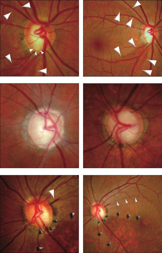
tions with the common feature of an optic neuropathy (i.e., that is open, closed or structurally abnormal. Classi-
glaucomatous optic neuropathy [GON]) characterized fication of the glaucoma on the basis of the appear-
by a distinctive loss of retinal nerve fibres and optic disc ance of the angle on gonioscopy will guide appropriate
changes. Loss of this neural tissue can lead to an irreversible management [Consensus ].
loss of visual field (VF), usually beginning paracentrally,
but becoming complete if the disease is unchecked. (See Primary open-angle glaucoma
Appendices B and C.) GON can develop under a num- Primary open-angle glaucoma (POAG) occurs when
ber of circumstances with varying contributions by several there is GON with or without elevated IOP. Population
known and as yet unidentified risk factors. The clinical surveys have shown that up to 61% of patients with POAG
term glaucoma is sometimes used when 1 risk factor, ele- have a single screening IOPCOS glaucoma clinical practice guidelines
the inferior and superior poles of the disc. There may be refers to a primary narrow angle without angle closure and
diffuse or focal loss of the retinal nerve fibre layer (RNFL), without elevated IOP. The anterior chamber depth is gen-
and hemorrhages can be found within the nerve fibre lay- erally not much shallower centrally compared to normal,
er on or adjacent to the disc margin. POAG is generally but very shallow peripherally.23 Plateau iris syndrome is
bilateral, but asymmetry of the disc findings is common. diagnosed when angle closure occurs either spontaneously
Documented progression of these disc findings confirms or with pharmacologic dilation in the presence of a patent
the diagnosis of glaucoma. iridotomy. Ultrasound biomicroscopy has shown that the
There are generally no symptoms of POAG until very ciliary processes are typically positioned forward of their
late in the disease when fixation is threatened.21 The VF normal position, bolstering the peripheral iris.24
damage follows the anatomy of the RNFL. The VF defects
of glaucoma occur in an arcuate distribution and align Secondary open-angle glaucomas
themselves along the horizontal raphe. They usually de- The most common open-angle glaucoma with an
velop more nasally than temporally, and often affect one identifiable cause is pseudoexfoliation (PXF) material in
hemifield (superior or inferior) more than the other. PXF glaucoma.25 It should also be recognized that ap-
proximately 9%–18% of eyes with PXF glaucoma have
Primary open-angle glaucoma suspects elevated IOP due to an angle-closure mechanism.26,27 Pig-
An individual is a suspect for POAG when he or she is ment dispersion is another frequent cause of ocular hyper-
found on history and clinical examination to have optic tension and open-angle glaucoma, particularly in young,
disc features suspicious for GON, suggestive VF defects myopic males.28,29 Other particulate material, such as red
or a constellation of risk factors for POAG that confer a or white blood cells, ghost cells, macrophages, and tu-
heightened probability of developing the disease. People mour cells may be responsible for plugging the trabecular
with elevated IOP (>21 mm Hg), but with no evidence meshwork. Young males are also the most frequent group
of GON or glaucomatous VF damage, would qualify as seen to develop angle-recession glaucoma following blunt
POAG suspects on the basis of having ocular hypertension. trauma.30 Prolonged exposure to topical or systemic corti-
costeroids is also a well-known cause of secondary open-
Primary angle-closure glaucoma angle glaucoma.31
Angle-closure glaucoma is considered primary when the
reason for the appositional or synechial closure of the angle Secondary angle-closure glaucomas
is due to pupillary block. The mechanism of the pupillary Neovascular glaucoma is a classic example of a second-
block must be in the context of normal anatomical struc- ary angle-closure glaucoma. It begins with new vessels
tures. Pupillary block secondary to identifiable causes such growing into and over the open trabecular meshwork;
as posterior synechiae or a subluxated lens is classified as during the cicatricial phase of the process, the iris root is
secondary angle closure. drawn up to the cornea to seal off the angle completely.32
On clinical examination, the anterior chamber appears Other well-accepted secondary causes of angle closure in-
shallow, and minimal to no angle structures are seen on clude a large or prolapsed/subluxated lens, suprachoroidal
gonioscopy. Appositional versus synechial angle closure can effusion/hemorrhage, aqueous misdirection, and anterior
be distinguished by indentation gonioscopy with a small displacement of the iris root due to a posterior pole tu-
surface area goniolens (e.g., Posner, Sussmann, or Zeiss). mour.15,16,33 Some causes of secondary angle-closure glau-
Primary angle-closure glaucoma (PACG) can be acute coma, such as uveitis and following intraocular surgery,
or chronic. If the iris root obstructs the outflow of aqueous can also cause a raised IOP and GON in the context of an
through the iridocorneal angle abruptly, acute angle closure open angle. Differentiation between the 2 mechanisms, to
is said to occur. In fact, unless the resulting high IOP causes explain the raised IOP and (or) glaucoma, is only possible
lasting damage to the optic nerve, GON may not develop. with careful gonioscopy.
Other mechanisms to explain the pathogenesis of angle
closure have recently been proposed.22 The damage to the Recommendations
optic disc and VF with PACG (whether acute or chronic)
may be indistinguishable from POAG. 2. As many secondary glaucomas (e.g., pseudoexfoliation,
neovascularisation, uveitis, and surgical trauma) can
Primary angle-closure suspect occur with angles that are either open or closed, careful
Patients may be considered suspect for developing PACG gonioscopy is required to clarify the pathogenesis and
if they have a potentially occludable angle as determined by management options [Consensus ].
gonioscopy. (See page S22.)
Mixed- and multiple-mechanism glaucomas
Plateau iris configuration and plateau iris syndrome The expression mixed-mechanism or combined glaucoma
In plateau iris the iris profile is flat, or like a plateau, with has been used to explain a variety of scenarios with little
the iris root crowding the angle. Plateau iris configuration agreement in the literature. Both open and closed angles
CAN J OPHTHALMOL—VOL. 44, SUPPL. 1, 2009 S9COS glaucoma clinical practice guidelines
are at play in the elevation of IOP. One reasonable appli- Pharmacoeconomics of glaucoma
cation of this term would be to describe an eye with ele- The societal and economic burden of living with and treat-
vated IOP and a closed angle that undergoes a peripheral ing glaucoma can be measured and modelled in a number
iridotomy to relieve pupillary block. With an angle that is of different ways. Commonly applied pharmacoeconomic
now clearly open, if the IOP were to go down partially, but models evaluate such endpoints as cost-effectiveness, cost-
remain outside the normal range, then a combined open- minimization, cost-consequence, and budget-impact an-
and closed-angle mechanism for the elevated IOP might alyses.43 The total costs of treatment, custodial care, and
be presumed.34 For practical purposes, mixed-mechanism special education are relatively easy to determine when the
glaucoma is a term that rarely would be used. data of major third-party payers (both private and public)
Multiple-mechanism glaucomas occur when the cause of can be accessed. In contrast, the determination of produc-
the elevated IOP is multifactorial with a number of pos- tivity losses by the patient, and nonremunerated caregivers,
sible influences of either, or both, open- and closed-angle is much more difficult to calculate. The pharmacoeconom-
mechanisms. A common example is the patient with iritis, ics of glaucoma management may become clearer once the
an elevated IOP, and an additional IOP response to the outcome measures of vision preservation and visual per-
corticosteroid treatment. Another scenario might involve a formance are better studied.
patient with known PXF glaucoma, or POAG, who suffers
a central retinal vein occlusion and subsequent neovascu- Screening for glaucoma
larisation of the anterior segment.
Due to the irreversible nature of GON, it might be ex-
Epidemiology and burden of blindness pected that intervention would be most effective if initiated
After cataract, glaucoma is the second leading cause of early. The asymptomatic nature of glaucoma would suggest
blindness worldwide. It is, however, the number one cause a routine screening program might serve to uncover the dis-
of irreversible vision loss.35 The prevalence of the various ease. Population screening for a disease must satisfy a few
types of glaucoma follows racial and ethnic boundaries. important criteria as outlined by Wilson and Jungner:44
PACG is more common in Asian people. As of 2001, it 1. The disease must have a known natural history, a pre-
was calculated that 4.5 million people were suffering from clinical phase that can be identified, and a progressive
PACG in China.36 Global projections estimate that by clinical course in which later stages lead to worsening
2010 there will be approximately 15.7 million people with of symptoms;
PACG, with half of them in China.37 2. The different tests used must be valid, reproducible,
POAG alone is believed to account for approximately acceptable, easy to perform, must have good sensitivity
12% of all blindness in the world and 32% of the blind- and specificity, a high positive predictive value, and do
ness in those of African descent.38 In the United States the so with an appreciable cost-benefit; and
prevalence of POAG in all people >40 years old was de- 3. The treatment must be available and effective.
termined to be 1.86%.39 It is suggested that only 10% of Canada has not instituted a population screening program
all glaucoma in the United States is PACG, but that up for glaucoma although it has been given serious considera-
to 20% might be secondary types.40 Presumably, common tion in the past.45 This has been reviewed comprehensively
secondary glaucomas, such as those due to PXF material, by Einarson et al.46 in light of newer understanding about
would feature significantly in this portion. This has been the nature of the disease, the risk factors amenable to screen-
inadequately documented. ing, and the effectiveness of treatment. We now know, for
There have been few epidemiologic studies on glaucoma example, that aggressively lowering IOP can reduce con-
and its subtypes in Canada. There may be approximately version of ocular hypertension to glaucoma47 and slow the
409 00041 people with glaucoma in this country. A meta- progression of the disease once established.11,12,48,49 This
analysis of 5 self-report surveys indicated that in 2002 and document will review some of the newer technologies that
2003 the prevalence of glaucoma was 2.7% for Canadians demonstrate high degrees of sensitivity, specificity, and
aged ≥40 years and 11% for those ≥80 years.41 This could, positive predictive values for early structural and functional
however, be an overestimate, as self-report surveys can in- damage. The cost-effectiveness of screening the general
clude individuals with ocular hypertension receiving treat- population, however, has not been established.50,51 It is like-
ment. One office-based survey of 962 charts in a small ly (and deserving of further study) that screening high-risk
British Columbia city showed that glaucoma was respon- populations would be cost-effective.52–55
sible for a relatively small percentage of the patients with Aside from the costs, another concern about screening
low vision and blindness (using World Health Organization programs is the additional strain on manpower that might
[WHO] criteria) compared to cataract, macular degenera- be generated. It may be more efficient to utilize and co-
tion, and other retinal diseases.42 Due to the asymptomatic ordinate the skills of a wide range of professionals including
nature of chronic glaucoma, it is surmised that up to 50% ophthalmologists, optometrists, ophthalmic technologists,
of those with glaucoma in the industrialized world are un- and other community healthcare providers. Telemedicine
aware of it and are not receiving care.9,10 may significantly reduce travel time and costs for patients.
S10 CAN J OPHTHALMOL—VOL. 44, SUPPL. 1, 2009COS glaucoma clinical practice guidelines
Finally, the media could assist in educating the public about Oculokinetic perimetry has been found to have different
individuals at risk (i.e., those to whom the screening pro- sensitivities (75%68 and 86%69) and specificities (56.1%,70
gram is directed). 65%,68 and 94%71) depending on the study. These values
The primary purpose of screening is to identify individ- indicate that neither is sufficient for use by itself in a glau-
uals who are at sufficient risk to require complete evalua- coma screening program.71
tion. In that regard, a patient’s risk factors (see below) may
be the most useful way to identify such patients, emphasiz- Structural tests
ing the need for public education. Subjective examination of the optic disc and RNFL
remains one of the most important steps in diagnosing
Risk factors, natural history and clinical course glaucoma. Recent technological advances, however, have
The primary risk factors for POAG are elevated IOP, ad- produced many new instruments that can objectively
vancing age, family history of glaucoma, and race. Tuck et quantify the optic nerve head (ONH) and the RNFL.
al.56 have described an increasing prevalence with increas- Confocal scanning laser ophthalmoscopy (CSLO), op-
ing age, especially after the age of 55. Their study showed tical coherence tomography (OCT), and scanning laser
a POAG prevalence of 1.2% in a Caucasian population polarimetry (SLP) may, in some cases, permit earlier
between ages 40–89 years. When divided into subgroups, glaucoma diagnosis. Therefore, their role in screening,
the over-80 age group had the highest POAG prevalence especially when coupled with telemedicine, appears
(4.3%) compared with that of the younger age groups. In- promising and remains to be studied. A summary of 3
vestigators in the Blue Mountains Eye Study,10 which exam- technologies is found in the section on diagnostic tests.
ined POAG prevalence in Australia, found an even higher (See page S15.)
prevalence in the same over-80 age group (8.17%). Other imaging systems, such as RNFL photography
and imaging cameras, have also been developed and are
Validity of glaucoma screening tests being investigated.72–75 Finally, telemedicine can contrib-
ute to glaucoma screening by allowing specialists who are
Tonometry not on location to participate in the screening process.76–78
Until recently, glaucoma diagnosis was based on the pres- This mode of screening is well received by patients as
ence of a combination of high IOP, optic nerve cupping, shown by Tuulonen et al.76 whose study showed that 96%
and VF damage. Nevertheless, up to 50% of patients with of patients preferred to have their next visit at a health
glaucoma may have a normal IOP.38 Moreover, high IOP clinic closer to home.
can also be seen in patients with ocular hypertension, the
majority of whom may never develop glaucoma.47,57 Treatment effectiveness
Glaucoma treatment satisfies another important screen-
Visual function tests ing criterion, namely that treatment given before symptoms
VF defects, detected with standard automated perim- develop is beneficial in reducing morbidity. If managed
etry (SAP), are the mainstay of demonstrating functional accordingly, with new medications, laser and surgical
glaucomatous damage. It is now suggested that up to 35%– procedures achieving a significant reduction in IOP, the
50% of nerve fibre axons may already be damaged before progression of glaucoma can be slowed in the majority of
SAP detects any functional VF loss.58 Frequency-doubling patients.11,12,48,49
technology (FDT) perimetry may offer certain advantages
for early glaucoma detection, in part due to its short patient Cost-effectiveness and implementation of a
examination time.59 FDT perimetry is thought to detect screening program in Canada
functional loss earlier than SAP by stimulating the specific Several studies have sought to estimate the impact of glau-
M-y ganglion cells of the magnocellular pathway, which are coma on various aspects and levels of healthcare costs,79–90 as
thought to be damaged in early glaucoma.60,61 Several inves- well as specifically in Canada,91 and have shown that costs
tigators have found that FDT perimetry has both sensitiv- are highest in the first year after diagnosis and increase with
ity and specificity in the 90th percentile range, making it a the severity of the disease on an annualized basis.
potentially useful screening tool.62–65
Short wavelength automated perimetry (SWAP), or blue- Recommendations
on-yellow perimetry, is another VF testing technique that
may also detect glaucomatous damage earlier than SAP.66 3. It is recommended that population screening can
However, FDT perimetry is a faster test and a study com- be considered for high-risk populations. Screening
paring all 3 methods (SAP, SWAP, and FDT perimetry) should include both structural and functional meas-
found FDT able to detect glaucomatous VF defects the ear- ures of the disease. Screening for IOP alone should be
liest.67 Other existing tools have been studied with regard avoided since it has low sensitivity, low specificity, and
to VF testing. The Damato campimeter has been found poor predictive value for the detection of glaucomas
to have a sensitivity of 50% and a specificity of 90%.58 [Level 1 8–10].
CAN J OPHTHALMOL—VOL. 44, SUPPL. 1, 2009 S11COS glaucoma clinical practice guidelines
Diagnosis of glaucoma gery etc.), a history of methanol poisoning, or an ischemic
or compressive neuropathy.93
Diagnosis Since family history of glaucoma in first-degree relatives
The essential elements of a comprehensive eye examina- is a recognized risk factor for the development of glaucoma,
tion form the basis of an examination for glaucoma.92 Glau- this history should be specifically sought, including details
coma by definition is an optic neuropathy and therefore such as age of onset and disease severity or visual loss from
specific attention should be directed to the evaluation of glaucoma.94–100
the optic nerve. In the diagnosis and management of glau- Subjective symptoms and impact of glaucoma and its
coma, it is also important to identify potential risk factors treatment on quality of life (QOL) need to be addressed in
for glaucoma, the possibility of secondary glaucomas, con- order to direct patients to appropriate community services.
comitant systemic diseases, medications, and finally sub- Where possible, information regarding previous glaucoma
jective symptoms. treatment, IOP, optic nerve appearance and VFs should
be obtained. (A sample referral letter is provided in Ap-
History pendix D.) For patients currently receiving treatment for
Table 292 lists the essential elements of a comprehensive glaucoma, information regarding the levels of IOP prior
glaucoma eye examination. When tailoring this examina- to treatment should be obtained and adherence to therapy
tion to the glaucoma patient, attention should be given to should be determined.
concomitant systemic diseases and medications that may
either influence disease (e.g., corticosteroid use) or affect Recommendations
treatment decisions (e.g., beta antagonists contraindi-
cated with asthma). In suspected cases of normal pressure 4. Specific information related to concomitant systemic
glaucoma (NPG), additional details such as vascular dys- diseases and medications that may influence glaucoma
function, including a history of migraines, Raynaud phe- treatment should be sought [Consensus ].
nomenon, and other antecedent events that could have 5. When considering the diagnosis of glaucoma, par-
resulted in cupping are important. NPG is a diagnosis of ticularly when IOPs are in the normal range, specific
exclusion. Pertinent antecedent events include a history of inquiry should be made with regard to antecedent
significant blood loss, major hypotension during any sur- events that could have resulted in cupping and (or)
gery, previous elevation in IOP (steroids, trauma, postsur- optic atrophy [Level 4 93].
Risk factors
Table 2—Essential elements of the comprehensive glaucoma
eye examination
A risk factor is a characteristic that is positively associated
Element Criteria
with development of a disease. As our knowledge of risk
History Patient name, date of birth, gender, and race
Driving status
Vocation and avocations
Table 3—Risk factors and signs for presence of open-angle
Chief complaint, if any (e.g., any perceived visual
glaucoma with level 1 evidence
handicap)
Current medication and allergies (ocular and systemic) Ocular risk factors and signs
Ocular history IOP
Medical history Elevated baseline IOP9,94,110
Medical and ocular family history (including family Optic disc
history of glaucoma) Deviation from the ISNT rule* 111–113
Directed review of systems Increased optic disc diameter114,115
Clinical examination Best corrected distance visual acuity with refraction Parapapillary atrophy116,117
and investigations documented Disc hemorrhage118,119
Pupillary reaction, relative afferent pupillary defect PXF120
Automated perimetry Thinner CCT20,121–123
Slit lamp examination of lids, lid margins, conjunctiva, Pigment dispersion124
cornea, anterior chamber (clarity and depth), lens
Myopia125,126
IOP and time of measurement
Decreased ocular perfusion pressure7
CCT101,102
Non-ocular risk factors
Gonioscopy103,104
Increasing age97,127–129
Dilated examination of:
African descent9,39,130–132
Lens
Hispanic ancestry40
Biomicroscopy of ONH and RNF105 including objective
documentation such as optic disc imaging Family history94–100
Fundus Genetics
Discussion with patient Discussion of findings with appropriate correction and Myocillin133
mitigating strategy Optineurin134
Counselling with respect to QOL issues (e.g. low Apolipoprotein135,136
vision rehabilitation, adherence) Migraine137–139
Follow-up recommendation Corticosteroids140,141
Adapted from the Canadian Ophthalmological Society clinical practice guidelines for the *ISNT rule; majority of normal optic discs with neuroretinal rims with descending order of
periodic eye examination in adults in Canada92,106–109 thickness—inferior, superior, nasal, temporal.
S12 CAN J OPHTHALMOL—VOL. 44, SUPPL. 1, 2009Vous pouvez aussi lire