Impact de la stimulation cérébrale transcrânienne sur l'apprentissage d'un jeu vidéo - CORTICO
←
→
Transcription du contenu de la page
Si votre navigateur ne rend pas la page correctement, lisez s'il vous plaît le contenu de la page ci-dessous
Impact de la stimulation cérébrale transcrânienne sur
                                          l’apprentissage d’un jeu vidéo

                   Chenot Q.1, Besson P.2, De Boissezon X.3, Scannella S.1, Perrey S.2
1
    ISAE-SUPAERO, Université de Toulouse, France.
2
    EuroMov, Univ. Montpellier, France.
3
    UMR 1214-Inserm/UPS-ToNIC, Toulouse NeuroImaging Center, Université de Toulouse, Hôpital Purpan, France.

Introduction

La tRNS (transcranial Random Noise Stimulation) est une technique de neuromodulation relativement récente
consistant à envoyer un faible courant alternatif d’intensité aléatoire au niveau du scalp afin de moduler la
plasticité cérébrale. Cette technique potentialise en théorie les effets d’entraînements cognitifs (Elmasry et al.,
2015). L’objectif de cette étude a été de tester les effets de la tRNS sur l’apprentissage d’un jeu vidéo mettant en
jeu les fonctions exécutives.

Méthode
Dix participants (23,8 ± 5,6 ans ; 4 femmes) ont été recrutés et répartis en deux groupes de 5 (tRNS vs SHAM).
Chaque participant a effectué 6 séances de 20 mn de jeu vidéo Space Fortress (Fig. 1, Mané & Donchin, 1989).
La performance était mesurée via le score obtenu à l’issue du jeu. Trois séances d’évaluation (référence, court-
terme et long-terme) et trois séances d’entraînement avec stimulation tRNS (1 mA) du cortex préfrontal
dorsolatéral droit ont été réalisées (Fig. 2).

Résultats
Nos résultats n’ont pas montré de différence entre les deux groupes pour l’apprentissage et pour l’évaluation à
court-terme. En revanche, le groupe tRNS a obtenu de meilleures performances lors de l’évaluation à long-terme
(p < 0,05 ; d de Cohen = 1,55 ; Fig. 3).

Discussion
Bien que notre échantillon constitue une limite, ces résultats préliminaires sont en faveur d’un apport de la tRNS
lorsqu’elle est appliquée pendant l’entraînement, avec un meilleur maintien des acquis de cet apprentissage sur le
long-terme. Ce résultat est en accord avec d’autres études ayant montré un gain dans le maintien des
apprentissages, aussi bien cognitifs et moteurs avec d’autres outils de stimulation cérébrale (Enriquez-Geppert et
al., 2013). En conclusion, la tRNS apparaît être une technique intéressante pour stimuler la plasticité cérébrale, et
nos résultats soulignent l’impact que pourrait avoir la tRNS sur les apprentissages longs.

Références

•     Elmasry, J., Loo, C., & Martin, D. (2015). A systematic review of transcranial electrical stimulation combined with
      cognitive training. Restorative neurology and neuroscience, 33(3), 263-278.
•     Enriquez-Geppert, S., Huster, R. J., & Herrmann, C. S. (2013). Boosting brain functions: Improving executive
      functions with behavioral training, neurostimulation, and neurofeedback. International Journal of
•     Mané, A., & Donchin, E. (1989). The space fortress game. Acta psychologica, 71(1-3), 17-22.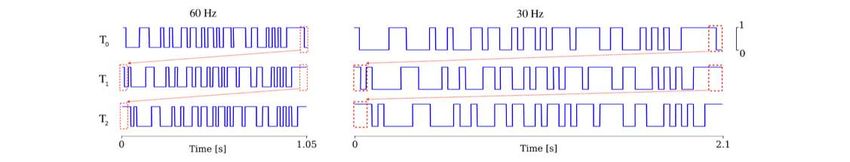
A Matlab GUI to optimize features selection with OpenViBE
              Juliana González Astudillo, Marie-Constance Corsi, Laurent Hugueville
                                   et Fabrizio De Vico Fallani.

In the last years many softwares have been developed to design EEG-based brain-computer interfaces (BCIs).
One of the most widely used is OpenViBE [1] , [2] due to its flexibility in handling the processing pipeline. In
motor imagery (MI)-based BCIs, the features selection is based on spatial patterns and frequency bands. To ensure
the best discrimination between mental states, those features must be adjusted to the target exercise and, most
importantly, to the subjects' specificity e.g. in terms of spectral patterns [3] and adopted strategies [4] .

The OpenViBE features selection block relies on two fixed channels (C3 and C4) within the α-β frequency range
[5] . Thus, we could miss relevant information since these features might not be the most appropriate for the target
subject. To address this issue, we developed a Matlab-based GUI to find the features that best differentiate two
mental states. For that purpose, the coefficient of determination, r2 , is computed between classes as a function of
frequency and channels to elicit the most suitable features.

We finally tested the usefulness of the developed GUI on an OpenViBE dataset and on EEG recordings from a
MI BCI experiment performed in our laboratory. For the first dataset, r2 values suggested that there was a possible
finer selection compared to the default one. Channel CP4 showed a more remarkable difference between
conditions. Notably, selecting its associated features threw better accuracy (70%) than the combination of C3 and
C4 (63%). We obtained similar results when we considered our experimental data. The strongest features differed
from the fixed ones, giving 92% accuracy for r2 selection (in this case, C3 and CP4) and 85% for the OpenViBE
selection. These results demonstrate the usability of our developed tool as an OpenViBE complement for MI
protocol.

References

. [1] Y. Renard et al. , “OpenViBE: An Open-Source Software Platform to Design, Test and Use Brain-
       Computer Interfaces in Real and Virtual Environments,” Presence Teleoperators Virtual Environ. , vol.
       19, no. 1, pp. 35–53, Apr. 2010.
. [2] F. Lotte, L. Bougrain, and M. Clerc, “Electroencephalography (EEG)-based Brain-Computer Interfaces,”
       p. 44, 2015.
. [3] V. Attina, E. Maby, R. Bouet, J. Gibert, J. Mattout, and O. Bertrand, “The importance of individual
       features for motor-imagery based BCI,” presented at the 4th International Brain-Computer Interface
       Workshop and Training Course, 2008.
. [4] C. Neuper, R. Scherer, M. Reiner, and G. Pfurtscheller, “Imagery of motor actions: differential effects of
       kinesthetic and visual-motor mode of imagery in single-trial EEG,” Brain Res. Cogn. Brain Res. , vol.
       25, no. 3, pp. 668–677, Dec. 2005.
. [5] G. Pfurtscheller and C. Neuper, “Motor imagery and direct brain-computer communication,” Proc. IEEE
, vol. 89, no. 7, pp. 1123–1134, Jul. 2001. 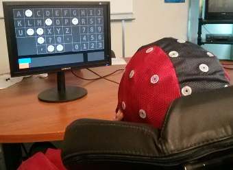
DESIGN AND PRELIMINARY STUDY OF A NEUROFEEDBACK
                    PROTOCOL TO REDUCE DROWSINESS

        Thibaut Monseigne1,3, Fabien Lotte1,2, Stephanie Bioulac3,4, Jean-Arthur Micoulaud-
                                   Franchi3,4, Pierre Philip3,4
1
    Inria Bordeaux - Sud-Ouest, Talence, France
2
    LaBRI - (CNRS / Univ. Bordeaux / Bordeaux INP), Talence, France
3
    SANPSY (CNRS / Univ. Bordeaux), USR 3413, Bordeaux, France
4
    Clinique du sommeil, Hôpital Pellegrin-Tripode, Bordeaux, France

E-mail: thibaut.monseigne@inria.fr

NeuroFeedback (NF) consists in using electroencephalographic (EEG) measurements to guide users to perform a
cognitive learning using information coming from their own brain activity, by means of a real- time sensory
feedback (e.g., visual or auditory)[4].
Many NF approaches have been studied to improve attentional abilities, notably for Attention Deficit
Hyperactivity Disorder [1, 2]. However, to our knowledge, no NF solution has been proposed to specifically
reduce drowsiness.
Thus, we propose a complete EEG-NF solution to train users to self-regulate an EEG marker of drowsiness. This
marker is based on a ratio of beta over theta/alpha power in Cz electrode. In addition to this EEG marker of
drowsiness, we also carefully selected and designed the duration, the sequencing, the objective evaluation metrics
and the visual and audio feedback to use in for each NF session.
Preliminary study with five healthy subjects showed that three of them could learn to self-regulate this EEG
marker with a relatively short number of NF sessions (up to 8 sessions of 40 min). Clinical trials with sleep-
deprived subjects are expected to begin in 2019 to study possible cognitive and clinical benefits of this self-
regulation. The implementation of this NF solution is available for free1, with the OpenViBE platform [3], under
the AGPL-3.0 license.

References

[1] Micoulaud-Franchi J.-A., Geoffroy P. A., Fond G., Lopez R., Bioulac S., Philip P. EEG neurofeedback
treatments in children with ADHD: an updated meta-analysis of randomized controlled trials. Frontiers in
Human Neuroscience. 2014;8.
[2] Micoulaud-Franchi J.-A., McGonigal A., Lopez R., Daudet C., Kotwas I., Bartolomei F. Electroen-
cephalographic neurofeedback: Level of evidence in mental and brain disorders and suggestions for good
clinical practice. Neurophysiologie Clinique/Clinical Neurophysiology. 2015;45(6):423–433.
[3] RenardY.et al.OpenViBE:AnOpen-SourceSoftwarePlatformtoDesign,TestandUseBrain-Computer Interfaces
in Real and Virtual Environments. Presence: Teleoperators and Virtual Environments. 2010;19(1):35– 53.
[4] Sitaram R. et al. Closed-loop brain training: the science of neurofeedback. Vol. 18. Nature Reviews
Neuroscience. Nature Publishing Group, 2016.
1
    https://github.com/tmonseigne/NEUROPERF
Technologie Interface Cerveau Ordinateur pour l’Autonomie:
Design ergonomique d’un casque EEG
Sofiane Guebba 2 - Violaine Guy 2,3 - Théodore Papadopoulo 1,2 Marianne Bruno 2,3 -
Maureen Clerc 1,2 - Marie-Hélène Soriani 2,3
1- Inra Sophia Antipolis-Méditerranée
2- Université Côte d’Azur
3- CRMR SLA et autres maladies du neurone moteur - Hôpital Pasteur 2 - CHU de Nice
Dans le cadre d’un axe de recherche transversal INRIA Sophia-Antipolis-CHU de Nice sur les ICO, il a récemment
été démontré qu’un prototype de P300 Speller développé par l’INRIA permet une communication alternative efficace chez
des patients en situation de handicap (SLA) [1]. Pour être adapté à une utilisation quotidienne répétée et/ou de longue durée
par des personnes en situation de handicap moteur lourd avec déficit du tonus cervical obligeant un appui occipital (voir
Fig.1), le prototype de P300 Speller doit évoluer vers un système d’acquisition simple et ergonomique [2]. Bien que divers
systèmes soient actuellement disponibles sur le marché, ils ne répondent pas ou tr ́es peu a` l’hétérogénéité des utilisateurs,
a` l’adaptabilité nécessaire aux capacités physiques et a` l’environnement parfois encombré d’appareils médicaux lourds
pouvant créer des interférences comme par exemple la ventilation sur trachéotomie. Le projet actuel a pour but de
développer un casque EEG sans fil, avec un nombre d’électrodes adapté [2]-[3] pour garantir a` la fois la qualité du signal
et le confort, utilisant des électrodes sèches (voir Fig.2) alliant ainsi une répartition homogène de la pression de contact
scalp-électrode [4] et une pénétration de différents types de cheveux afin de minimiser les artefacts présents sur les
technologies d’ électrodes sèches. Au cœur des objectifs du design se trouve l’alliance de fonctionnalité et d’esthétisme,
soit être léger, confortable et personnalisable, nécessitant ainsi l’utilisation de matériaux adaptés pour un bon maintien sur
différentes morphologies de tête et une pression optimale sur les électrodes [5]. Ce projet justifie donc une approche
transversale regroupant plusieurs domaines de compétence : mécanique des matériaux et électronique (ingénieur
mécatronicien), analyse du signal et adaptation des logiciels (INRIA) et expertise dans le domaine du handicap (CHU).
Figure 1: Test étude préliminaire, textile waveguard original caps ANT B.V (Enschede, Netherlands) et électrodes à gel, plis induits
par posture SLA
Figure 2: Illustration d’une électrode sèche multi-pins, polyurethane dureté Shore A98 (ANT B.V), avec dimensionsReferences
. [1] Violaine Guy, Marie-Helene Soriani, Mariane Bruno, Theodore Papadopoulo, Claude Desnuelle, and Maureen
Clerc. “Brain computer interface with the P300 speller: usability for disabled people with amyotrophic lateral
sclerosis”. In: Annals of physical and rehabilitation medicine 61.1 (2018), pp. 5–11.
. [2] Michael T McCann, David E Thompson, Zeeshan H Syed, and Jane E Huggins. “Electrode subset selection
methods for an EEG-based P300 brain-computer interface”. In: Disability and Rehabilitation: Assistive
Technology 10.3 (2015), pp. 216–220.
. [3] Yuki Ijichi and Hisaya Tanaka. “Electrodes arrangement on brain-computer interface for the ALS’s posture”. In:
2016 IEEE International Conference on Systems, Man, and Cybernetics (SMC). IEEE. 2016, pp. 004712–004715.
. [4] Patrique Fiedler et al. “Contact pressure and flexibility of multipin dry EEG electrodes”. In: IEEE Transactions on
Neural Systems and Rehabilitation Engineering 26.4 (2018), pp. 750–757.
. [5] Dani ̈el Lacko. “The application of 3D anthropometry for the development of headgear: a case study on the design
of ergonomic brain-computer interface devices”. PhD thesis. University of Antwerp, 2017. Leveraging BCI performances with the integration of connectivity
and local features
1,2 2 1 4,5,6
Tiziana Cattai , Stefania Colonnese , Marie-Constance Corsi , Danielle S. Bassett ,
2 1
Gaetano Scarano , Fabrizio De Vico Fallani
1
ARAMIS Lab, ICM, Inserm U1127, CNRS UMR 7225, Sorbonne Université, Inria, Paris, France
2
Dept. Of Information Engineering, Electronics and Telecommunication, Sapienza University of Rome
3
Dept. of Bioengineering, University of Pennsylvania, Philadelphia, PA, 19104, USA
4
Dept. of Electrical and Systems Engineering, University of Pennsylvania, Philadelphia, PA, 19104, USA
5
Dept. of Physics & Astronomy, University of Pennsylvania, Philadelphia, PA, 19104, USA
6
Dept. of Neurology, Hospital of the University of Pennsylvania, Philadelphia, PA, 19104, USA
In brain-computer interfaces (BCI), the detection of different mental states is a key element. In Motor Imagery
(MI)-based BCIs, the considered features typically rely on the power spectral density (PSD) of the control brain
signals, but alternative features can be explored looking for better performance. One possibility is the integration
of functional connectivity (FC) [1]. These features quantify the interactions between different brain areas that
could represent a valuable tool to detect differences between two mental conditions. Here, we investigated the
behavior of coherence-based FC features and PSD features, alone and in combination [2].
For a better comparison, we characterized the network centrality of each brain area by computing the weighted
node degrees from the estimated FC networks [3].To classify the subjects’ mental state, we used the linear
discriminant analysis (LDA) method Our classifier operates on single subject, single frequency bin and single
channel.For each subject, we selected the best value in terms of classification performances (e.g. accuracy)
comprised in the central left area. The best accuracy value is associated with a given (electrode; frequency bin)
couple.Our results showed that alpha and beta frequency bands are usually more involved during the task. In
fact, the accuracy obtained in those bands are higher. Another interesting result is that the fusion between
connectivity feature and a local one gives higher accuracy for the majority of the subject, for 12 over 15. This
means that the integration of features reflecting different brain mechanisms can be useful for the discrimination
of different mental states [4].
Références
[1]Hamedi, Mahyar, Sh-Hussain Salleh, and Alias Mohd Noor. "Electroencephalographic motor imagery brain
connectivity analysis for BCI: a review." Neural computation 28.6 (2016): 999-1041.
[2]Ruta, Dymitr, and Bogdan Gabrys. "An overview of classifier fusion methods." Computing and Information systems
7.1 (2000): 1-10.
[3]De Vico Fallani, Fabrizio, et al. "Graph analysis of functional brain networks: practical issues in translational
neuroscience." Philosophical Transactions of the Royal Society B: Biological Sciences 369.1653 (2014): 20130521.
[4]Corsi, M. C., et al. "Integrating EEG and MEG Signals to Improve Motor Imagery Classification in Brain-Computer
Interface." International journal of neural systems (2018): 1850014-1850014.Towards Measuring states of curiosity through Electroencephalography
and body sensors responses
Aurélien Appriou, Jessy Ceha, Edith Law, Pierre-Yves Oudeyer et Fabien Lotte.
The neurophysiological mechanisms underlying curiosity and intrinsic motivation are currently not well
understood. However, being able to identify objectively, from neurophysiological signals, the curiosity level of a
user, would bring a very useful tool both to neuroscientists and psychologists, to understand curiosity deeper, as
well as to designers of human-computer interaction, in order to trigger curiosity or to adapt an interaction to the
curiosity levels of its users. A first step to do that, is to collect neurophysiological signals during known states of
curiosity, in order to develop signal processing/machine learning tools to recognize those states from such signals.
We propose an experimental protocol, that has been designed but has not been tested so far, in order to
measure both brain activity through Electroencephalography (EEG) and physiological responses (heart rate, skin
conductance, Electrocardiogram) when subjects are induced into different states of curiosity. During the
experiment, fun facts will be presented to subjects to induce different levels of curiosity. We obtained those fun
facts using the Google functionality "I’m feeling curious" as well as crowdsourcing. A subject will be able to
choose a fun fact that makes him curious, and push forward with a 4-to-10 questions chain on this theme. For
each question on a given theme, a subject will be able to reveal the answer (interpreted as a curious state) or to
skip it (interpreted as a non-curious state). Skipping an answer will automatically break the chain and will point
the subject to the next fun fact. Neurophysiological signals will be collected between a question and the choice
of revealing the answer. Then the subject will grade the question on a 1-to-7 curiosity level scale.
Neurophysiological measures during these states of curiosity will be recorded and we expect to find biological
markers of curiosity by analyzing such information.Towards a low-cost EEG band to induce Lucid Dream.
Morgane Hamon, Emma Chabani, Philippe Giraudeau
Lucid dreaming (LD) is a phenomenon during which the person is aware that he/she dreaming
and is able to control the dream content (Laberge, 1985). Studies have shown that only 20%
of people can experience lucid dreams on a regular basis (Snyder & Gackenbach, 1988).
However, LD frequency can be increased through induction techniques (LaBerge 1988).
External stimulation technique relies on the ability to integrate external information into the
dream content (Dement and al. 1958). The aim is to remind the sleeper that she/he is dreaming.
If this type of protocol is not fully efficient, it demonstrates how sensorial stimuli can be easily
incorporated into people's dreams (Paul and al. 2014). The objective of our project was to
replicate this induction technique using material less expensive and more portable. This
material could simplify experimental procedures. Participants could bring the material home,
then have a more ecological night. First, we used the OpenBCI cython, a low-cost EEG signal
acquisition board in order to record and manually live- score sleep. Then, we designed a mask
containing two LEDs, connected to a microcontroller to flash visual stimulation during sleep.
We asked two volunteers to sleep for 2 hours in a dedicated room. One of the participants
declares having a dream during which the blue lights diffused by the mask were embedded
into the dream environment. The other participant woke up during the visual stimulation.
These results are congruent with previous studies (Paul and al. 2014). This work marked the
first step of a larger project. Our ongoing research includes the use of an online sleep stage
scoring tool and the possibility to automatically send stimuli according to the sleep stage. We
will also investigate other types of stimulus induction in the future such as vibro-tactile
stimulation that showed great potentials (Stumbrys and al. 2012).
Keywords: Lucid Dream, Induction techniques, Sleep, Visual stimulation, Low-cost EEGTHE USE OF HAPTIC FEEDBACK IN BRAIN COMPUTER INTERFACES
AND NEUROFEEDBACK
M. Fleury 1, A. Lecuyer 2, C. Barillot 3
1
Univ Rennes, Inria, CNRS, Inserm, IRISA, EMPENN ERL U1228, Hybrid Project Team, F-35000, Rennes, France
2
Univ Rennes, Inria, CNRS, IRISA, Hybrid Project Team, F-35000, Rennes, France
3
Univ Rennes, Inria, CNRS, Inserm, IRISA, EMPENN ERL U1228, F-35000, Rennes,
E-mail: mathis.fleury@gmail.com
KEYWORDS: Neurofeedback, BCI, Haptic Feedback, EEG, fMRI, Multisensory, BMI, touch.
BACKGROUND
NF and BCI are promising approaches in different areas. These approaches are based on the recording of the cerebral activity
associated with the requested task and the presentation of a feedback. This feedback can be in a visual, auditory [1][2] or
tactile form [3]. Today, the use of visual feedback in a BCI/NF study is very common, but its use may seem questionable.
For example, a visual feedback is not suitable for impaired visual system or during a mental motor imagery task; in this
case, a haptic feedback would seem more appropriate. It has also been reported to feel more engaging than visual feedback
[4], which could improve the sense of agency. Haptic BCI/NF could be a promising alternative for the design of the feedback
and potentially improve the clinical efficacy of NF.
OBJECTIVE
The first aim of this survey is to provide a status report regarding advances in haptic based BCI/NF. The second goal is to
recognise problematics that require further investigation and to recommend directions for future research in this area. We
reviewed 15 articles from January 2007 to December 2018.
METHODS
We provide an overview of the existing design of haptic based BCI/NF in various applications. Applications related to
haptic feedback are multiple: one of the most common uses of applying haptic in a clinical BCI/NF setting is rehabilita- tion
training in patients with stroke.
CONCLUSION
Since the first haptic BCI/NF study in 2007 by Cincotti [4] and the first pilot study with patients by Buch [5] in 2008, several
studies haptic based BCI/NF have been conducted. A critical review of these studies could contribute to gain knowledge
and converge on the effectiveness of haptic feedback in general. It is important to highlight the evolution of the BCI/NF
community on the interest of different sensory feedback other than visual.
REFERENCES
[1] Nijboer F, Furdea A, Gunst I, et al. An auditory brain–computer interface (BCI). Journal of Neuroscience Methods.
2008;167(1):43–50.
[2] Robineau F, Saj A, Neveu R, Van De Ville D, Scharnowski F, Vuilleumier P. Using real-time fMRI neurofeedback to
restore right occipital cortex activity in patients with left visuo-spatial neglect: proof-of-principle and preliminary results.
Neuropsychological Rehabilitation. 2017:1–22.
[3] Murguialday A, . . . VA, 2007 I, 2007 U. Brain-computer interface for a prosthetic hand using local machine control
and haptic feedback. ieeexplore.ieee.org. 2007.
[4] Cincotti F, Kauhanen L, Aloise F, et al. Vibrotactile feedback for brain-computer interface operation. Computational
intelligence and neuroscience. 2007;2007:48937.
[5] Buch E, Weber C, Cohen LG, et al. Think to move: a neuromagnetic brain-computer interface (BCI) system for
chronic stroke. Stroke. 2008;39(3):910–7.Towards a new passive Brain-computer interface to detect accidental
awareness during general anesthesia
Sébastien Rimbert, Nathalie Gayraud et Laurent Bougrain.
Introduction : Accidental Awareness during General Anesthesia (AAGA) occurs in 1-2% of high-risk practice
patients and is responsible for severe psychological trauma, termed post- traumatic stress disorder (PTSD) [1, 2].
Currently, monitoring techniques have a limited accuracy in the prediction or detection of AAGA [3]. Since the
first reflex for a patient experiencing AAGA is to move, a passive Brain-Computer Interface (BCI) based on the
detection of an intention of movement would be conceivable to alert the anesthetist and prevent this phenomenon
[4]. However, the way in which the propofol affects the motor brain activity and is reflected by the
electroencephalographic (EEG) signal has been poorly investigated and is not clearly understood.
Methods : We analyzed the EEG signal (128 sensors) of 4 healthy volunteers during several motor tasks and
under 3 propofol concentration (0 μg.ml, 0.5 μg.ml, and 1 μg.ml).
Results : The main objective of this study was to investigate how the EEG signal of the motor cortex was
modulated with increasing sedation of propofol. Results indicated few variations in terms of ERDs and ERSs for
each motor task, suggesting that intention to move can be detected under propofol.
The second goal was to verify that a passive BCI could detect the intention of movement, even when the subject
is under propofol. Our results confirm that a state-of-the-art BCI can discriminate MI vs Rest under propofol.
Indeed, classification accuracies are better for 3 out of 4 subjects.
We also proposed to use a median nerve stimulation as a routine procedure and classify MNS vs MNS+MI to
detect an intention of movement during a general anesthesia. Our results are consistent with those previously
announced: the classification is not impacted by propofol sedation, highlighting that this technique can be used to
detect accidental awareness during general anesthesia.
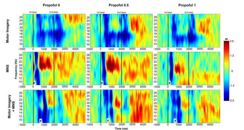
Figure 1 Time-frequency grand average analysis (ERSP) for MI, MNS and MI+MNS motor tasks under 3 propofol
concentrations (0 μg.ml, 0.5 μg.ml and 1 μg.ml) for electrodeC3. Black lines indicate when the motor task started
and finished. Red color corresponds to a strong ERS and blue to a strong ERD.References [1] P. Sebel, T. Bowdle, M. Ghoneim, I. Rampil, R. Padilla, T. Gan, and K. Domino, “The incidence of awareness during anesthesia: a multicenter united states study.” Anesth Analg , vol. 99, no. 3, pp. 833– 9, 2004. [2] K. MacGregor, “A waking nightmare: how can we avoid accidental awareness during general anaesthesia?” J Perioper Pract , vol. 23, no. 9, pp. 185–90, 2013. [3] S. Tasbighou, M. Vogels, and A. Absalom, “Accidental awarenessduring general anaesthesia - a narrative review,”Anaesthesia, vol. 73,no. 1, pp. 112–122, 2018. [4] Y. Blokland, J. Farquhar, J. Lerou, J. Mourisse, G. J. Scheffer, G.-J. van Geffen, L. Spyrou, and J. Bruhn, “Decoding motor responses from the eeg during altered states of consciousness induced by propofol,” Journal of Neural Engineering , vol. 13, no. 2, p. 026014, 2016.
Adaptive stimulus parameter setting for c-VEP BCI
                                        Federica Turi - Maureen Clerc

                      Inria Sophia Antipolis-Méditerranée, Université Côte d’Azur, France

A code-modulated visual evoked potential (c-VEP) BCI allows for spelling from a keyboard of flashing characters
(see Fig.1). All characters flash according to a predefined pseudo-random binary sequence, circular-shifted by a
character-dependent time lag. For a given character, the binary sequence evokes a VEP in the EEG of the subject
[1], which can be used as a template. This template is obtained during a calibration phase. A c-VEP BCI can
potentially achieve a very high-speed communication level [1] and the setting of stimulus parameters is
fundamental to obtain a high performing BCI. Many studies investigate different stimuli layout parameters such
as the size, color, proximity of the stimuli, different length sequences, different lags between adjacent stimuli [2]
and the stimulus presentation rate [3]. Analyzing these studies is clear that it is not possible to define an universal
optimal stimulus parameter setting suitable for each BCI user. In this work we design a subject-dependent c-VEP
BCI, with four different stimulus presentation rates of 15 Hz, 20 Hz, 30 Hz and 60 Hz (see Fig. 2), in which it is
possible to find the optimal stimulus presentation rate per each subject thanks to an adaptive setting parameter
phase. This phase takes place at the beginning of each session, it replaces the longer traditional calibration [1],
and allows to define the stimulus parameters that are used during the spelling phase. The objective is to find the
optimal presentation frequency to obtain a pleasant stimulus per each subject, reaching a high performance and
keeping the flash duration unchanged even when stimulus presentation rate is lower of 60 Hz, which is the most
common value of frequency rate used in c-VEP BCI. We acquired data from 4 subjects in two sessions. The
results obtained for the offline spelling show the variability between subjects and therefore the importance of
subject-dependent adaptation of c-VEP BCI.

Figure 1: Virtual keyboard. If the bit in the corresponding binary stimulation sequence is 0 the character flickers
in light grey, if it is 1 in black.

Figure 2: Illustration of the circular-shift process for the stimulus sequence for the first 3 targets, respectively
“A”, “B” and “C” in the virtual keyboard. On the left, the stimulus sequences have a frame rate of 60 Hz and on
the right, a frame rate of 30 Hz. The figure shows the stimulation sequence for the target T0, for the target T1,
circularly shifted with a time lag τs with respect to T0 and for the target T2, circularly shifted with a time lag τs
with respect to T1.The red dash boxes indicate the time lag τs that depends on the stimulation rate.
References
. [1] G. Bin, X. Gao, Y. Wang, Y. Li, B. Hong, and S. Gao. “A high-speed BCI based on code modulation
VEP”. In: Journal of neural engineering 8.2 (2011), p. 025015.
. [2] Q. Wei, S. Feng, and Z. Lu. “Stimulus specificity of brain-computer interfaces based on code modulation
visual evoked potentials”. In: PloS one 11.5 (2016), e0156416.
. [3] H. Nezamfar, S.S.M. Salehi, and D. Erdogmus. “Stimuli with opponent colors and higher bit rate enable
higher accuracy for C-VEP BCI”. In: 2015 SPMB. IEEE. 2015, pp. 1–6. Vous pouvez aussi lire