Cardiopathies acquises - Dr Daniela Laux Cardiopédiatre associée - UE3C -Paris Médecin hospitalier temps partiel - Arcothova

La page est créée Monique Pichon
 
CONTINUER À LIRE
Cardiopathies acquises - Dr Daniela Laux Cardiopédiatre associée - UE3C -Paris Médecin hospitalier temps partiel - Arcothova
Cardiopathies acquises

           Dr Daniela Laux

 Cardiopédiatre associée – UE3C –Paris

    Médecin hospitalier temps partiel
  Centre Chirurgical Marie Lannelongue
          Le Plessis Robinson
Cardiopathies acquises - Dr Daniela Laux Cardiopédiatre associée - UE3C -Paris Médecin hospitalier temps partiel - Arcothova
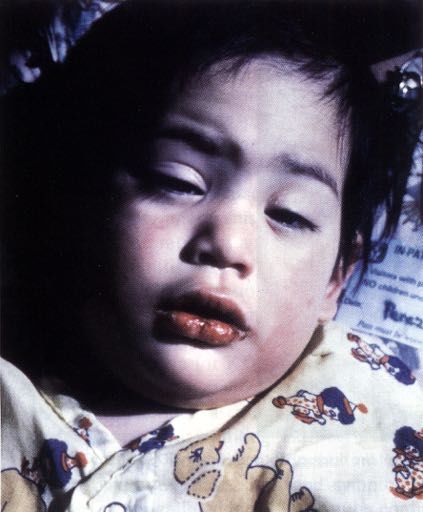
Plan du cours

• Maladie de Kawasaki

• Endocardite infectieuse

• Myocardite
Cardiopathies acquises - Dr Daniela Laux Cardiopédiatre associée - UE3C -Paris Médecin hospitalier temps partiel - Arcothova
Maladie de Kawasaki
Cardiopathies acquises - Dr Daniela Laux Cardiopédiatre associée - UE3C -Paris Médecin hospitalier temps partiel - Arcothova
Kawasaki – Les points clés

• Maladie décrite en 1967 – seulement 50 ans de recul !
• Vascularite systémique qui touche essentiellement les
  artères de moyen calibre avec un tropisme électif pour les
  artères coronaires (gravité de la maladie)
• Les complications coronaires surviennent dans 15 à 25 % des
  cas chez les enfants non traités
• L'administration précoce d'immunoglobulines humaines par
  voie intraveineuse a transformé le pronostic en diminuant
  par 5 le risque d'anévrisme coronaire
• Risque de mortalité 0,015 % (Japon), surtout entre le 15-
  45eme jour (thrombocytose concomittante avec la
  vascularite)
Cardiopathies acquises - Dr Daniela Laux Cardiopédiatre associée - UE3C -Paris Médecin hospitalier temps partiel - Arcothova
Epidémiologie
• Première cause de cardiopathie acquise de l'enfant dans les
  pays développés

• Tous les âges pédiatriques (80 % des cas avant 5 ans)
• Les patients de moins de 1 an ou de plus de 8 ans sont rares
  mais ont un risque plus élevé d'anévrisme coronaire

• Formes de l'adulte: première fois décrite en 1977
• symptômes majeurs décrits identiques

• Kawasaki atteint chaque année:
   – 265/100.00 enfants < 5 ans au Japon
   – extrapolation française: 600 nouveaux cas par an en
     France
                                         Newburger JACC 2016
Cardiopathies acquises - Dr Daniela Laux Cardiopédiatre associée - UE3C -Paris Médecin hospitalier temps partiel - Arcothova
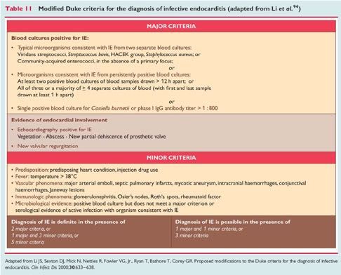
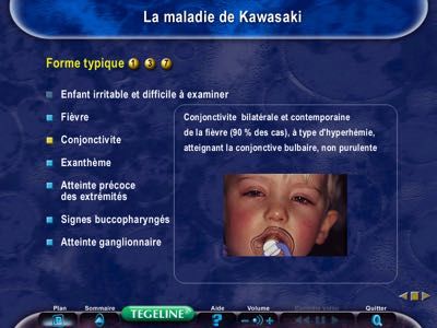
Caractéristiques cliniques: critères majeurs

    La fièvre de plus de 5 jours et au moins 4 critères suivants

    - La conjonctivite bulbaire non purulente

    - L’atteinte muqueuse : la pharyngite, la chéilite, la langue framboisée, la stomatite

    - L'exanthème polymorphe du tronc

    - L'atteinte des extrémités : un érythème des paumes des mains et/ou des plantes
    des pieds, l'œdème palmo-plantaire, la desquamation palmo-plantaire secondaire

    - L'atteinte unilatérale des ganglions cervicaux, de plus de
     1.5 cm de diamètre

Critères majeurs définis par l'American Heart Association Committee On Rheumatic Fever, Endocarditis, and Kawasaki Disease
Cardiopathies acquises - Dr Daniela Laux Cardiopédiatre associée - UE3C -Paris Médecin hospitalier temps partiel - Arcothova
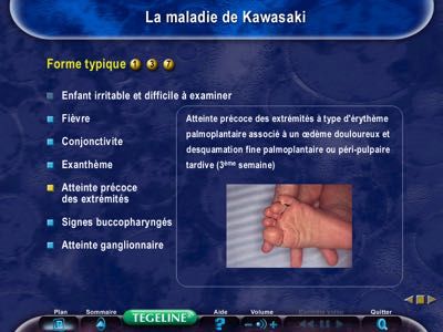
Faciès typique d’un enfant avec
   une maladie de Kawasaki
Cardiopathies acquises - Dr Daniela Laux Cardiopédiatre associée - UE3C -Paris Médecin hospitalier temps partiel - Arcothova
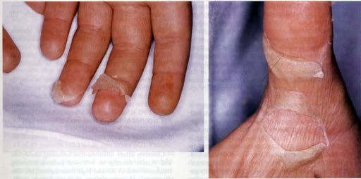
Atteinte muqueuse et desquamation palmo-plantaire
Cardiopathies acquises - Dr Daniela Laux Cardiopédiatre associée - UE3C -Paris Médecin hospitalier temps partiel - Arcothova
Formes rares: forme psoriasiforme/ BCGite
Atteinte cardiovasculaire

• Pas d’atteinte coronaire dans 75% des cas !
• Anomalie ECG ou échocardiographique:
   – Dilatation des artères coronaires (20%)
   – Anévrismes coronaires
   – Infarctus
   – Myocardite avec possible insuffisance ventriculaire gauche
     sévère
   – Péricardite, épanchement péricardique
   – Fuites valvulaires par inflammation des valves cardiaques et
     particulièrement la valve mitrale (1 %)
   – Troubles conductifs et troubles du rythme par inflammation
     du tissu de conduction
Les anévrismes coronaires
• Entre le 10ème et le 25ème jour d'évolution
• 5 % lorsque le traitement est fait précocement
        = < 10 jours du début des symptômes
• Souvent multiples et siègent habituellement dans la partie proximale
  des artères coronaires

• Pronostic cardiaque dépend essentiellement de leur taille.
• Nouveauté 2016: plus la taille en mm mais le Z score !

  Selon les guideslines américaines:
  Dilatation coronaire                 Z score > 2 < 2,5
  Petit anévrysme coronaire            Z score > 2,5  10 ou > 8 mm

                                                            Newburger JACC 2016
Formes atypiques
Tableau clinique dominé par un symptôme inhabituel:

« convulsions, œdème pulmonaire, diarrhée sanglante, ascite,
obstruction des voies aériennes supérieures, épiglottite,
adénopathies cervicales compressives ou hémolyse et
défaillance multi-viscérale, syndrome néphrotique,
hyponatrémie…. »

Formes de l'adulte
•             Troubles digestifs, atteinte hépatique, signes
      articulaires et encéphalites sont plus fréquent
Les formes incomplètes
Patients ayant eu une fièvre depuis au moins 5 jours et
au moins deux critères cliniques de Kawasaki, sans cause évidente,
et des critères biologiques en faveur d'une inflammation systémique

• Différent de la « forme atypique »

• Manque un ou plusieurs des cinq critères diagnostiques majeurs

• Plus fréquentes chez les enfants les plus jeunes, à risque
  d'anomalies coronaires

Diagramme décisionnel proposé par l'American Academy
of Paediatrics pour aider à la prescription d’IgG dans les formes
incomplètes

                                                   Newburger JW et al., Circulation. 2004
Critères cliniques et biologiques supplémentaires
Cardiovasculaires : dilatation des artères coronaires, anévrismes coronaires,
infarctus, myocardite avec possible insuffisance cardiaque congestive, péricardite,
épanchement péricardique, fuites valvulaires, troubles conductifs et troubles du
rythme, anévrismes des vaisseaux du cou, des artères rénales, spléniques,
hépatiques, pancréatiques, génitales, gangrènes distales et pseudo-Raynaud
Digestifs : diarrhées, vomissements, douleurs abdominales, hydrocholécyste,
dysfonction hépatique
Respiratoires : toux et rhinorrhée
Neuro-méningés : troubles de la conscience avec irritabilité, apathie, état grognon,
hypoacousie
Articulaires : arthrite, arthralgies
Autres : uvéite, érythème au niveau de la cicatrice de BCG, desquamation de l’aine

Albumine < ou égal 3g/dl
Anémie pour l’âge
Plaquettes ³ à 450 000/ mm³ à J7
Globules blancs ³ à 15 000 / mm³
ECBU ³ à 10 globules blancs/ champ
                                                   D’après Newburger JW et al., Circulation. 2004
einte unilatérale des ganglions cervicaux, de plus de 1,5 cm de diamètre, ferme

                                                                 Formes incomplètes
émentaires
                                              Fièvre de plus de 5 jours et 2 ou 3 critères cliniques
cardite, état de choc,                                                 ou
 e, anévrismes extra-                 Fièvre de plus de 7 jours sans cause retrouvée (enfants ≤ 6 mois+++)
 eaux du cou, artères
créatiques, génitales),
 ot aortique                                                 Faire un bilan biologique
missements, douleurs
sfonction hépatique,
atite, hydrocèle
                             CRP < 30 mg/l et VS < 40 mm/h                    CRP ≥ 30 mg/l et/ou VS ≥ 40 mm/h
nchique et interstitiel,
res
 cience avec irritabilité
 ie faciale, hypoacousie     Réexaminer et contrôler le bilan               Au moins 3 critères biologiques ou plus :
 yperleucocytose du           biologique si la fièvre persiste
)                                                                                     Anémie pour l’âge
 e la cicatrice de BCG,       Échocardiographie en cas de                         Plaquettes ≥ 450 000/mm3
                                                                      Non
on rétropharyngé             desquamation en doigt de gant                            Albumine ≤ 30 g/l
                                        typique                                       ALAT augmentées
                                                                                Globules blancs ≥ 15 000/mm3
                                                                               ECBU ≥ 10 globules blancs/champ
 différentiels                                                        Oui                       ou
 irus, EBV, rougeole                                   Traiter                     Échocardiographie positive
 phylococcique
oxique                                                    Échocardiographie positive
 enne
dicaments                                          Z-score IVA ou CD ≥ 2,5
Johnson                                            Ou anévrisme coronaire (Z-score ≥ 2,5)
 ile                                               Ou plus de 3 critères :
                                                   - dysfonction ventriculaire gauche
                                                   - fuite mitrale
                                                   - épanchement péricardique
ire élevé si score ≥ 5)                            - Z-score IVA ou CD compris entre 2 et 2,5
                                                                                                                Bajolle et al. 2018 EMC
Evolution naturelle

1 ) La phase aiguë (J0-J10) : atteinte cardiaque rare

2 ) La phase subaiguë (J10-J20) : diagnostic de complication
coronaire

3 ) La phase de convalescence (J20-J70) : constatation
d’anévrysmes et de sténoses cicatricielles en cas de complication
coronaire à la deuxième phase
Exemples d’atteinte coronaires
Dilatation anévrysmale des artères coronaires

  Aorte         IVA =6.9 mm
                                                  Thrombus
                                        Aorte     dans l’IVA

          (A) IVA=interventriculaire antérieure
          (B) thrombus dans l’IVA
Dysfonction ventriculaire gauche sévère en
              échographie

 Etat de choc dans 7% des cas de maladie de Kawasaki

             VG
            dilaté
                                    VG
                                   dilaté
Dilatation anévrysmale des artères
       coronaires au scanner

   CD =4.7 mm

                Aorte

                    IVA =6.9 mm
Dilatation anévrysmale de l’IVA en
            chapelet au scanner

Aorte
                            Aorte

    Chapelet anévrysmal
                               Chapelet anévrysmal
Anévrysme coronaire avec sténose
coronaire au cathétérisme cardiaque

         Sténose      Anévrysmes et sténoses
         coronaire    multiples des coronaires
Atteinte diffuse des axes vasculaires

                                       A                                                         B

                                                                  Foie

                                                        Dilatation
                                                        fusiforme

                 Aorte

(A) épaississement pariétal hyperéchogène de l’aorte, de l’artère mésentérique supérieure et du tronc
coeliaque en échographie
(B) dilatation fusiforme de l’artère mésentérique supérieure, du tronc caeliaque au scanner
Le traitement de            1ère   intention

Immunoglobulines intraveineuses:
• 2g/kg en 8 à 12 heures, à posologie progressive

En association à de l’acide acétylsalicylique (AAS) :
• à fortes doses pour ses effets anti-inflammatoires et anti-
  thrombotiques (30-50 mg/kg/j en 4 fois) à la phase aiguë
• puis à dose anti-aggrégante plaquettaire rapidement 48-72 h
  après l’apyrexie (3-5 mg/kg/jour)

                                   De Graeff et al 2019, Newburger 2016
CE NTRAL ILL USTRATIO N Management of Kawasaki Disease

                                 Diagnosis of Kawasaki disease according to American Heart Association criteria*

                 Anti-inflammatory therapy with intravenous immunoglobulin (IVIG) and ASA (acetylsalicylic acid or aspirin).
                                    Echocardiogram to assess coronary arteries and determine Z score.

      Defervescence with no coronary dilation (Z score
Recommandations américaines
TABLE 1 Principles in Acute Management of KD

1. The goal of therapy is to reduce systemic and tissue-level inflammation as rapidly as possible. For this reason, patients should be treated as soon as
   diagnosis can be confidently established.
2. All patients within the first 10 days of fever onset should be treated with IVIG. Patients diagnosed after 10 days should receive IVIG treatment if
   they are still febrile, have markedly elevated inflammatory parameters, or have coronary artery dilation.
3. Recrudescent fever at least 36 h after the end of IVIG infusion without other explanation is a marker for persistent inflammation and should prompt
   immediate and aggressive anti-inflammatory therapy
  a. Antibody-mediated hemolysis has become common in KD patients who have received IVIG retreatment and have type A or B blood; rescue
     therapies other than IVIG (e.g., infliximab, corticosteroids) should be considered.
4. Patients with coronary artery dilation (z-score >2.0) should be followed with a repeat echocardiogram at least twice a week until dimensions
   stabilize; additional anti-inflammatory therapy should be considered.
5. Patients with giant aneurysms should have frequent echocardiograms in the first 3 months of illness for thrombus surveillance, even after
   dimensions stabilize.
6. Infants under 6 months of age are at extremely high risk of aneurysm formation, even with timely therapy. They require echocardiograms every few
   days until dimensions have stabilized.
7. Patients with giant CAA (z-score $10) are at highest risk for thrombosis during the first 3 months after fever onset
  a. Systemic anticoagulation together with an antiplatelet agent should be administered until coronary dimensions improve.
  b. Low-molecular-weight heparin is easier to regulate than warfarin in infants, as well as in patients of any age, during the acute phase of illness or
     until hsCRP normalizes.

CAA ¼ coronary artery aneurysm; hsCRP ¼ high-sensitivity C-reactive protein; IVIG ¼ intravenous immunoglobulin; KD ¼ Kawasaki disease.

                                                                                                                                         Newburger JACC 2016
 The progression of aneurysm formation in some                                       Children With Kawasaki Disease and Coronary Artery
Surveillance à court terme
Pour les patients sans complication coronaire:
• 1 echo entre 1-2 semaines et 1 echo entre 4-6 semaines (Classe 1)

Pour les patients avec Z score coronaire > 2,0 à la phase aigue:
• 2 echos par semaines jusqu’à l’arret de la progression

Pour les patients avec anévrismes géants:
• 2 échos par semaine tant que les lésions progressent
• 1 écho/sem pdt 45 jours puis
• 1 écho/mois pendant 3 mois (Classe IIA)

                                                         Newburger JACC 2016
Antiagrégation et anticoagulation
Patients sans atteinte coronaire:
• Aspirine pendant 6 semaines (Cl 1)

Patients avec atteinte coronaire d’aggravation rapide:
• Hospitalisation pour mise sous heparine (AntiXa 0,5-1)
• Arret si stabilisation; Z score < 10 ou 8 mm
• Aspirine 12 mois

Patients avec anévrysmes géants:
• Hospitalisation pour Heparine et relais AVK (INR 2-3)
• Aspirine à vie
Que faire en cas de persistance de la fièvre
          après une première cure d’IgIV?
• Résistants: 15 à 20 % des cas
• Associée à un risque plus élevé d'atteinte coronaire

• Deuxième dose d’IVG
• associée à un bolus de corticoides (20-30 mg/kg/jour IV
  methylprednisolone)

• Discuter: prednisolone p.o. plus prolongé 2-3 semaines
• Alternative: infliximab
• En cas d’échec: ciclosporine

                                          Newburger JW et al., Circulation. 2004
                                                   Egami K et al., J Pediatr 2006
Qui sont les patients à haut risque?
         Comment identifier les résistants ?
         • Score issu de la littérature de patients japonais!!

Score d’Egami (2006)            Score de Kobayashi (2006)        Score de Sano (2007)

Age ≤ 6 mois (2 points)         Age ≤ 12 mois (1 point)          Bilirubine totale ≥ 0.9mg/dL (1
                                                                 point)
≤ 4 jours de fièvre (1 point)   Traitement dans les 4 premiers CRP ≥ 7mg/dL (1 point)
                                jours de fièvre (2 points)
Plaquettes ≤ 300.109/L (1 Plaquettes          ≤   300.109/L   (1 ASAT ≥ 200 U/L (1 point)
point)                          point)
CRP ≥ 8mg/dL(1 point)           CRP ≥ 10mg/dL (1 point)
ALAT > 100 U/L (1 point)        ASAT ≥ 100 U/L (1 point)
                                ≥ 80% neutrophiles (2 points)
                                Na+ ≤ 133 mmol/L (2 points)

Haut risque si ≥ 3 points       Haut risque si ≥ 5 points        Haut risque si ≥ 2 points

Scores issus de la population japonaise, spécifique mais non sensible
3. In non-Japanese patients, the Kobayashi criteria may indicate risk of IVIG resistance if ‘positive’ (score                                                                          2A                                                                                                  C
                   Original article
           54) but may not reliably exclude IVIG resistance if ‘negative’ (score
Quelle est l’histoire naturelle des complications
                  coronaires de la MK?

• Disparition complète dans plus de 50% des cas même
  en cas d’anévrysme (sauf géant) dans les 2 ans

• Occlusion coronaire; sténoses localisées ou multiples
  parfois très tardives…

• Gravité des lésions tardives car multiples et chirurgie
  difficile
Anévrysme géants (1%)
• Mortalité et morbidité +++
• Survie à 30 ans: 88-90%

•   Cardiac event free à 30 ans : 30%
•   26% infarctus myocardique            Adapted with permission from Newburger et al. (99).

•   Risque accru dans les 2 ans après le diagnostic
•   50% de bypass coronaire à 30 ans
Surveillance à long terme
19-1800 ! Maladie de Kawasaki

                                                       Immunoglobulines IV à 2 g/kg sur 12 h
                                     Aspirine à dose anti-inflammatoire 60 mg/kg/j jusqu’à disparition de la fièvre
                                                            Puis relais par AAP 3–5 mg/kg/j

               75 %                    20 %                                         4%                                              1%

                                                                                                                         Anévrisme géant
              Coronaires                                    Petit anévrisme                Anévrisme moyen
                                        Dilatation                                                                    Z-score ≥ 10 ou ≥ 8 mm
               normales                                     2,5 ≤ Z-score < 5               5 ≤ Z-score < 10
                                     2 ≤ Z-score < 2,5                                                                      AAP à vie
              Z-score < 2                                       AAP 1 an                       AAP à vie
                                     AAP 6 semaines                                                                      AVK pour INR 2-3
            AAP 6 semaines                                 Cs à M3, M6, M12                Cs à M3, M6, M12
                                     Cs à 6 semaines                                                               Cs à M1, M2, M3, M6, M9, M12
            Cs à 6 semaines                                Coroscanner à 1 an              Coroscanner à 1 an
                                                                                                                      Coronarographie à 1 an

                                                                                         Même                Si         Normali-           Si
                         Normali-           Si
             Arrêt                                        Normali-         Si               si          persistance      sation       persistance
                          sation       persistance
            aspirine                                       sation     persistance     normalisation      Aspirine       Aspirine     Aspirine à vie
                           Arrêt        Maintien
            Clôture                                         Arrêt      Maintien       Aspirine à vie       à vie          à vie      Maintien AVK
                         aspirine       aspirine
              du                                          aspirine     aspirine         ± double         ± double       ± double     ± double AAP
                        Clôture du       Cs/2–5
            dossier                                      Cs/1–3 ans     Cs/an             AAP              AAP            AAP            ± BB
                         dossier           ans
                                                                                         Cs/an            Cs/an          Cs/an         Cs/6 mois

                                                             Prévention des FDRCV pour tous !

Figure 2. Arbre décisionnel. Prise en charge proposée par le centre de référence Malformations Cardiaques Congénitales Complexes (M3C) Necker. IV : par
voie intraveineuse ; AAP : aspirine à dose antiagrégante plaquettaire ; Cs : consultation ; AVK : antivitamine K ; INR : international normalized ratio ; FDRCV :
facteurs de risques cardiovasculaires.

! Formes incomplètes                                                                 lines est le plus souvent excellente. La recommandation est une
                                                                                     perfusion lente de 12 heures pour éviter tout effet secondaire
  Lorsqu’on a une forme incomplète de maladie de Kawasaki                            (flush, hypertension, malaise,Bajolle     et al.etc.).
                                                                                                                     hypotension,      2018 Les EMC
                                                                                                                                                immuno-
orth    ionizing radiation. Children too young to exercise
 pe-
eas-                     Devenir à long terme
late     T A B L E 2 Principles in the Long-Term Management of Patients With KD
sta-
         1. On the basis of available data, patients with no demonstrated coronary artery
ays-         dilation by echocardiogram with excellent visualization of all arterial segments
rs of        during the first weeks of illness appear to have normal cardiovascular status in
             early adulthood.
         2. Remodeling (so-called regression) of aneurysms, especially if moderate or large,
             to normal internal lumen diameter is often accompanied by luminal
             myofibroblastic proliferation and abnormal vascular reactivity.
         3. Patients with persistent CAA are at lifelong risk of progressive coronary artery
             stenosis or occlusion and worsening ischemia.
ong      4. Patients with CAA documented at any stage require lifelong cardiovascular
e to         surveillance tailored to disease severity and age.
         5. Testing should minimize exposure to ionizing radiation whenever possible.
dial
         6. Sedentary life-style should be avoided.
  2).
         7. Women with coronary aneurysms can carry pregnancy successfully, but should
 the         have reproductive counseling.
ting     8. Monitoring and counseling regarding traditional CV risk factors is appropriate to
            reduce the likelihood of later atherosclerosis.
pon                                                                                Newburger JACC 2016
Définition de MIS-C: multisystem
    inflammatory syndrome in children
Eur J Pediatr (2021) 180:307–322                                                                                                                309

Table 1   Case definitions for SARS-CoV-2-associated multisystem inflammatory syndrome in children

Royal College of Paediatrics and Child           Centers for Disease Control and Prevention         World Health Organization (WHO)
Health, UK                                       (CDC), USA

A child presenting with persistent fever         An individual aged < 21 years presenting           Children and adolescents 0–19 years of age
  (> 38.5 °C), inflammation (neutrophilia,         with fever*, laboratory evidence of                 with fever ≥ 3 days;
  elevated CRP, and lymphopenia) and               inflammation**, and evidence of clinically       AND two of the following:
  evidence of single or multiorgan                 severe illness requiring hospitalization,        1. Rash or bilateral non-purulent
  dysfunction (shock, cardiac, respiratory,        with multisystem (≥ 2) organ involvement            conjunctivitis or muco-cutaneous
  kidney, gastrointestinal, or neurological        (cardiac, renal, respiratory, hematologic,          inflammation signs (oral, hands or feet).
  disorder) with additional features*.             gastrointestinal, dermatologic or                2. Hypotension or shock.
This may include children fulfilling full or       neurological);                                   3. Features of myocardial dysfunction,
  partial criteria for KD.                       AND                                                   pericarditis, valvulitis, or coronary
Exclusion of any other microbial cause,          No alternative plausible diagnoses;                   abnormalities (including echo findings or
  including bacterial sepsis, staphylococcal     AND                                                   elevated troponin/NT-proBNP),
  or streptococcal shock syndromes,              Positive for current or recent SARS-CoV-2          4. Evidence of coagulopathy (by PT, PTT,
  infections associated with myocarditis such      infection by RT-PCR, serology, or antigen           elevated d-dimers).
  as enterovirus (waiting for results of these     test, or COVID-19 exposure within                5. Acute gastrointestinal problems (diarrhea,
  investigations should not delay seeking          4 weeks prior to the onset of symptoms.             vomiting, or abdominal pain).
  expert advice).                                *Fever ≤ 38 °C for ≥ 24 h, or report of            AND
SARS-CoV-2 RT-PCR test results may be              subjective fever lasting ≥ 24 h.                 Elevated markers of inflammation such as
  positive or negative.                          **Including, but not limited to, one or more of       ESR, C-reactive protein, or procalcitonin.
*Additional features:                              the following: an elevated CRP, ESR,             AND
Clinical:                                          fibrinogen, procalcitonin, d-dimer, ferritin,    No other obvious microbial cause of
Most: oxygen requirement, hypotension              LDH, or IL-6, elevated neutrophils,                 inflammation, including bacterial sepsis,
Some: abdominal pain, confusion,                   reduced lymphocytes and low albumin.                staphylococcal or streptococcal shock
  conjunctivitis, cough, diarrhea, headache,     Additional comments:                                  syndromes.
  lymphadenopathy, mucus membrane                Some individuals may fulfill or partial criteria   AND
  changes, neck swelling, rash, respiratory        for KD but should reported if they meet the      Evidence of COVID-19 (RT-PCR, antigen
  symptoms, sore throat, swollen hands and         case definition for MIS-C;                          test or serology positive), or likely contact
  feet, syncope vomiting;                        Consider MIS-C in any pediatric death with            with patients with COVID-19.
Laboratory:                                        evidence of SARS-Cov-2 infection.                Consider this syndrome in children with
All: abnormal fibrinogen, high D-dimers,                                                               features of typical or atypical KD or toxic
  high ferritin, hypoalbuminemia;                                                                      shock syndrome
Some: acute kidney injury, anemia,
  thrombocytopenia, coagulopathy, high
  IL-10, high IL-6, proteinuria, high CK,
  high LDH, high TG, high troponin,
  transaminitis;
Imaging:                                                                                                                     Sperotto et al. 2021 Review
Echo and ECG: myocarditis, valvulitis,
MIS-C
• Tableau clinique: fièvre, asthénie, rash cutanée,
  symptômes digestifs+++, signes de choc

• Covid-19 +: PCR (20-50%) ou sérologie ou contact
  familial -> phénomène immunologique tardif ?
• Délai infection Covid- symptômes: jours à mois
• Biologie: CRP, VS, PCT, ferritine, IL-6, fibrinogène
  élévé++, leucocytose, Hb et plaq normal ou diminué
• BNP et troponine très élevée

• Atteinte cardiaque:
   – Dysfonction myocardique: 35-100% ->inotropes, VM, ECMO
   – Dilatation coronaire: 6-24%, rares anévrismes
   – Arythmies: 7-60%-> anomalies ST, QT, BAV , fibrillation
                         Sperotto et al. 2021 Review   European Journal of Pediatrics (2021) 180:307–322
                                                       https://doi.org/10.1007/s00431-020-03766-6
MIS-C et Kawasaki

               MIS-C       Ka                Kawasaki

•   Enfants plus âgés : moy 8-9 ans
                                                    0-5 ans
•   Symptômes digestifs au premier plan
                                        Atteinte coronaire 25%
•   plus souvent en choc                Kawasaki choc syndrome: 7%
•   Atteinte cardiaque plus fréquente
    50%
•   BNP et troponine plus élevée

                          Cardiomyopathie
                              septique
Endocardite
Endocardite infectieuse

Def: Infection/inflammation de l’endocarde = valves cardiaques

Dg: Echographie trans-thoracique voire ETO

         - EI des VAV: sur le versant auriculaire

         - EI des valves sigmoïdes: sur le versant ventriculaire

•   Hémocultures: au moins 3!!!!!!!!!!! (au mieux 6)

•   Pas d’ATB à l’aveugle

•   Scanner total body (cérébral, thoracique et abdominal)
•   Examen ophtalmologique, bandelette urinaire
•   Recherche porte d’entrée: examen dentaire, ORL, cutané, digestif,
    urinaire, KTC…
Endocardite: germes
Endocardite: Traitement médical
Endocardite: Traitement chirurgical
Endocardite: prévention

- Bonne hygiène dentaire quotidienne
- Consultation dentaire tous les 6 mois

               Indispensable pour diminuer le risque d’endocardite
POPULATION CONGENITALE - ENFANTS

 34 279 enfants avec CC suivis de 0 à 18 ans
Incidence annualisée = 4.1 / 10 000 pt-année

                                               Rushani et al. Circulation
                                                                    2013
POPULATION CONGENITALE - ADULTES

              Registre CONCOR (14 224 patients>18 ans)
                  Incidence EI : 1.33/1000 pt-years
               Prothèse valvulaire: HR=3.57(2.58–5.36)

                                              Kuijpers et al. Eur Heart Jour
                                                                       2017
INCIDENCES COMPARATIVES

Valve Melody : 0.8 – 3% pt-année

Valves/conduits pulmonaire chir : 0.5 - 3% pt-année

TAVI: 0.67 – 2.1% pt-année

Valves Ao/mitrale chir : 0.3 – 1.2% pt-année

Dispositifs electroniques implantables : 1.9/1000 device-année

Patients avec CC: 0.4 – 1.33 / 1000 pt-année
                                                                 Miranda et al. Eur Heart Jour 2016
                                                                           Wang et al. JAMA 2007
                                                                     Rushani et al. Circulation 2013
Population générale : 30 -100/ million pt-année                    Habib et al. Eur Heart Jour 2015
                                                                          Dayer et al. Lancet 2015
VALVES PERCUTANÉES VS CHIRURGICALES

          p=0.1
            3

134 chir et 208 percut (33 Sapien)    195 chir et 93 percut (0 Sapien)         631 chir et 107 percut (0 Sapien)

Incidence IE: 0.5 vs 1.5 %pt/années   Incidence IE: 1.2 vs 3.9 %pt/années      Incidence IE: 0.8 vs 2.7 vs 3% %pt/années

           Lluri et al. CCI 2017          Malekzadeh-Milani et al. JTCS 2014                   Van Dijck et al. Heart 2014
SUBSTRAT VALVULAIRE
  Author     Year     n       Substrate           EI      EI Annualized    Median
                                             Cumulative     Incidence     Follow-up
                                              incidence    (% pt-year)     (years)

 Albanesi    2014   12/106     Contegra         11.3                         7.6

Malekzadeh   2014   5/190    Homografts         2.6            1.2           2
                              Contegra

Ramanan      2015   6/115      Freestyle        5.4             -            4.3

  Mery       2016   23/586    Homograft          4              -            7
                               Contegra
                             Porcine valve
  Ugaki      2016   21/298    Contegra           7              -            3.4
                              Homograft

 Tous les dispositifs valvulaires sont susceptibles d’être le
                        siège d’une EI
      Avec une incidence variable mais significative                                             Albanesi et al. EJCTS 2014
                                                                                       Ramanan et al. Ann Thorac Surg
                                                                                                                      2015
                                                                                      Ugaki et al. Ann Thorac Surg 2016
                                                                                                     Mery et al. JTCS 2016
SUBSTRAT VALVULAIRE

• EI plus fréquente chez les patients avec VJB
• Quelle que soit la techniqued’implantation (i.e. Contegra et
   Melody)
• Comparés aux homogreffes RR=8.7 and 9.7 pour Melody et Contegra
Malekzadeh-Milani et al. JTCS 2014                                                Mery et al. JTCS 2016
Van Dijck et al. Heart 2014                                         Ugaki et al. Ann Thorac Surg 2015
SUBSTRAT VALVULAIRE
                                      Méta-analyse sur IE chez les patients avec RVP chirurgical ou
                                      percutané
                                      7063 patients
                                      Incidence cumulative globale = 2.5%
                                      VJB vs autres substituts : 5.4% vs 1.2%; p < 0.0001

Sharma et al. JACC Int. 2017
VALVE SAPIEN
                                                                                                                                                                        JACC: CARDIOVASCULAR INTERVENTIONS                                                                          VOL. 10

                                                                                                                                                                        ª 2017 BY THE AMERICAN COLLEGE OF CARDIOLOGY FOUNDATION                                                  ISSN 1936

                                                                                                                                                                        PUBLISHED BY ELSEVIER                                                                http://dx.doi.org/10.1016/j.jc

    512           Hascoet et al.                                                                                  JACC: CARDIOVASCULAR INTERVENTIONS VOL. 10, NO. 5, 2017

                  Endocarditis After PPVI: Melody Versus Sapien                                                           Infective Endocarditis Risk After                 MARCH 13, 2017:510–7

                                  JACC: CARDIOVASCULAR INTERVENTIONS VOL. 10, NO. 5, 2017
                                                                                                                          Percutaneous Pulmonary             Hascoet et al.Valve513
                                  MARCH 13, 2017:510–7                                                                       Endocarditis After PPVI: Melody Versus Sapien
                                                                                                                          Implantation With the Melody
                                                                                         and from phone calls to the patients and to their
      TABLE 1        Patient Demographics, Procedural Data, and Post-Procedural Outcomes                                  and Sapien Valves
                                                                                         cardiologists and general practitioners. For cases of
                                             PPVI With                 PPVI With                                IE, every effort was made to obtain
                                                                                                                                               Sebastieninformation
                                                                                                                                                         Hascoet, MD,a Luciaon
                                                                                                                                                                            Mauri, MD,a Caroline Claude, MD,a Emmanuelle Fournier, MD,a Julie Lou
                                            Melody Valve              Sapien Valve          Standardized                                                                                        c                     c
                                                                                                                the Duke criteria, clinical and microbiological
                                                                                                                                                   Jean-Yves Riou, MD, details,
                                                                                                                                                                       Philippe Brenot, MD,                               Jérôme Petit, MDa
                                              (n ¼ 32)                  (n ¼ 47)             Difference

      Age (yrs)
                                    (73 of19.979;(15.8–28.9)
                                                    92.4%). A single   stent was sufficient
                                                               26.3 (18.9–39.9)    0.58*
                                                                                           in all
                                                                                              medical and surgical strategies, and outcome.
                                                                                                                           TABLE 1 Continued
      Weight (kg)                   Melody56.5
                                            group
                                               ! 13.5patients, 65.8
                                                               whereas
                                                                    ! 17.6 in the Sapien
                                                                                    0.59* group 4
                                                                                                                                                                          ABSTRACT
                                                                                                   STATISTICAL ANALYSIS. Statistical analyses were
      Male (%)                               53.1                         66.0           0.26                                         PPVI With                          PPVI With
                                    patients required 2 to 4             stents. Pre-stenting was not
                                                                                                   performed using Stata 11.2 software    (StataCorp,
                                                                                                                                     OBJECTIVES                 Col- the risk of infective endocarditis (IE) after percutaneous pulmonary valve
                                                                                                                                                    This study compared
      Genetic syndrome (%)                       18.8                     10.6          -0.23                                        Melody    Valve                   Sapien   Valve             Standardized
                                                                                                                                     implantation (PPVI) with the Sapien and Melody valves.
      History of severe infectiousperformed
                                          9.4in          6 patients      with
                                                                           8.5 valve-in-valve
                                                                                        -0.03 implan-
                                                                                                   lege Station, Texas). Continuous data   were
                                                                                                                                       (n ¼ 32)      described            (n ¼ 47)                 Difference
            disease (%)
      History of endocarditis (%)
                                    tation. Balloon
                                             6.3
                                                    post-dilation
                                                               2.1
                                                                   was performed
                                                                            -0.20
                                                                                  in 19 as
                                                                                        of meanInfective
                                                                                                  ! SD if normally distributed
                                                                                                         endocarditis during
                                                                                                                                 and asThemedian
                                                                                                                             BACKGROUND
                                                                                                                                25.0
                                                                                                                                          incidence of IE after PPVI is estimated at 3% per year with the Melody valve. The Sapien v
                                                                                                                                                              0.0                         -0.80*
                                                                                     (interquartile range [IQR])                                                      more recently Categorical
                                                                                                                                                                  otherwise.        marketed valve used for PPVI.
      Pacemaker/defibrillator (%)79       patients
                                               6.3 (24.1%) and was
                                                               10.6 more common
                                                                            0.16 in the          follow-up (%)
                                                                                     variables were described as                                                   number  (%).
                                                                                                                                                                      METHODS      Bivariate
                                                                                                                                                                              We retrospectively included consecutive patients who underwent PPVI at a single center between 20
      Congenital heart diseases (%)                                                       Pulmonary valve replacement     25.0                               4.3                                                                     -0.59*
                                    Melody group (46.9% vs. 6.5%, respectively). Pro-
                                                                                   analyses with calculation of standardized       differences
                                                                                                                      2016. IE was diagnosed using the modified DUKE criteria.
          Conotruncal malformation               81.3                      68.1                                            during follow-up (%)
          Ross procedure            cedure duration,
                                            9.4             21.3 time, and irradiation
                                                     fluoroscopy                     were                             performed to compare variables
                                                                                                                        Percutaneous
                                                                                                                                                                 between the
                                                                                                                                                            3.1 PPVI was performed in 79 patients
                                                                                                                                                      RESULTS                                     2.1 (Melody valve, 40.5%;-0.06
                                                                                                                                                                                                                               Sapien valve, 59.5%). Median age was 24
                                                                                                               two valve types and between patients   (range 18.1 towith
                                                                                                                                                                     34.6). IEversus
                                                                                                                                                                               occurred in 8 patients (10.1%) at a median of 1.8 years (minimum: 1.0; maximum: 5.6)
          TGA
                                    were higher
                                             3.1
                                                 in the Sapien 0.0
                                                               valve group.                                             Surgical                           21.9
                                                                                                                                                      surgery.                                    2.1
                                                                                                                                                               Causative organisms were methicillin-sensitive   Staphylococcus-0.63*
                                                                                                                                                                                                                               aureus (n ¼ 3), Staphylococcus epidermidis
          PA-IVS/PVS                              3.1                      4.3                                 without IE during follow-up.
          DORV                         Severe 3.1
                                               procedural complications
                                                              6.4       occurred in 2                         pa- Kaplan-Meier
                                                                                                                      Death during curves
                                                                                                                                   follow-up of
                                                                                                                                             (%) the cumulative
                                                                                                                                                            3.1
                                                                                                                                                      Streptococcus  mitis (n ¼ 1),
                                                                                                                                                                            IE inci-
                                                                                                                                                                                    Aerococcus    2.1
                                                                                                                                                                                               viridans (n ¼ 1),             -0.06
                                                                                                                                                                                                                 Corynebacterium  striatum (n ¼ 1), and Haemophilus influ
                                                                                                                                                                          (n ¼ 1). All 8 cases occurred after Melody PPVI (25.0% vs. 0.0%). The incidence of IE was 5.7% (95% confidence i
      RVOT (%)                      tients (2.5%). One patient died of massive hemo-
                                                                                  dence were plotted using the date of  PPVI
                                                                                                                     2.9%        asperthe
                                                                                                                          to 11.4%)         entry
                                                                                                                                       person-year after Melody PPVI. The Kaplan-Meier cumulative incidence of IE with Melody P
          Native RVOT                             3.1                      25.5                                           Values are time
                                                                                                                                     median since
                                                                                                                                            [interquartile range]
                                    thorax due  to perforation                       date
                                                                  of a distal pulmonary                                 and   the                    PPVI     as orthe
                                                                                                                                                                    %. Standardized
                                                                                                                                                                    24.0%time        difference
                                                                                                                                                                                 scale.
                                                                                                                                                                          (95% confidence           computed
                                                                                                                                                                                            The12.2%
                                                                                                                                                                                         interval:            as the
                                                                                                                                                                                                       to 43.9%)  afterdifference
                                                                                                                                                                                                                        4 years and in means
                                                                                                                                                                                                                                    30.1% (95%orconfidence interval: 15.8% to
          Bioprosthesis                     9.4              23.4                                                          proportions divided by the SE. *Significant after
                                                                                                                                                                       imbalance.
                                                                                                                                                                            6 years, compared with 0.0% with the Sapien PPVI after 4 years (p < 0.04 by log-rank test). There was
                                                                                                                right censor was the date of IE, valve      replacement,
          Homograft                 branch     during
                                                25.0       Sapien    valve
                                                                         31.9PPVI     over a Lunderquist                                          toward a higher incidence of IE in the first 20 patients with Melody PPVI (who received prophylactic antibiotics du
                                                                                                                             DORV ¼ double-outlet right ventricle; PA-IVS ¼ pulmonary atresia with intact ventricular septum; PPVI ¼
          Conduits                               62.5                      19.2
                                                                                       heart transplantation,          death, or follow-up
                                                                                              percutaneous pulmonary valve implantation; procedure only) andcomple-
                                                                                                                                            PVS ¼ pulmonary  in patients who had percutaneous interventions, dental care, or noncardiac surgery after P
                                                                                                                                                                  valve stenosis; RVOT ¼ right ventricle outflow
      RVOT lesion (%)
                                    guidewire. In the other patient, who had a mechanical
                                                                                       tion. Differences      in incidence
                                                                                              tract; TGA ¼ transposition of the greatwere
                                                                                                                                      arteries.evaluated using
                                                                                                                                                                          CONCLUSIONS IE after PPVI may be less common with the Sapien compared with the Melody valve.
          Stenosis                  aortic valve,
                                             84.4 a large femoral
                                                             50.0 hematoma developed
                                                                                  the log-rank test. The Kaplan-Meier   method
                                                                                                                    (J Am               was
                                                                                                                          Coll Cardiol Intv    also © 2017 by the American College of Cardiology Foundation.
                                                                                                                                            2017;10:510–7)
          Regurgitation                          0.0                       35.7                                 used to assess the cumulative incidences of pulmo-
          Mixed                                  15.6                      14.3                                 nary valve replacement and of death, with the date of
      Pre-stenting (%)
Hascoet   et al.
     1-stage PPVIJACC
                  (%) Int. 2017
      No. of stents (%)
                                                 90.6
                                                 87.5
                                                                          93.6
                                                                           87.2
                                                                                                0.11
                                                                                                0.01

                                        FIGURE 1 Kaplan-Meier Cumulative Incidences of Death and Pulmonary Valve
                                                                                                                PPVI as the entry date and the time since
                                                                                                  time scale and with the right censor set
                                                                                                                 Replacement and Kaplan-Meier
                                                                                                                                       outflow
                                                                                                                                             geryasforthe
                                                                                                                                               Cumulative
                                                                                                                                               tract
                                                                                                  valve replacement, heart transplantation, death,   (RVOT).
                                                                                                                                                                       P
                                                                                                                                                             PPVI as the
                                                                                                                                                                                ercutaneous pulmonary valve implantation
                                                                                                                                                      (PPVI) has emerged as an alternative to sur-
                                                                                                                                                            date ofthe right ventricular (Edwards SAPIEN pulmonic transcatheter h
                                                                                                                                                       reconstructing
                                                                                                                                                               Incidences
                                                                                                                                                              PPVI  was firstofdescribed
                                                                                                                                                                    or          Infective
                                                                                                                                                                                        in Endocarditis
                                                                                                                                                                                            Edwards Lifesciences, Irvine, California)
                                                                                                                                                                                                                                       certification in 2006 and Food and Drug A
                                                                                                                                                                                                                                       tion approval in 2010 for PPVI. The Sap
VALVE SAPIEN

Edwards SAPIEN XT Transcatheter Heart Valve with the NovaFlex1Deliv-
ery System. Vol. 2016. https://www.accessdata.fda.
gov/cdrh_docs/pdf13/p130009s037d.pdf.
PREVENTION - EDUCATION

                                                                PROCEDURE
 PRE-IMPLANTATION
                                                                 Salle hybride
    Patient et famille
                                                             Optimisation gradient
Depistage foyer infectieux
                                                                    résiduel
  Bilan dentaire et ORL
                                                           Antibioprophylaxie per +/-
                                                                post procédure

                                POST-IMPLANTATION
               Education patient, parents, médecin traitant, dentiste
                             Anti-aggrégants au long cours
                                Antibioprophylaxie à vie
FACTEURS AGGRAVANTS

                   Portes d’entrée évitables

                       Manque observance

                      Déficiences mentales

                 Education - Prophylaxie EI

Bauer et al. Int Jour Cardio
2017
Buber et al. Circ Intrv 2013
Cas clinique: Pierre Hervé, né le 26/06/1995
• Septembre 2011: épisode fébrile d'allure grippale

• Octobre 2011: arthralgies et réapparition de la fièvre à 39 C en
  plateau avec des myalgies et éruption cutanée.

• Décembre en Tunisie: aggravation des symptômes avec fièvre
  asthénie, arthralgies des genoux, coudes, chevilles, poignets et
  doigts bilatérales et altération de l'état général avec une perte de
  7 kg en un mois.

• Janvier 2012 : hospitalisation
   – Syndrome inflammatoire.
   – Myélogramme normal pour une suspicion de maladie de Still
     devant cette fièvre prolongée
   – transfert…
Résultats
Clinique:         Pouls amples, TA: 108/33,
                  souffle systolique et diastolique
ECG:              BAV I
ECHO:
        • végétation hyperéchogène mobile de 10 x 8 mm sur la grande valve
          mitrale.
        • perforation de la grande valve avec une fuite.
        • bicuspidie aortique.
        • végétation de 5 mm sur la sigmoïde aortique postérieure. Il n'y a pas de
          perforation vue sur les sigmoïdes aortiques. Il n'y a pas d'abcès du trigone
          aortique vu.
        • La fuite aortique avec pression diastolique basse et reflux diastolique au
          niveau de l'isthme aortique supérieur à 0.35 m/s. La fuite aortique est
          très excentrée.
        • épanchement péricardique circonférentiel de 10 mm non compressif.
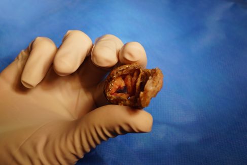
        • VG dilaté
Attitude
– Hémocultures: positives à Streptococcus mutans sensible à la Ceftriaxone.
– Consultation stomato, ORL: RAS
– Cutanée: éruption cutanée maculo-papuleuse érythémateuse sur la jambe
  et les membres supérieurs et une lésion purpurique sur l'index droit.
– Fond d'œil: taches de Roth en périphérie et para-maculaires et un signe de
  Tyndall vitréen.
– Scanner thoraco-abdomino-cérébral a montré un abcès sur le pôle inférieur
  de chaque rein et également des images suspectes d'emboles cérébraux
– IRM cérébrale : plusieurs emboles de petite taille dans différents territoires
  droits et gauches notamment capsulo-lenticulaires droit et dans le
  pédoncule cérébral droit. Ces emboles sont d'âges différents. Il y a
  également une lacune du corps calleux de la substance blanche frontale
  interne évoquant une lacune anoxique plus ancienne.

– Que faites-vous?
Attitude

– Traitement initialement probabiliste par Claforan,
  Gentamycine et Fosfomycine.
– Puis arrêt Fosfomycine
– Indication opératoire rapide (J5 ATB):
   • Abcès du trigone, perforation mitrale et Ao
   • Plastie mitrale et Bentall avec homogreffe aortique
   • 6 semaines ATB

                    Byrne et al., Ann Thorac Surg 2011; 91:2012-9)
Byrne et al., Ann Thorac Surg 2011; 91:2012-9)
Léo – VDDI réparé à 1.5 ventricules

10/10/12: Consultation aux urgences NEM: syndrome fébrile
  ressemblant à un syndrome grippal.

« DS: Je l’ai surtout vu pour m’assurer qu’il n’y avait pas de
   végétations quelque part dans ce cœur : il n’y en a pas. Il a
   simplement un syndrome grippal. «

24 après: choc septique à staphylocoque doré

Porte d’entrée?
Léo
•   08/12/2012
•   KONNO BENTALL.
•   REMPLACEMENT DU TUBE VD/AP.
•   DÉBRIDEMENT DES ABCÈS CARDIAQUES.

• Durée de CEC : 335 mn (quasi 6 h!!!!)
• Durée de clampage aortique : 146 mn + 23 mn
J10 post op
J10 post op
J10 post op
Léo
• réapparition des végétations pulmonaires à J10
• collection autour du tube de Ven Pro.

• Le scanner réalisé le 20/12/2012 montre des collections rétro-
  sternales probablement abcédées.

• Inscription sur liste le 23.12.2012
• Transplantation cardiaque le 30/12/12
Myocardites aiguës
Généralités
• Série autopsique: identification d’une myocardite dans 8,6% à
  12% en cas de mort subite
• Evolution vers la CMD possible et non exceptionnelle
• Physiopathologie

                                                Kindermann, JACC 2012
Kociol et al
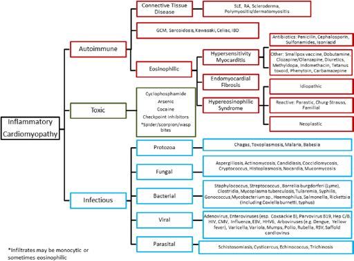
                                          Etiologies des myocardites                                                       Recognition and Management of Fulminant Myocarditis

                                                                                                                                                                                  CLINICAL STATEMENTS
                                                                                                                                                                                     AND GUIDELINES
        Downloaded from

Kociol et al              Figure 5. Causes of lymphocytic myocarditis.                                                         Recognition and Management of Fulminant Myocarditis
                          Diagram demonstrating the primary causes and associated subcategories of lymphocytic myocarditis. GCM indicates giant cell myocarditis; IBD, inflammatory
                                                                                                                                     99
Diagnostic positif
• Clinique évocatrice:
   – douleur thoracique, fièvre (30%), tachycardie (58%),
      dyspnée (68%)
   – Choc cardiogénique (Forme fulminante, 5-10/1 mill
      d’habitants/an)
   – Mort subite (TDR ou TDC)
• Biologie:                  Troponine, BNP ou N-proBNP
•   ECG:                    infarctus du myocarde
•   Echocardiographie:      dysfonction modérée à sévère
•   IRM et/ou biopsie endomyocardique (BEM)
•   Sérologies virales      peu utiles en pratique clinique
Critères de Dallas historiques (1986)

Infiltration lymphocytaire
Signe de nécrose non ischémique

               Magnani JW et al. Circulation 2006   Cooper LT et al. Circulation 2007
Problèmes des critères de Dallas
• Myocardite avec atteinte hétérogène du myocarde -
  > Biopsies multiples > 5
• Geste invasif: mortalité 0,5%, complications 5%:
  perforation cardiaque, hémopéricarde, tamponnade
• Geste plus risqué chez le nourrisson
• Variabilité d’interprétation même entre experts
                                      1916       Cooper et al.
                                                 Endomyocardial Biopsy in Cardiovascular Disease

                                      Table 1. Risks Associated With Endomyocardial Biopsy in      failure,

• « Goldstandard » mais discutée ++
                                      546 Procedures                                               potentia
                                      Overall 33 complications (6%)
                                      Sheath insertion 15 (2.7%)                                   Analy
                                        12 (2.0%) arterial puncture during local anesthesia
                                        2 (0.4%) vasovagal reaction                                EMB P
                                        1 (0.2%) prolonged venous oozing after sheath removal
                                                                                                   Sample
                                                                                                   ventricu
                                      Biopsy procedure 18 (3.3%)
                                                                                                   range fr
                                        6 (1.1%) arrhythmia
                                                                                                   and eac
                                        5 (1.0%) conduction abnormalities                          must be
                                        4 (0.7%) possible perforation (pain)                       ferred f
                                        3 (0.5%) definite perforation (pericardial fluid)          formali
                                           2 of 3 patients with definite perforation died          (21,22)
                                        Data derived from Deckers et al (20).                      prevent
                                                                                                      The
Indication d’une BEM
                      Kociol et al                                                                Recognition and Management of Fulminant Myocarditis
CLINICAL STATEMENTS
   AND GUIDELINES

                                                                                                        Figure 3. Indications for endomyocardial
                                                                                                        biopsy (EMB).
                                                                                                        Guideline-based algorithm for whether EMB
                                                                                                        is indicated. COR indicates Class of Recom-
                                                                                                        mendation; LOE, Level of Evidence; and MRI,
                                                                                                        magnetic resonance imaging. *Usually a dilated
                                                                                                        cardiomyopathy. Fulminant myocarditis may
                                                                                                        have normal end-diastolic diameter with mildly
                                                                                                        thickened walls. Exclude ischemic, hemodynam-
                                                                                                        ic (valvular, hypertensive), metabolic, and toxic
                                                                                                        causes of cardiomyopathy as indicated clinically.
                                                                                                        Reprinted from Bozkurt et al.3 Copyright ©
                                                                                                        2016, American Heart Association, Inc.

                                                                               despite normalization of cardiac enzymes and biomark-
                      EMB, CORONARY ANGIOGRAPHY,                               ers.78 EMB can be considered the primary diagnostic
                      AND INVASIVE
                            Circ 2020
                                      HEMODYNAMICS                             strategy76,79 when     magnetic resonance imaging is not
                      Kociol et al                                                            Recognition and Management of Fulminant Myocarditis
                      In the setting of cardiogenic shock, right-sided heart   possible (eg, shock, presence of metal devices) if expe-
                                                                               rienced operators and cardiac pathologists are readily
Lake Louise Criteria: IRM

Trois séquences IRM contributives:
• 1. Œdème en T2
• 2. rehaussement précoce du myocarde
• 3. rehaussement tardif du myocarde

• Diagnostic positif si > 2 critères :
   – Hypersignal T2
   – Ratio Signal myocarde / muscle périph augmenté après injection de
      Gadolinium
   – Hypersignal en rehaussement tardif

• Refaire IRM à 1-2 semaines si:
   – 0 critère mais symptômes trop récents, forte suspicion clinique
   – 1 seul critère présent

                                              Friedrich MG et al. JACC 2009
Traitement en fonction de la forme clinique

• Myocardite segmentaire focale: Repos
• Myocardite aiguë diffuse chez l’enfant
   – Surveillance +/- assistance circulatoire (HNF)
   – Traitement d’attaque:
      • Immunomodulateurs, immunosuppresseurs, Anti-
        inflammatoire, immunoadsorption
• Myocardite fulminante
   – PEC du choc cardiogénique
   – (Traitement spécifique en fonction du type histologique)

• Myocardite chronique active
   – Discuter immunosupresseurs
Traitement
ce qui est admis …
Traitement de l’insuffisance cardiaque
• Selon les guidelines
• Selon la classe fonctionnelle NYHA
   – IEC
   – Diurétiques
   – B-bloquant
   – ARA II
• Formes sévères: Prise en charge en réanimation
   – Traitement « agressif »
   – Assistance circulatoire (> 60 à 80 % survivants et
     récupération ad integrum possible)
   – Drogues Inotropes positives et héparine!
                                          Kindermann, et al., JACC 2012
                                          Amabile et al. Heart 2006
Traitement de la myocardite fulminante
                Kociol et al                                                                                            Recognition and Management of Fulminant Myocarditis

                                                                                                                                                                                 CLINICAL STATEMENTS
                                                                                                                                                                                    AND GUIDELINES
Downloaded fr

                                                                                                                                                                     Circ 2020
                Figure 4. General approach to initial stabilization of patients in cardiogenic shock.
                ACS indicates acute coronary syndrome; CABG, coronary artery bypass grafting procedure; ECMO, extracorporeal membrane oxygenation; IABP, intra-aortic bal-
Traitement
ce qui est discuté …

  Immunoglobulines
  Anti inflammatoires
       Antiviraux
 Immunosuppresseurs
Immunosuppressive Treatment for
           Myocarditis in the Pediatric
                              He et al.                                                                                                             Immunosuppressive Treatment for Myocarditis in the Pediatric

           Population: A Meta-Analysis
                              TABLE 1 | Characteristics of studies included in the meta-analysis.

                              Study                N       Age                 Study       IMSA                IMSA dosage, time of            Follow-up             Observed             Inclusion criteria
                                                                               methodology                     IMSA start                                            variables
           Bing He* , Xiaoou Li and Dan Li
                              Camargo et al.       50      5 months−15         PNCT             P, CyA         P & A: 2.5 mg/kg/d, 1           8.4±1.2 months        LVEDD, LVEF,         Active myocarditis based
           Department (9)of Pediatrics, Renmin
                                         years Hospital of Wuhan University,     Wuhan,
                                                                  week; 2.0 mg/kg/d, 3  China                                                                        PWP, CI, HR          on EMB findings
                                                                                                               weeks; 1.5 mg/kg/d, 4
                                                                                                               weeks
                                                                                                               Cy: 1.5 mg/kg/d, 1 week;
           The use of immunosuppressants in the treatment of myocarditis in children remains                   1.0 mg/kg/d, 7 weeks;
                                                                                                               0.5 mg/kg/d, 1 week
           controversial.
                    Aziz et al. (6)
                                      The
                                        68
                                                 aim of RCTthis meta-analysis
                                                3.7 ± 2.9                           P               2 mg/kg/d, 1 month
                                                                                                                            is to  15.1±9.2
                                                                                                                                             summarize    LVEDD, LVESD,
                                                                                                                                                                           the            current empirical
                                                                                                                                                                             Duration of symptoms
           evidence for immunosuppressive       years                                  treatment for myocarditis                   months                 LVEF in the for
power to detect a significant difference in the treatment effect.    in pediatric population with myocarditis. Although this meta-
                                  Moreover, we were able to ascertain publication bias in only         analysis reported beneficial outcomes with immunosuppressive
                              The four
                                    use    ofstudies,
                                                immunosuppressants
                                                       which means only four of sixin     thedata  treatment
                                                                                                       therapy, theof    myocarditis           in children         remains
                    Immunosuppressive Treatment for
                                       of six
                              controversial.        The aimDueofto this
                                  findings of this meta-analysis.
                                                                                       studies’
                                  could be merged, which may have impacted the analysis of the
                                                                                  meta-analysis
                                                                          the included studies’ lack
                                                                                                                     results have to be interpreted cautiously because
                                                                                                       one RCT was included in this meta-analysis; more large-scale
                                                                                                          is to summarize the current empirical
                                                                                                       RCTs are required in the future.
                                                                                                                                                                       only

                    Myocarditis in the Pediatric
                              evidence      for follow-up
                                  of long-term    immunosuppressive                 treatment
                                                            (only two studies had median    follow- for myocarditis in the pediatric population.
                                  up > 1 year), their inferences can only be applied to short-         AUTHOR CONTRIBUTIONS
                              We term
                                  searched         PubMed,          MEDLINE,          and Embase            for articles to identify studies analyzing
                    Population: A Meta-Analysis
                                        outcomes. In  this meta-analysis,

                                          didn’t report
                                                                          we couldn’t provide
                                  data of viral genome and histologic type, because the included
                              the studies
                                   efficiency        of these
                                                           immunosuppressive
                                                                data even if this information
                                                                                                exact

                                                                                             treatment
                                                                                                 was
                                                                                                       BH is responsible for the provision of the overall idea and writing
                                                                                                       articles. in   the data
                                                                                                                 XL collects  pediatric
                                                                                                                                 and modifiespopulation.            Pooled
                                                                                                                                                the paper. DL is responsible
                                  important to the therapy and prognosis. RCTs in the future           for statistical analysis.
                    Bing estimates wereLigenerated
                                          and Dan using                                      fixed- or random-effect models. Heterogeneity within
                       • Groupe d’enfants avec immunosuppresseurs
                         He* , Xiaoou
                       studies
                 Department         was assessed
                             of Pediatrics,
                                                   Li

                                                      Renmin using    Hospital    Cochran’s
                                                                                     of Wuhan University,Q and I2Wuhan,  statistics.  ChinaFunnel plots and Begg’s rank

                 The were
                         amélioration significative:
                            REFERENCES
                       correlation
                        use 1. also
                               of
                                           method were constructed to evaluate
                                       immunosuppressants                                       in potential
                                Sagar S, Liu PP, Cooper LT Jr. Myocarditis. Lancet. (2012) 379:738–7.
                                                                                                       the treatment
                                                                                                                               publication
                                                                                                                        detection
                                                                                                                                          ofheterogeneity.
                                                                                                                                                          bias.
                                                                                                                                    of viruses in myocardial
                                                                                                                                                  myocarditis After
                                                                                                                                                                        Sensitivity
                                                                                                                    11. Camargo PR, Okay TS, Yamamoto L, et al. Myocarditis in children and
                                                                                                                                                               tissue: implications             analyses
                                                                                                                                                                                     for immunosuppressive
                                                                                                                                                                                    in children
                                                                                                                        therapy. Int J Cardiol. (2011) 14:148:204–8. doi: 10.1016/j.ijcard.2009.
                                         conducted to evaluate the
                                doi: 10.1016/s0140-6736(11)60648-x                                                   sources
                                                                                                                        11.002            of                                                 a detailedremains
                 controversial.
                       screening  – Fraction d’éjection VG
                            2. Dancea AB.
                                          ofThe          aim
                                                 Myocarditis
                                               159Paediatrics
                                the paediatrician.        studies,    of
                                                                 in infants   this
                                                                              six
                                                                       and Child
                                                                                          meta-analysis
                                                                                and children:
                                                                                     separate
                                                                                                 A review for
                                                                                                          studies were
                                                                                     Health. (2001) 6:543–5.
                                                                                                                            is to
                                                                                                                    12. Richardson           summarize
                                                                                                                                       P, McKenna
                                                                                                                                     identified,
                                                                                                                        J, et al. Report
                                                                                                                                                     W, Bristow M, Maisch
                                                                                                                                           of the 1995 worldwith health 181
                                                                                                                                                                              the         current
                                                                                                                                                                                 B, Mautner
                                                                                                                                                                                   patients
                                                                                                                                                                                               B, O’Connell empirical
                                                                                                                                                                         organization/international    in   the
                                                                                                                                                                                                       society
                 evidence for immunosuppressive treatment for myocarditis in the pediatric population.
                                doi: 10.1093/pch/6.8.543                                                                and   federation  of cardiology  task  force on  the definition  and classification of
                       immunosuppressive                         treatment             group,          and      199 incardiomyopathies.
                                                                                                                           the conventional   Circulation. (1996) treatment                group. The
                                  – Diamètre télédiastolique VG
                            3. Burch M. Immunosuppressive           treatment in pediatric    myocarditis  Scanty                                                   93:841–2. doi: 10.1161/01.CIR.93.5.841
                 We searched    Evidence. PubMed,
                                           Heart. (2000) 90:1103–4.MEDLINE,                      and Embase
                                                                       doi: 10.1136/hrt.2004.034082                 13. Higgins forJPT, articles             to identify
                                                                                                                                         Green S (eds). Cochrane                          studies
                                                                                                                                                                        Handbook for Systematic       Reviewsanalyzing
                       immunosuppressive
                            4.
                 the efficiency
                                Liu PP, Mason  JW.  Advances    treatment group showed
                                                               in
                                             of immunosuppressive
                                (2001) 104:1076–82.
                                                                  the understanding
                                                      doi: 10.1161/hc3401.095198
                                                                                      of myocarditis. Circulation.  a significant
                                                                                                            treatment
                                                                                                                        of   Interventions
                                                                                                                        Collaboration
                                                                                                                                                improvement
                                                                                                                                              Version
                                                                                                                                     in (2011).
                                                                                                                                             the
                                                                                                                                                       5.1.0    [updated
                                                                                                                                                        pediatric
                                                                                                                                                  Available
                                                                                                                                                                               inpopulation.
                                                                                                                                                                           March     left ventricular Pooled
                                                                                                                                                                                     2011].  The    Cochrane
                                                                                                                                                              online at: http://www.cochrane-handbook.

                                  – Diminution décès et transplantation
      Edited by:
eppe Limongelli,
                       ejection
                 estimates             fraction         (LVEF) [mean
                                                generated
                                   al. Current state                          using
                                                     of knowledge on aetiology,
                                                                                          difference
                            5. Caforio AL, Pankuweit S, Arbustini E, Basso C, Gimeno-Blanes J, Felix SB,
                                etwere                                                       fixed-
                                                                                    diagnosis, management,or
                                                                                                                 1.10;  org
                                                                                                                           95% CI: 0.41,
                                                                                                              and random-effect
                                                                                                                    14. Wells GA, Shea B, O’Connell     models.  1.79]        and         significantly within
                                                                                                                                                                  D, Peterson Heterogeneity
                                                                                                                                                                                  J, Welch V, Losos M,
                                therapy of myocarditis: a position statement of the European Society of                 et2al. The Newcastle–Ottawa Scale (NOS) for Assessing the Quality of
 of Naples, Italy      decreased
                 studies    was Cardiology left
                                        assessed   ventricular
                                             Working    Group on using       end-diastolic
                                                                     Myocardial  Cochran’s               dimension
                                                                                  and Pericardial Diseases.             I (LVEDD)
                                                                                                           QEurand Nonrandomised
                                                                                                                               statistics.        [mean
                                                                                                                                            Studies      Funnel    difference
                                                                                                                                                    in Meta-Analyses.        plots
                                                                                                                                                                           Ottawa,          andHospital
                                                                                                                                                                                      ON:−0.77
                                                                                                                                                                                            Ottawa        mm, rank
                                                                                                                                                                                                        Begg’s
                                Heart J. (2013) 34:2636–48. doi: 10.1093/eurheartj/eht210                               Research Institute (2019). Available online at: http://www.ohri.ca/programs/
 Reviewed by:    correlation
                       95%6. CI:    method
                                Aziz KU,−1.35          towere
                                          Patel N, Sadullah  T,−0.20
                                                                         constructed
                                                                Tasneem H, mm]Thawerani when
                                                                                                         toviralevaluate
                                                                                                       compared
                                                                                          H, Talpur S. Acute                to the   publication
                                                                                                                                             conventional
                                                                                                                        clinical_epidemiology/oxford.asp
                                                                                                                                                                     bias.          Sensitivity
                                                                                                                                                                             treatment               group.    analyses
   Marco Merlo,  wereFurthermore,
                         also myocarditis:
                                 conducted       the20:509–15.
                                Cardiol Young. (2010)      riskto doi:evaluate              the potential
                                              role of immunosuppression: a prospective randomised study.
                                                                       of10.1017/S1047951110000594
                                                                              death and                                  sources                 of    heterogeneity.                         Afterwas
                                                                                                                    15. Okada R, Kawai S, Kasyuya H. Non-specific myocarditis: a statistical and
                                                                                                       heart transplant clinicopathologicalin study
                                                                                                                                                  conventional
                                                                                                                                                       of autopsy cases. Jpn treatment
                                                                                                                                                                                   Circ J. (1989) 53:40–8.
                                                                                                                                                                                                             a detailed
y of Trieste, Italy
 Meena Nathan,         • MAIS: 1 seule étude RCT, effectifs faibles,
                 screening        of 159
                            7. Drucker
                       significantly
                                          NA, Colanstudies,
                                Gamma-globulin
                 immunosuppressive
                                              higher
                                                      SD, Lewis AB, Beiser
                                                   treatmentthan          in
                                                                            six
                                                               of acute myocarditis
                                                            treatment
                                Circulation. (1994) 89:252–7.
                                                                                    separate
                                                                              AS, Wessel
                                                                                the
                                                                                          DL, Takahashi M,studies
                                                                                         immunosuppressive
                                                                                     in the
                                                                                                             et al.
                                                                                            pediatric population.
                                                                                      group, and 199in in
                                                                doi: 10.1161/01.CIR.89.1.252
                                                                                                                        doi:were
                                                                                                                    16. Waller BF,
                                                                                                                            2007the
                                                                                                                                            identified, with 181 patients in the
                                                                                                                              10.1253/jcj.53.40
                                                                                                                                   treatment
                                                                                                                                      Catellier MJ, Clark group
                                                                                                                                            conventional
                                                                                                                                    consecutive
                                                                                                                                                           MA, Hawley DA,  [relative
                                                                                                                                                  forensic autopsies. Clin treatment
                                                                                                                                                                                              risk
                                                                                                                                                                                Pless JE. Cardiac
                                                                                                                                                                                Cardiol. (1992) 15:760–5.
                                                                                                                                                                                                         (RR):
                                                                                                                                                                                                   pathology
                                                                                                                                                                                                          group. The
                         follow-up court
                       4.74; 95%           CI: 2.69,treatment  8.35].         No significant               heterogeneity                across the               studies was in              observed.
Medical School,             8. Bhatt GC, Sankar J, Kushwaha KP. Use of intravenous immunoglobulin                       doi: 10.1002/clc.4960151014

   United  States
                 immunosuppressive
                                compared with standard                                group
                                                                   therapy is associated        with showed
                                                                                                        improved    17. a   significant
                                                                                                                        Klugman     D, Berger JT, Sableimprovement
                                                                                                                                                        CA, He J, Khandelwal SG, Slonim        AD.left      ventricular
                                                                                                                                                                                                     Pediatric
ed by:
                       Therefraction
                 ejection       was no evidence
                                complicated by (LVEF) myocarditis. [mean
                                                                        of publication
                                clinical outcomes in children with acute encephalitis syndrome
                                                                       Pediatr Cardiol.  difference    bias whenCardiol.
                                                                                             (2012) 33:1370–6. 1.10;
                                                                                                                           assessed
                                                                                                                                 95%              bydoi:Begg’s
                                                                                                                                                CI:
                                                                                                                                  (2010) 31:222–8.        0.41,            test.
                                                                                                                        patients hospitalized with myocarditis: a multi-institutional analysis. Pediatr
                                                                                                                                                                           1.79] and significantly
                                                                                                                                                          10.1007/s00246-009-9589-9
ongelli,
rrespondence:        doi: 10.1007/s00246-012-0350-4                                          18. Arola A, Pikkarainen E, Sipilä JO, Pykäri J, Rautava P, Kytö V. Occurrence and
s, Italy   decreased
       Bing He
                        leftPR,ventricular
                Conclusions:
                  9. Camargo        Snitcowsky R, da Luz end-diastolic
                                                 There may be a possible Assoc.       dimension
                                                         PL, Mazzieri R, Higuchi ML, Rati M,     features of(LVEDD)
                                                                                                         benefit,                [mean
                                                                                                                                     a nationwidedifference
                                                                                                                            in the short term, to the
                                                                                                             childhood myocarditis:                                       −0.77
                                                                                                                                                   study in Finland. J Am Heart                  mm,
ed by:
0701@sina.com
 Merlo,
                     et al. Favorable effects of immunosuppressive therapy in children with dilated
             95% CI: −1.35
                     cardiomyopathy to
                     addition
                                               −0.20         mm]
                                     of immunosuppressive therapy in the
                                         and active myocarditis.
                              doi: 10.1007/BF00796819
                                                                          when
                                                                 Pediatr Cardiol.  (1995) compared
                                                                                          16:61–8.              toBillingham
                                                                                                    19. Aretz HT,      the ME,   conventional
                                                                                                                 management of myocarditis in the       Immunosuppressive Trea
                                                                                                              (2017) 6:e005306. doi: 10.1161/JAHA.116.005306
                                                                                                                                                             treatment
                                                                                                                                     Edwards WD, Factor SM, Fallon JT, Fenoglio
                                                                                                           JJ, et al. Myocarditis: a histopathologic definition and classification. Am J
                                                                                                                                                                                              group.
                Furthermore,              the risk of death and heart transplant                                                   in conventional treatment was
 e, Italy
ecialty
 athan,section:
                          10. Gagliardi MG, Bevilacqua M, Bassano C, Leonardi B, Boldrini R, Camassei
                     pediatric
                significantly
                                     population.
                              FD, ethigher
                                       al. Long term than     However,
                                                      follow up ofinchildren
                                                                               further       prospective
                                                                        thewithimmunosuppressive
                                                                                myocarditis treated by
                                                                                                           Cardiovasc Pathol. (1986) 1:3–14.
                                                                                                                  investigation
                                                                                                                          treatment
                                                                                                       20. Cooper LT, Baughman               is AM,
                                                                                                                                   KL, Feldman
                                                                                                                                                        Myocarditis in the Pediat
                                                                                                                                                 warranted
                                                                                                                                                    group
                                                                                                                                                                        to
                                                                                                                                                                    [relative
                                                                                                                                                     Frustaci A, Jessup       validate
                                                                                                                                                                        M, Kuhl   U, et al.risk (RR):

 as             4.74;this
     submitted to
 chool,
 States
                       95%
                              immunosuppression and of children with dilated cardiomyopathy. Heart.
                          finding.
                                CI:
                              (2004)      2.69,
                                      90:1167–71.      8.35]. No significant heterogeneity
                                                  doi: 10.1136/hrt.2003.026641                             disease: a scientificacross
                                                                                                                                 statement fromthe      Population: A Meta-Analy
                                                                                                           The role of endomyocardial biopsy in the management of cardiovascular
                                                                                                                                                         studies
                                                                                                                                                 the American               wastheobserved.
                                                                                                                                                                 Heart Association,
atric Cardiology,
                There   was no evidence of publication bias when assessed by Begg’s test.
on of the journal
 ence:               Keywords: immunosuppressive treatment, myocarditis, pediatric, cardiac function, meta-analysis                                     Bing He*, Xiaoou Li and Dan Li
 ng  He
ers in          Conclusions:
       Pediatrics                                There may
                      Frontiers in Pediatrics | www.frontiersin.org                       be        a     possible
                                                                                                             8                   benefit,            in      the       short
                                                                                                                                                             November 2019 | Volume 7 |term,
                                                                                                                                                                                       Article 430     to      the
                                                                                                                                                        Department of Pediatrics, Renmin Hospital of Wuhan University, Wuhan, China
Vous pouvez aussi lire