GDR CNRS 3751 B2i Bio-Ingénierie des Interfaces - PARIS - Campus Pierre et Marie Curie - FEMTO-ST events
←
→
Transcription du contenu de la page
Si votre navigateur ne rend pas la page correctement, lisez s'il vous plaît le contenu de la page ci-dessous

GDR CNRS 3751 B2i
Bio-Ingénierie des Interfaces
9ème journée thématique
Biofilms Marins et Bactériens
25 mars 2022
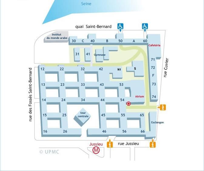
PARIS - Campus Pierre et Marie Curie
Programme
9h30-10h00      Accueil – Amphithéâtre Durand & Cave Esclangon, Bâtiment Esclangon

10h00-10h50     Introduction

                Conférence invitée
                Alain Dufour, Laboratoire de Biotechnologie et Chimie Marines
                « Batailles navales : biofilms, protéines antibiofilm et interactions hôte-pathogène »
10h50-12h10     Contribution 1 : Exploration des interactions dans l’holobionte algal pour la découverte de
                nouveaux antifoulings éco-compatibles
                Emilie ADOUANE, Raphaël LAMI, Soizic PRADO

                Contribution 2 : Marine Antibiofouling Properties of TiO2 and Ti-Cu-O Films Deposited by
                Aerosol-Assisted Chemical Vapor Deposition
                Lisa Deblock, Caroline Villardi de Oliveira, Marianne Weidenhaupt, Fabienne Faye, Carmen
                Jimenez

                Contribution 3 : Activité antibiofilm d’exopolysaccharide bactérien
                Marie CHAMPION, Isabelle LINOSSIER, Emilie PORTIER, Karine REHEL, Xavier MOPPERT,
                Claire HELLIO, Fabienne FAŸ

                Contribution 4 : Marine Biofouling from the Molecular Perspective by Atomic Force
                Microscopy
                G. Julius VANCSO
12h10-13h30     Pause déjeuner – Patio 56
13h30-14h20     Conférence invitée
                Pascal Thébault, Laboratoire Polymères, Biopolymères et Surfaces
                « Différentes approches d'élaboration de surfaces prévenant la formation de biofilms
                bactériens »
14h20-15h00     Contribution 5 : Grafting phosphonic acid polymers onto titanium implant by UV irradiation.
                Caroline Pereira, Jean-Sébastien Baumann, Vincent Humblot, Céline Falentin-Daudré

                Contribution 6 : Hybrid photoactivatable and antibacterial coatings from bioressources
                synthesized by photochemistry.
                Fanny SCHNETZ, Marc Presset, Isabelle Navizet, Yamin Leprince-Wang, Davy-Louis Versace
15h00-16h00     Pause café – POSTERS
16h00-17h00     Contribution 7 : Initial bacterial retention on polydimethylsiloxane of various stiffnesses: the
                relevance of modulus (mis)match.
                Viktoriia Drebezghova, Hubert Gojzewski, G. Julius Vancso, Corinne NARDIN

                Contribution 8 : Revêtements SLIPS (Slippery Liquid Infused Porous Surface) Résistant à la
                Déplétion de Lubrifiant pour Applications Antibactériennes
                David Riassetto, Hong-Huy Tran, Céline Ternon, Michel Langlet

                Contribution 9 : New solid-state NMR approaches to decipher at high resolution the
                molecular organization of bacterial biofilms.
                Antoine Loquet

                Mot de clôture

              9ème Journée thématique du GDR B2i - Vendredi 25 mars 2022 – Paris
Programme Posters
How to characterize the liquid/organic layer/solid interface at the nanometric scale? Application to
silane monolayers.
Adonis Steve Takala, Solène Lecot, Antonin Lavigne, Frédéric Dubreuil, Thomas Gehin, Yann
Chevolot, Christelle Yeromonahos, Magali Phaner-Goutorbe
Phospholipid-porphyrin supramolecular assemblies with photodynamic and photothermal activities
against bacteria and their biofilms.
Ali Makky, Paul Cressey, Louis-Gabriel Bronstein, Christophe Regeard
Développement du greffage de polymères bioactifs sur des copolymères d'oléfines cycliques pour
améliorer l'hydrophilie.
Jean-Sébastien Baumann, Emile Jubeli, Angelo Morocho, ,Nadjet Yagoubi, Céline Falentin-Daudré
Elaboration d’un patch par electrospinning pour le traitement de la myéloméningocèle.
Khaoula Benabdderrahmane, Jean-Sébastien Baumann, Salah Ramtani, Julien Stirnemann, Céline
Falentin-Daudré
Development of an Artificial Bioresorbable Ligament Based on pNaSS Grafted PCL.
Maya Abdallah, Céline Falentin-Daudré
Surface functionalization of non-woven polypropylene (PP) using plasma treatment: chemical and
biological approaches to induce antimicrobial properties.
Tianyue Wei, Caroline Aymes-Chodur, Christophe Regeard, Nadine Aubry-Barroca, Philippe Roger
Greffage de polymères bioactifs sur des implants en silicone afin d'améliorer la réponse biologique.
Anna Wozniak, Mylan Lam, Vivien Moris, Vincent Humblot, Céline Falentin-Daudré
Bacteria detection by surface plasmon resonance imaging of antimicrobial peptides arrays.
Charlène Pleniere, Raphaël Mathey, Arnaud Buhot, Loïc Leroy, Sandrine Boisset, Didier Boturyn,
Yoann Roupioz
9ème Journée thématique du GDR B2i - Vendredi 25 mars 2022 – ParisRÉSUMÉS COMMUNICATIONS ORALES 9ème Journée thématique du GDR B2i - Vendredi 25 mars 2022 – Paris
9ème Journée thématique du GDR B2i - Vendredi 25 mars 2022 – Paris
Conférence invitée

        Batailles navales : biofilms, protéines antibiofilm et
                    interactions hôte-pathogène
Alain DUFOUR1,*, Albane Jouault1, Amandine Morot1,2, Angélique Gobet3, Émilie Portier1,
Sophie Rodrigues1, Yannick Fleury4, Christine Paillard2, Alexis Bazire1

1
 Laboratoire de Biotechnologie et Chimie Marines (LBCM), EA 3884, Université de Bretagne-Sud,
IUEM, 56100 Lorient.
2
 Laboratoire des Sciences l’Environnement Marin (LEMAR), UMR 6539 CNRS, IRD, Ifremer, Université
de Bretagne Occidentale, IUEM, 29280 Plouzané.
3
 MARBEC, Ifremer, IRD, Université de Montpellier, CNRS, 34200 Sète.
4
 Laboratoire de Biotechnologie et Chimie Marines (LBCM), EA 3884, Université de Bretagne
Occidentale, IUEM, 56100 Quimper.

Les biofilms sont des communautés microbiennes constituées de cellules vivantes attachées
à un substrat, une interface ou les unes aux autres. Le mode de vie « biofilm » est considéré
comme prédominant pour les bactéries dans l’environnement et constitue une étape de
nombreux processus infectieux. Dans le milieu marin, les bactéries appartenant aux genres
Pseudoalteromonas et Vibrio sont particulièrement répandues. Les Pseudoalteromonas sont
connus pour leur capacité à secréter une variété de molécules bioactives et peuvent vivre en
association avec d’autres organismes marins tels que des macroalgues, tandis que les Vibrio
sont souvent des pathogènes d’animaux marins. Seront présentés des exemples de protéines
(AlpP et altérocine) à activité antibiofilm secrétées par des Pseudoalteromonas, ainsi que
d’interactions hôte-pathogènes où des Vibrio infectent des mollusques (palourdes, ormeaux)
et pour lesquels les rôles des biofilms sont examinés. Enfin, l’action antibiofilm d’une souche
de Pseudoalteromonas contre une souche de Vibrio pathogène sera présentée.
Les travaux du LBCM dans ce domaine sont financés par la Région Bretagne, les fonds
européens FEDER, le programme IDEALG (ANR-10-BTBR-04), et l’EUR ISblue (ANR-17-EURE-
0015).

Mots-Clés : biofilm, bactéries marines, Pseudoalteromonas, Vibrio, altérocine

Biographie
Après un doctorat à l’Université de Rennes 1, et deux postdoctorats à l’UTHSC à San Antonio (USA) et
à l’Université de
Cambridge (UK), Alain Dufour a intégré l’Université de Bretagne-Sud en 1996 en tant qu’enseignant-
chercheur (MCF puis PR). Ses recherches en microbiologie ont porté sur les molécules
antimicrobiennes, la régulation de la réponse aux stress, et se focalisent depuis 2005 sur les biofilms
bactériens. Il a été directeur du LBCM (2011-16), est responsable d’une mention de Master en
Biotechnologies, et a organisé le 4th International Symposium on Antimicrobial Peptides (AMP2014) en
2014.

*Correspondance : alain.dufour@univ-ubs.fr

                 9ème Journée thématique du GDR B2i - Vendredi 25 mars 2022 – Paris
Exploration des interactions dans l’holobionte algal pour la
découverte de nouveaux antifoulings éco-compatibles
Emilie ADOUANE1,2, Raphaël LAMI1,*, Soizic PRADO2
1
Sorbonne Université, CNRS, Laboratoire de Biodiversité et Biotechnologies Microbiennes (LBBM),
USR3579, Observatoire Océanologique de Banyuls, 66650 Banyuls-sur-Mer.
2
Unité Molécules de Communication et Adaptation des Microorganismes (MCAM), Muséum National
d’Histoire Naturelle (MNHN), Centre National de la Recherche Scientifique (CNRS), CP 54, 75005 Paris.
Les biofilms sont des communautés complexes de microorganismes en contact avec une
surface et incluses dans une matrice qu’elles sécrètent. Ces biofilms sont omniprésents dans
les environnements et ont un rôle majeur dans le fonctionnement des écosystèmes et les
cycles géochimiques. En milieu marin, il se développent sur des surfaces immergées
(phénomène appelé « biofouling »), à l’origine de nombreux problèmes pour les activités
marines militaires et civiles. La plupart des composés antifouling utilisés actuellement sont
des biocides ayant un effet toxique sur l’environnement. Aucune alternative durable n'a été
développée à grande échelle pour résoudre ce problème environnemental majeur en raison
du manque de composés disponibles présentant une faible écotoxicité. Cependant, ces
composés chimiques existent dans la nature : de nombreuses espèces marines ont développé
des stratégies pour se protéger du biofouling. C'est le cas de certaines macroalgues qui
sécrètent des molécules ciblant le quorum sensing, un type de communication bactérien clef
dans la formation des biofilms, limitant ainsi leur colonisation par des bactéries pathogènes.
Une stratégie innovante pour trouver de nouveaux composés naturels durables dotés
d’activité antifouling est donc de cibler les principaux mécanismes responsables de la
formation de biofilms mais qui n’affectent pas viabilité cellulaire. En ce sens, l'application de
composés à activité quorum quenching, capables d'inhiber le quorum sensing sans effet
toxique, pourrait constituer une stratégie prometteuse contre le biofouling.
Notre connaissance des holobiontes algaux a révélé l’existence et les rôles écologiques d’un
microbiote qui est capable d’impacter l’état de santé de l’algue hôte. L’épimicrobiote algal est
le siège d’une communication chimique intense et dynamique, multidirectionnelle et
multipartenaires, qui régule l’organisation des biofilms en surface de l’algue. Les mécanismes
clefs mis en jeu dans la formation de ces biofilms ont été très peu étudiés. Nous avons
cependant démontré que l'épimicrobiote de l'algue Saccharina latissima, espèce clé des côtes
du nord de l'Europe, produit des médiateurs chimiques capables d'inhiber le quorum sensing
bactérien et par extension d'inhiber la formation de biofilms. Le dialogue moléculaire du
micro-écosystème hébergé par l'algue apparaît ainsi comme une cible bio-inspirée efficace
pour trouver de nouveaux composés naturels durables, respectueux de l'environnement et à
activité antifouling.
Mots-Clés : Quorum sensing, holobionte algal, antifouling
*Correspondance : raphael.lami@obs-banyuls.fr
9ème Journée thématique du GDR B2i - Vendredi 25 mars 2022 – ParisMarine Antibiofouling Properties of TiO2 and Ti-Cu-O Films
Deposited by Aerosol-Assisted Chemical Vapor Deposition
Lisa Deblock1,*, Caroline Villardi de Oliveira1, Marianne Weidenhaupt1, Fabienne Faye2,
Carmen Jimenez1
1
Laboratoire des matériaux et du génie Physique, Univ. Grenoble Alpes, CNRS, Grenoble INP*, 38000
Grenoble, France.
2
LBCM - Laboratoire de Biotechnologie et Chimie Marines, Université Bretagne Sud.
Since 2003, the use of tributyltin (TBT) in antifouling coatings is prohibited because of its
toxicity towards marine environments(1). The last decade has seen the emergence of a new
research area: environmentally-friendly antifouling coatings. Combined with a non-toxic, time
and corrosion-resistant character, the photocatalytic properties of TiO2 make this oxide a good
eco-friendly antibiofouling alternative for marine environments(2). But even if it is showing
promising results in in vitro experiments (3), the limited exposure to sunlight in the sea and
the restricted absorbance range (UV) of TiO2 in the solar spectrum are important issues. To
enhance this range, the wide band gap of TiO2 (3,2eV) must be reduced.
In this context, two materials were synthesized(4) :
First, TiO2 films have been synthesized using AA-MOCVD deposition. Using precise
parameters, microflower-like structures are obtained, enhancing the reactive surface of the
interface and the photocatalytic activity of the material.
TiO2-Cu2O composite materials (with a Cu:Ti ratio of 1:3 and 1:1) were also synthesized.
According to Zhang et al.(5) , the substitution of Ti4+ by Cu2+/Cu1+ in the TiO2 lattice induces
the creation of oxygen vacancies and additional impurity bands right above the valence band
maximum of pure TiO2, which results in a significant reduction of both band gap and the
recombination rate of photoelectron–hole pairs, improving the photocatalytic performance
and visible light absorption of TiO2.
The film morphology, composition and photocatalytic performances were investigated, as
well as their antifouling properties during in vitro experiments and marine immersion.
Références :
(1) Vellwock, A. and Yao, H., 2021. Biomimetic and bioinspired surface topographies as a green strategy for
combating biofouling: a review. Bioinspiration & Biomimetics, 16(4), p.041003.
(2) Qi, K., Cheng, B., Yu, J. and Ho, W., 2017. A review on TiO 2 -based Z-scheme photocatalysts. Chinese Journal
of Catalysis, 38(12), pp.1936-1955.
(3) Selim, M., El-Safty, S., El-Sockary, M., Hashem, A., Elenien, O., EL-Saeed, A. and Fatthallah, N., 2016. Smart
photo-induced silicone/TiO2 nanocomposites with dominant [110] exposed surfaces for self-cleaning foul-
release coatings of ship hulls. Materials & Design, 101, pp.218-225.
(4) Caroline Villardi de Oliveira, Julie Petitbois, Fabienne Fay, Frédéric Sanchette, Frédéric Schuster,et al..
Marine Antibiofouling Properties of TiO2 and Ti-Cu-O Films Deposited by Aerosol-AssistedChemical Vapor
Deposition. Coatings, MDPI, 2020, 10 (8), pp.779. 10.3390/coatings10080779 hal-02927595
(5) Zhang, H.; Yu, X.; McLeod, J.A.; Sun, X. First-principles study of Cu-doping and oxygen vacancy effects
onTiO2for water splitting.Chem. Phys. Lett.2014,612, 106–110
Mots-Clés : marine biofouling, material interface, nanostructure, photocatalysis ; TiO 2/TiO2-Cu-O thin films
*Correspondance : lisa.deblock@grenoble-inp.fr
9ème Journée thématique du GDR B2i - Vendredi 25 mars 2022 – ParisActivité antibiofilm d’exopolysaccharide bactérien
Marie CHAMPION1,*, Isabelle LINOSSIER1 , Emilie PORTIER1, Karine REHEL1, Xavier
MOPPERT2, Claire HELLIO3, Fabienne FAŸ1
1
Laboratoire de biotechnologie et de chimie marines, 56230, Lorient.
2
Pacific Biotech, 289, Arue Tahiti.
3
Institut Universitaire Européen de le Mer, 29280, Plouzané.
La formation de biofilm sur toute surface immergée entraîne de nombreux problèmes dans
les domaines marins et médicaux. Les solutions actuelles, telles que les antibiotiques et les
biocides, sont de plus en plus confrontées à des problèmes d’antibiorésistance et de
pollution(1). Il est alors nécessaire de trouver de nouveaux moyens de lutte.
Les exopolysaccharides (EPS) sont des polymères produits par les bactéries comme moyen de
communication cellulaire, d’interactions cellules-cellules, d’interactions cellules-surfaces et
de protection. Il a été montré que des EPS ont la capacité de modifier des surfaces biotiques
ou abiotiques, moduler l’expression de certains gènes ou encore perturber la formation d’un
biofilm ou le détruire(2). Toutes ces capacités font des EPS de bons candidats comme
alternatives aux antibiotiques et aux biocides.
Dans ce travail, un EPS, MO245 a été étudié. Cet EPS, produit par une bactérie marine du genre
Vibrio isolée en Polynésie française, a des similitudes structurelles avec l’acide hyaluronique,
connu pour avoir des propriétés antiadhésives sur de nombreux modèles bactériens (3,4).
L’étude de MO245 s’est concentrée d’une part sur ses propriétés antiadhésives et antibiofilms
sur deux modèles bactériens (Pseudomonas aeruginosa sp. PaO1 et Vibrio harveyi DSM
19623). Les résultats montrent un effet antiadhésif et antibiofilm significatif sur les deux
souches sans avoir d’effet biocide.
D’autre part, et au vu des résultats, il semble important de comprendre les mécanismes
d’action de MO245. L’impact de MO245 sur la motilité, la production de HSL et de
rhamnolipides chez Pseudomonas aeruginosa et sur les voies du quorum sensing de Vibrio ont
été étudiées. Cependant, l’action de MO245 semble reposer sur ses capacités de modification
de la physico-chimie des surfaces, faisant de lui un bon candidat dans la protection des
surfaces immergées.
Références :
(1)
Khatoon Z, McTiernan CD, Suuronen EJ, Mah T-F, Alarcon EI. Bacterial biofilm formation on implantable
devices and approaches to its treatment and prevention. Heliyon. déc 2018;4(12):e01067.
(2)
Rendueles O, Kaplan JB, Ghigo J-M. Antibiofilm polysaccharides: Bacterial antibiofilm polysaccharides.
Environ Microbiol. févr 2013;15(2):334‑46.
(3)
Martin-Pastor M, Ferreira AS, Moppert X, Nunes C, Coimbra MA, Reis RL, et al. Structure, rheology, and
copper-complexation of a hyaluronan-like exopolysaccharide from Vibrio. Carbohydrate Polymers. 15 oct
2019;222:114999.
(4)
Romanò CL, Vecchi ED, Bortolin M, Morelli I, Drago L. Hyaluronic Acid and Its Composites as a Local
Antimicrobial/Antiadhesive Barrier. Journal of Bone and Joint Infection. 1 janv 2017;2(1):63‑72.
Mots-Clés : exopolysaccharide, antibiofilm, bactéries, biofilm
*Correspondance : marie.champion@univ-ubs.fr
9ème Journée thématique du GDR B2i - Vendredi 25 mars 2022 – ParisMarine Biofouling from the Molecular Perspective by
Atomic Force Microscopy
G. Julius VANCSO*
University of Twente, Sustainable Polymer Chemistry, Enschede, the Netherlands
& Nanyang Technological University, Materials Science and Engineering, Singapore
The first steps of forming a biofilm at objects immersed in marine environments involve
protein deposition. This can occur by spontaneous adsorption from seawater, or by directed
attachment by marine organisms, like barnacle cyprid larvae, in their search of a suitable
location for settlement. Atomic Force Microscopy (AFM) allows one to image proteins from
single molecule to ensemble levels, enables measurements of molecular adhesion forces, and
makes it possible to image the surface desorption process of proteins. In this presentation we
shall summarize some of the key results in this field, obtained in the author’s laboratory,
focusing on marine fouling by the barnacle Balanus amphitritae.
We first discuss observations on the morphology of 'footprints' of a putative adhesive
secretion deposited by cyprids during surface exploration. AFM provided images across the
length scales and enabled correlating protein deposit morphology, substrate surface
chemistry, and results of field settlement studies. We then turn our attention to discussing
molecular forces measured by AFM based molecular force spectroscopy and establish the
molecular basis for the sacrificial bond model for cyprid temporary protein adhesives. (1)
Protein charge at various pH and isoelectric point (pI) values are important in
understanding protein function. Here, we show a method based on AFM to determine pI using
minute quantities of proteins, with conclusions for surface fouling by cyprid settlement. (2)
Finally, we turn our attention to preventing marine fouling by surface chemical engineering.
Our results demonstrate the efficient suppression of protein adsorption on zwitterionic
pSBMA brushes. Pull-off forces by AFM force spectroscopy were hardly detectable in AFM
adhesion measurements using fibronectin-modified AFM tips that were used as a model for a
strongly nonspecifically adhering protein. (3)
References:
(1)
Phang, I.Y., Aldred, N., Clare, A.S., Vancso, G.J. ; Towards a nanomechanical basis for temporary adhesion in
barnacle cyprids (Semibalanus balanoides)
Journal of the Royal Society Interface (2008), 5 (21), pp. 397-401.
(2)
Guo, S., Zhu, X., Jańczewski, D., Lee, S.S.C., He, T., Teo, S.L.M., Vancso, G.J. ; Measuring protein isoelectric
points by AFM-based force spectroscopy using trace amounts of sample
Nature Nanotechnology (2016), 11 (9), pp. 817-823.
(3)
Schön, P., Kutnyanszky, E., ten Donkelaar, B., Santonicola, M.G., Tecim, T., Aldred, N., Clare, A.S., Vancso,
G.J. ; Probing biofouling resistant polymer brush surfaces by atomic force microscopy based force
spectroscopy, Colloids and Surfaces B: Biointerfaces (2013), 102, pp. 923-930.
Keywords : Marine fouling, adhesion proteins, molecular force spectroscopy, nanoscale imaging by AFM.
*Correspondance : g.j.vancso@utwente.nl
9ème Journée thématique du GDR B2i - Vendredi 25 mars 2022 – ParisConférence invitée
Différentes approches d'élaboration de surfaces prévenant
la formation de biofilms bactériens
Pascal THEBAULT*
Université de Normandie Unirouen, Laboratoire CNRS UMR6270 laboratoire Polymères Biopolymères
Surfaces (PBS), F-76825 Mont-Saint-Aignan.
Les contaminations microbiennes sont à l’origine de multiples pathologies dans des domaines
très divers. En effet, la colonisation de la surface des matériaux par les bactéries est un
véritable fléau tant dans le domaine de la santé publique (infections nosocomiales, plaies
chroniques, rejets d’implants,...) que dans le secteur industriel (phénomènes de biocorrosion,
occlusions des circuits de refroidissement,...) par exemple. Il est aujourd’hui admis que tout
micro-organisme favorise une vie sous la forme fixée (dite sessile) plutôt qu’en suspension
(dite planctonique). Dans la plupart des cas, ceci entraîne la formation d’un biofilm permettant
aux bactéries de persister et de résister à un environnement défavorable. Il a été montré que
ce « mode de vie » rend les bactéries jusqu’à 1000 fois plus résistantes aux traitements
antibiotiques ou autres bactéricides que leurs équivalentes planctoniques. Cette résistance
élevée rend la lutte contre les biofilms très problématique. Dans ce contexte, la prévention
semble préférable à tout traitement. Cette stratégie préventive implique notamment
l’élaboration de surfaces antibactériennes. Ces surfaces peuvent être passives afin
d’empêcher l’adhésion des bactéries sur la surface ou actives grâce à l’immobilisation de
molécules ayant des propriétés bactériostatiques ou bactéricides permettant d’inhiber la
croissance bactérienne voire entraîner la lyse des bactéries. Au sein du Laboratoire PBS de
l’Université de Rouen, nous avons développé différentes approches de surfaces
antibactériennes, de la conception à l’évaluation de l’activité antibactérienne en passant par
la caractérisation.
Mots-Clés : Biofilms, surfaces, antibactériennes, antiadhésives, modification
Biographie
Pascal Thébault a soutenu son doctorat de chimie de l’Université de Nice-Sophia Antipolis en 2007,
portant sur « l’élaboration de surfaces métalliques à visée antibactérienne ». Il a ensuite effectué deux
post-doctorats, l’un à l’Université Pierre et Marie Curie de Paris et l’autre au Commissariat à l’énergie
atomique (CEA). En 2010, il a obtenu un poste de maître de conférences au sein du laboratoire
polymères, biopolymères, surfaces de l’Université de Rouen. Il a soutenu son HDR en 2015 et il est
depuis 2017 le responsable de l’axe Polymères et Vivant. Il est auteur de plus de 25 articles dans le
domaine des surfaces fonctionnelles à visée biologique.
*Correspondance : pascal.thebault@univ-rouen.fr
9ème Journée thématique du GDR B2i - Vendredi 25 mars 2022 – ParisGrafting phosphonic acid polymers onto titanium implant by
UV irradiation
Caroline Pereira1, Jean-Sébastien Baumann1, Vincent Humblot2, Céline Falentin-Daudré1,*
1
LBPS/CSPBAT, UMR CNRS 7244, Institut Galilée, Université Sorbonne Paris Nord, 99 avenue JB-
Clément, 93430, Villetaneuse, France.
2
FEMTO-ST Institute, UMR 6174 CNRS, Université Bourgogne Franche-Comté, 15B avenue
Montboucons, 23030 Besançon Cedex, France.
Titanium (Ti) and its alloys are among the most effective metals for structural implantable
device applications. These materials have excellent physical and chemical properties such as
good corrosion resistance, acceptable biocompatibility, and mechanical properties close to
the human bone (1). However, Ti-based materials are passively integrated into the bone,
resulting in a purely mechanical attachment. Consequently, the loss of osseointegration often
leads to implant failure (2). Therefore, enhancing bone formation surrounding the implant is
primordial. Previous studies assessed in our laboratory have shown that grafting bioactive
polymers such as poly (sodium styrene sulfonate) (polyNaSS) favors osteoblast cell adhesion
and differentiation (3). In this context, this contribution proposes to functionalize Ti with a
phosphonic acid-based polymer, poly (vinyl benzyl phosphonic acid) (poly(VBP)). A two-step
UV-initiated grafting polymerization was developed to covalently graft VBP onto Ti surfaces.
The Ti substrates were evaluated for cell viability, spreading, alkaline phosphatase activity,
and calcium nodules formation using MC3T3-E1 osteoblast cells. Ti grafted samples showed
significantly better interaction with osteoblast cells and enhanced cell/surface interaction.
Together, these findings demonstrated that the presence of poly(VBP) grafted on Ti surfaces
improved osteoblasts' early cell adhesion and spreading processes (4), which is crucial for
applications aiming at improving osseointegration.
References:
(1)
Long, M.; Rack, H.J. Titanium alloys in total joint replacement—a materials science perspective. Biomaterials
1998, 19, 1621.
(2)
Windler, M.K.; in Titanium in medicine, ed. D. M. Brunette, T.P. Tengvall, M. Textor and P. Thomsen.
Springer, Berlin, 2001, p. 703.
(3)
Anagnostou, F.; Debet, F.; Pavon-Djavid, G.; Goudaby, Z.; Helary, G.; Migonney, V. Osteoblast functions on
functionalized PMMA-based polymers exhibiting Staphylococcus aureus adhesion inhibition. Biomaterials 2006,
27, 3912.
(4)
Pereira, C.; Baumann, J.S.; Humblot, V.; Falentin-Daudré, C. Biological effect of direct grafting by ultraviolet
irradiation of vinyl benzyl phosphonic acid onto titanium surfaces, Reactive and Functional Polymers (2022
under review).
Keywords: Titanium, Poly(vinyl benzyl phosphonic acid) grafting, Osseointegration, Bioactive, UV.
*Correspondence: falentin-daudre@univ-paris13.fr
9ème Journée thématique du GDR B2i - Vendredi 25 mars 2022 – ParisHybrid photoactivatable and antibacterial coatings from
bioressources synthesized by photochemistry
Fanny SCHNETZ1,*, Marc Presset1, Isabelle Navizet2, Yamin Leprince-Wang3, Davy-Louis
Versace1,*
1
Institut de Chimie des Matériaux Paris Est UMR 7182 CNRS-UPEC, 2-8 rue Henri Dunant, 94320
Thiais, France.
2
Université de Paris-Est, Laboratoire de Modélisation et Simulation Multi Échelle, UMR 8208 CNRS-
UPEM, 5 boulevard Descartes, 77420 Champs sur Marne, France.
3
Université de Paris-Est, Laboratoire Électronique, Systèmes de Communication et Microsystème,
UMR 9007 CNRS-UPEM, 5 boulevard Descartes, 77420 Champs sur Marne, France.
Photoactive polymers have shown increasing interest in recent years. Indeed, these materials
contain photoactive molecules able of producing cytotoxic species under visible light such as
reactive oxygen species (ROS). This allows to kill bacteria in contact with the surface
autonomously when irradiated.
These polymers would be synthesized by photopolymerization, which is generally considered
as a more economical and safer chemistry. Indeed, photochemical reactions occur in minutes,
at low energy cost, under mild conditions and with few organic solvents. Many types of
photoinitiators have already been developed to enable radical or cationic
photopolymerization (1), but recently there has been a growing interest in new structures that
absorb wavelengths in the visible-light or near infrared range, such as porphyrin derivatives (2,
3). Using visible light allows the use of harmless and economic lamps, such as LEDs.
New molecules derived from porphyrin would be synthesized to initiate photopolymerization
in the visible range. After polymerization, these photoinitiators would remain trapped in the
matrix. Upon irradiation, they react with oxygen to produce ROS causing bacteria’s death. This
method seems promising against nosocomial infections, since it is very unlikely that bacteria
will develop resistance (4).
Références :
(1)
Y. Yagci, S. Jockusch et N. J. turro, Photoinitiated Polymerization: Advances, Challenges, and Opportunities,
Macromolecule, 2010, vol. 43, pp. 6245-6260.
(2)
A. Al Mousawi, C. Poriel, F. Dumur, J. Toufaily, T. Hamieh, J. P. Fouassier et J. Lalevée, Zinc
tetraphenylporphyrin as high performance visible light photoinitiator of cationic photosensitive resins for LED
projector 3D printing applications, Macromolecules, 2017, vol. 50, pp. 746-753.
(3)
H. Marcille, J.-P. Malval, M. Presset, N. Bogliotti, A. Blancha-Grzechnik, V. Brezova, Y. Yagci et D.-L. Versace,
Diphenyl functional porphyrins and their metal complexes as visible-light photoinitiators for free-radical,
cationic and thiol–ene polymerizations, Polymer Chemistry, 2020, vol. 11, pp. 4237-4249.
(4)
T. Maisch, «Resistance in antimicrobial photodynamic inactivation of bacteria,», Photochem. Photobiol. Sci.,
2015, vol. 14, p. 1518.
Mots-Clés : Antibactérien - Photochimie - Porphyrines - Bioressources
*Correspondance : versace@icmpe.cnrs.fr, fanny.schnetz@u-pec.fr
9ème Journée thématique du GDR B2i - Vendredi 25 mars 2022 – ParisINITIAL BACTERIAL RETENTION ON POLYDIMETHYLSILOXANE
OF VARIOUS STIFFNESSES:
THE RELEVANCE OF MODULUS (MIS)MATCH
Viktoriia Drebezghova1, Hubert Gojzewski2, G. Julius Vancso2*, Corinne NARDIN1,*
1
Université de Pau et des Pays de l’Adour, E2S UPPA, CNRS, IPREM, 64053, Pau, France.
2
Materials Science and Technology of Polymers, Faculty of Science and Technology, University of
Twente, 7500AE, Enschede, The Netherlands.
Controlling the colonization of materials by microorganisms is crucial in a wide range of
industrial and clinical settings. Owing to continuous advances in polymer chemistry, polymer-
based devices have become increasingly used to reduce chronic infection and medical device
failure. However, the underlying mechanisms that govern the interactions of bacteria with
material surfaces remain to be established, which limits the ab initio design and engineering
of biomaterials to control bacterial attachment and further biofilm growth. Besides, the
material properties, which alter bacterial attachment, are intricately entangled and include
chemistry, hydration, surface charges, topography and mechanical properties. In order to
shed further light on the complex issue of cell-substrate adhesion and the impact on it by
surface elasticity, we investigated the initial bacterial retention of Escherichia Coli on model
poly(dimethysiloxane) (PDMS) surfaces utilizing variations of substrate stiffness without
changing other parameters that impact adhesion.
Our reference PDMS system was designed such that out of the main parameters, that
determine bacterial retention, only one was systematically varied, i.e. the mechanical
stiffness. Initial bacterial retention could thus be monitored depending on a single parameter,
i.e. materials Young’s modulus. Bulk moduli were assessed by dynamic mechanical analysis.
Substrates were engineered such that the surface modulus values, as measured by atomic
force microscopy, ranged between 0,7 and 9 MPa. Following crosslinking, we performed
Soxhlet extraction to rule out the effect of non-crosslinked, free chains. The number of
bacteria retained, as assessed by initial retention studies, decreases with the increase of both
the bulk and surface mechanical stiffness values down to a value corresponding to the Young’s
modulus of the bacterial cells. For higher values than this threshold, the number of adherent
bacteria remains constant. We tentatively explain this observation by considering conformal
overlay of bacterial and material surfaces, which might be addressed by considering contact
mechanics between bacteria and a soft elastomer surface.
Mots-Clés : bacterial retention, PDMS, mechanical stiffness, atomic force microscopy, surface modulus
*Correspondance : corinne.nardin@univ-pau.fr
9ème Journée thématique du GDR B2i - Vendredi 25 mars 2022 – ParisRevêtements SLIPS (Slippery Liquid Infused Porous Surface)
Résistant à la Déplétion de Lubrifiant pour Applications
Antibactériennes
David Riassetto1,*, Hong-Huy Tran2, Céline Ternon1, and Michel Langlet2
1
Université Grenoble Alpes, Grenoble INP, 38016, Grenoble.
2
Université Grenoble Alpes, CNRS, 38016, Grenoble.
La superhydrophobie est une caractéristique d'une surface qui caractérise sa capacité à ne pas
être mouillée par l’eau. Cette caractéristique dépend de manière synergique de la chimie et
de la morphologie de la surface. Bien que ce type de surface soit étudié depuis de nombreuses
années, maintenir cette caractéristique sur le très long terme est toujours un enjeu de taille.
Une solution afin de maintenir une surface déperlante est de combler les porosités à l’aide
d’un liquide qui va servir de lubrifiant et induire le glissement des gouttes d’eau sur la surface.
C’est que l’on nomme les revêtements SLIPS (pour Slippery Liquid Infused Porous Surfaces).
Toutefois ces surfaces présentent l’inconvénient majeur de perdre leur lubrifiant au cours du
temps les transformant ainsi en surfaces quelconques.
Lors de cette présentation, je vous présenterai d’abord les raisons principales de cette perte
de lubrifiant. Puis, je vous présenterai l’approche expérimentale que nous avons mise au point
pour synthétiser des réseaux de nanofils (NFs) de ZnO, et le lien entre la morphologie de ces
réseaux de NFs et leur aptitude à retenir plus ou moins bien le lubrifiant (1). Enfin, je vous
présenterais de premiers résultats montrant les qualités antibactériennes de nos revêtements
SLIPS en contact statique ou dynamique avec des bactéries et leur milieu de culture.
Références : (1) H.H. Tran et al., Advanced Materials Interfaces 2021, 8, 2002058.
Mots-Clés : Revêtement SLIPS, Durable, Antibactérien
*Correspondance : David.Riassetto@grenoble-inp.fr
9ème Journée thématique du GDR B2i - Vendredi 25 mars 2022 – ParisNew solid-state NMR approaches to decipher at high
resolution the molecular organization of bacterial biofilms
Antoine Loquet*
CNRS, Chemistry and Biology of Membranes and Nanoobjects (CBMN) , Institut Europeen de Chimie et
Biologie (IECB), University of Bordeaux, France.
Bacteria have a wide range of adaptive behavior to overcome extreme environmental
conditions. Amongst such responses, biofilm formation remains one of the most fascinating
today as it implies the development of complex bacterial communities which are surface-
associated. A common feature of bacterial biofilms is the production of an extracellular matrix
composed mainly of exopolysaccharides (EPS)
In Bacillus subtilis, a sporulating gram-positive bacterial species, such gigantic organization
also involves the production of two proteins, TasA and TapA, which are cell wall-associated
and have been proven crucial for intact biofilm development. eps-deleted mutants show
drastically different morphologies when grown in biofilm-forming conditions. It is unclear how
the molecular organization of the B. subtilis cell wall and its biofilms is related to the
expression and co-expression of eps, tasA and tapA genes
Gaining structural information on how eps, tasA and tapA impact the cell wall and biofilm
molecular organization is currently a technical challenge, since the molecular composition of
such bacterial surfaces (including both the cell wall and biofilms) is highly complex.
Here we will present the development of new approaches using magic-angle spinning solid-
state nuclear to decipher the molecular composition and organization of bacterial cell wall
and biofilms at high resolution. Our results encompass the analysis of the entire bacterial
biofilm including the extracellular matrix and the bacteria, and the effects of biofilm-related
gene deletions (eps, eps/tasA and eps/tapA) on such compositions. Illustrated here on Bacillus
species, our approach will open an avenue to investigate at high resolution the biofilm
organization of other micro-organisms.
Mots-Clés : biofilm, polysaccharides, NMR, high-resolution, cell wall
*Correspondance : a.loquet@iecb.u-bordeaux.fr
9ème Journée thématique du GDR B2i - Vendredi 25 mars 2022 – Paris9ème Journée thématique du GDR B2i - Vendredi 25 mars 2022 – Paris
RÉSUMÉS POSTERS

9ème Journée thématique du GDR B2i - Vendredi 25 mars 2022 – Paris
9ème Journée thématique du GDR B2i - Vendredi 25 mars 2022 – Paris
How to characterize the liquid/organic layer/solid interface
    at the nanometric scale ? Application to silane monolayers
Adonis Steve TAKALA1, Solène LECOT1, Antonin LAVIGNE1, Frédéric DUBREUIL 2, Thomas
GEHIN3, Yann CHEVOLOT3, Christelle YEROMONAHOS1,*, Magali PHANER-GOUTORBE1,*
1
 Univ Lyon, Ecole Centrale de Lyon, CNRS, INSA Lyon, Université Claude Bernard Lyon 1, CPE Lyon, INL,
UMR5270, 69134 Ecully Cedex, France.
2
 Université Lyon, CNRS, ECL, LTDS, UMR5513, 69130 Ecully, France.
3
 Univ Lyon, CNRS, Ecole Centrale de Lyon, INSA Lyon, Université Claude Bernard Lyon 1, CPE Lyon, INL,
UMR5270, 69134 Ecully Cedex, France.

A large number of phenomena occur at solid/liquid interfaces, in the first nanometers of the
liquid layer in contact with the solid surface: catalysis reactions, bio-recognition in biosensors,
wetting, corrosion processes, tribology, fretting, biofilm formation, flow into a lab-on-chip
system, smart surfaces elaboration, etc … For some of these applications, the solid surface in
contact with the liquid is covered by an organic self-assembled or grafted monolayer. The
effectiveness of this layer as a sensing layer or as a protecting strongly depends on the vertical
and lateral organization of the molecules composing it at the solid/liquid interface.
 In this study, we used Atomic Force Microscopy (AFM) in its spectroscopic mode [1] and
Molecular Dynamics Simulation [2] to characterize the impact of a PBS solution on the
organization of different silane monolayers deposited on a silicon solid surface.
 It was shown that the structure of the silane monolayers and the charge of the silane molecule
head group played a major role on the organization and dynamics properties of the ions and
water molecules on the PBS/silane/ silicon interface [3].
The advantages and limits of the two techniques of characterization will be discussed in the
framework of the development of the « 3D-AFM » method that has allowed for the first time
to generate atomically resolved 3D-images of the organization of atoms, ions and molecules
at solid-liquid interfaces[5]. However, the method has not been used to investigate a layered
system composed on interfacial water, an organic monolayer and the substrate. It remains
therefore unknown how fluctuating molecules and interfacial water can be visualized and
what information is obtained regarding their influence on the structure and functions of the
whole system.

Références :
[1]
    F. Zuttion, C. Ligeour, O. Vidal, M. Wälte, F. Morvan, S. Vidal, J.J. Vasseur, Y. Chevolot, M. Phaner-Goutorbe,
H. Schillers, Nanoscale, 2018, 10, 12771-12778.
[2] F. Zuttion, Doctoral Thesis, University of Lyon, Ecole centrale de Lyon 2016.
[3]
    S. Lecot, Doctoral thesis, Université de Lyon,Ecole centrale de Lyon 2021.
[4]
    S. Lecot, A. Lavigne, Y. Chevolot, M. Phaner-Goutorbe, C. Yeromonahos, Langmuir 2021, 18, 5563-5572.
[5]
    T. Fukuma, R. Garcia, ACS Nano, 2018, 12, 11785-11797.

Mots-Clés : Liquid/solid interface, Silane monolayer, Atomic Force Microscopy, Molecular Dynamic Simulation

*Correspondance : magali.phaner@ec-lyon.fr for AFM experiments
*Correspondance : christelle.yeromonahos@ec-lyon.fr for Molecular Dynamic simulations

                  9ème Journée thématique du GDR B2i - Vendredi 25 mars 2022 – Paris
Phospholipid-porphyrin supramolecular assemblies with
photodynamic and photothermal activities against bacteria
and their biofilms
Ali Makky1,*, Paul Cressey1, Louis-Gabriel Bronstein1, Christophe Regeard2
1
Université Paris-Saclay, CNRS, Institut Galien Paris-Saclay, 92296 Châtenay-Malabry, France.
2
I2BC - Institut de Biologie Intégrative de la Cellule, 91198 Gif sur Yvette cedex, France.
Phospholipid-porphyrin conjugates (Pl-Por) are amphiphilic scaffolds that consist of porphyrin
derivatives grafted to a lysophosphatidylcholine backbone (1-4). Owing to their structural
similarities with phospholipids, several Pl-Por conjugates have shown to be able to self-
assemble into liposome-like assemblies exhibiting unique photophysical properties compared
to their monomeric counterparts. For these reasons Pl-Por conjugates are considered
nowadays as versatile backbones to design supramolecular assemblies with multifunctional
properties and thus their application in photodynamic therapy, photothermal therapy,
photoacoustic imaging and photo-triggerable release properties (1-3). However, little is known
about the impact of their structure on their assembling properties, their optical properties as
well as on their photothermal and photodynamic activities.
In this work, we synthesized six new Pl-Por conjugates exhibiting different alkyl chain lengths
in the sn2 position of C16 Lysophosphatidylcholine and linked via peptidic bond to two types
of porphyrin derivatives; either pheophorbide-a (PhxLPC) or pyropheophorbide-a (PyrxLPC) (5).
By combining a variety of experimental techniques with molecular dynamics simulations, we
investigated the thermodynamic, the optical properties and the structure of the Pl-Por either
self-assembled or when incorporated in lipid bilayer membranes which exhibit different
fluidity. Finally, the photothermal and photodynamic efficiencies of these assemblies were
assessed on planktonic bacteria and their biofilms.
Our results demonstrated that all of the conjugates can form supramolecular assemblies with
bilayers structures but exhibit different photothermal and photodynamic activities against
Gram + and Gram - bacterial planktonic cultures and their biofilms depending on their
chemical structure of the Pl-Por conjugates.
Références:
(1)
J. F. Lovell, C. S. Jin, E. Huynh, H. Jin, C. Kim, J. L. Rubinstein, W. C. Chan, W. Cao, L. V. Wang and G. Zheng, Nat
Mater, 2011, 10, 324-332.
(2)
J. Massiot, W. Abuillan, O. Konovalov and A. Makky, Biochimica et Biophysica Acta (BBA) - Biomembranes,
2022, 1864, 183812.
(3)
J. Massiot, V. Rosilio, N. Ibrahim, A. Yamamoto, V. Nicolas, O. Konovalov, M. Tanaka and A. Makky, Chemistry
– A European Journal, 2018, 24, 19179-19194.
(4)
J. Massiot, V. Rosilio and A. Makky, Journal of Materials Chemistry B, 2019, 7, 1805-1823.
(5)
L.-G. Bronstein, P. Cressey, W. Abuillan, O. Konovalov, M. Jankowski, V. Rosilio and A. Makky, J Colloid Interf
Sci, 2022, 611, 441-450.
Mots-Clés : phospholipid-porphyrin conjugates, photodynamic therapy, photothermal therapy, supramolecular
assemblies, biofilm
*Correspondance : ali.makky@universite-paris-saclay.fr
9ème Journée thématique du GDR B2i - Vendredi 25 mars 2022 – ParisDéveloppement du greffage de polymères bioactifs sur des
copolymères d'oléfines cycliques pour améliorer
l'hydrophilie
Jean-Sébastien Baumann1, Emile Jubeli2, Angelo Morocho1,2,Nadjet Yagoubi2, Céline
Falentin-Daudré1,*
1
LBPS/CSPBAT, UMR CNRS 7244, Institut Galilée, Université Sorbonne Paris Nord Villetaneuse, France.
2
Laboratoire Matériaux et Santé EA 401, UFR de Pharmacie, Université Paris-Sud, 92290 Châtenay-
Malabry, France.
Les copolymères d'oléfines cycliques (COC) sont des matériaux relativement nouveaux par
rapport aux polymères synthétiques établis tels que les dérivés de polyoléfine, de polystyrène,
de polyuréthanne et de polyacrylate. Cette nouvelle famille de polymères présente des
propriétés physico-chimiques intéressantes, ce qui en fait des matériaux très prometteurs
pour de nombreuses applications pharmaceutiques et médicales. Les COC présente une
surface hydrophobe rendant difficile son intégration dans les systèmes biologiques. Afin
d’améliorer l'hydrophilie des COC, une méthode simple et rapide utilisant deux étapes avec
irradiation ultraviolette a été développée afin de greffer un polymère bioactif sur ces surfaces.
Le poly(sodium styrène sulfonate) un polymère bioactif2,3 a été greffé en deux étapes sur la
surface du copolymère d'oléfine cyclique. Le procédé consiste à activer la surface à l'ozone et
à greffer sous irradiation UV en présence de styrène sulfonate de sodium. La présence de
polymère sur les surfaces a été caractérisée par angle de contact avec l'eau, spectroscopie
infrarouge à transformée de Fourier microscopie électronique à balayage avec spectroscopie
à dispersion d'énergie et la quantité de polymère greffé a été déterminée par une méthode
colorimétrique. Tout d'abord, le temps d'irradiation UV pour le greffage a été étudié.
Deuxièmement, l'influence de la présence d'additif sur le greffage a été étudiée. le degré de
greffage est significativement réduit par rapport à un échantillon sans additif Nous avons
développé une méthode simple et rapide pour greffer un polymère hydrophile et bioactif de
manière covalente sur une surface COC.4
Références :
1
Bernard, M.; Jubeli, E.; Bakar, J.; Tortolano, L.; Saunier, J.; Yagoubi, N. Journal of Biomedical Materials
Research Part A 2017, 105 (12), 3333–3349.
2
Alcheikh, A.; Pavon-Djavid, G.; Hélary, G.; Petite, H.; Migonney, V.; Anagnostou, F. ,J. Mater. Sci.: Mater. Med.
2013, 24, 1745.
3
Chouirfa, H.; Evans, M. D. M.; Bean, P.; Saleh-Mghir, A.; Crémieux, A. C.; Castner, D. G.; Falentin-Daudré, C.;
Migonney, V. ACS Appl. Mater. Interfaces 2018, 10 (2), 1480–1491.
4
Baumann, J.-S.; Jubeli, E.; Morocho, A.; Migonney, V.; Yagoubi, N.; Falentin-Daudré, C. IRBM 2021
Mots-Clés : Cyclic olefin polymer, greffage de polymère bioactif, irradiation UV
*Correspondance : falentin-daudre@univ-paris13.fr
9ème Journée thématique du GDR B2i - Vendredi 25 mars 2022 – ParisElaboration d’un patch par electrospinning pour le
traitement de la myéloméningocèle
Khaoula Benabdderrahmane1, Jean-Sébastien Baumann1, Salah Ramtani1, Julien
Stirnemann2, Céline Falentin-Daudré1,*
1
Laboratoire des Biomatériaux Pour laSanté (LBPS), institut Galilée, Université Sorbonne Paris Nord, CSPBAT
UMR CNRS 7244, 93430, Villetaneuse.
2
Service d’obstétrique et de médecine Fœtale, Hopital Necker-Enfants Malades, AP-HP et EA7328,
Université de Paris, 75015, Paris.
Le myéloméningocèle (MMC) est une anomalie congénitale du système nerveux central, caractérisée par
la protrusion des méninges et de la moelle épinière à travers les arcs vertébraux ouverts. Bien qu'il soit
démontré que la réparation chirurgicale par fœtoscopie améliore les symptômes neurologiques cognitifs1,
une approche moins invasive applicable plus tôt dans la gestation est souhaitable, pour l’amélioration des
symptômes neurologiques moteurs, ainsi que la réduction des risques maternels et fœtaux. Plusieurs
études ont proposé une approche alternative et attrayante à la chirurgie prénatale, basée sur la
couverture du défaut grâce à la mise en place d’un patch pour protéger le tissu neural exposé 2. Les
matériaux utilisés dans la fabrication de ces patchs sont soit d'origine naturelle biorésorbables mais avec une
faible résistance mécanique comme le collagène3, soit d’origine synthétique avec une meilleure résistance
mécanique mais non dégradables, ce qui peut nécessiter une chirurgie de récupération secondaire2. Les
travaux antérieurs du LBPS ont permis de développer des membranes bioactives à base de Polycaprolactone
(PCL) avec d’excellentes propriétés mécaniques et biologiques4-5. En se basant sur ces travaux, l’objectif sera de
développer, en collaboration avec l’Hôpital Necker (Pr J. Stirnemann), un patch imperméable et
biodégradable, destiné à couvrir et protéger la moelle épinière exposée in utero, tout en permettant la
prolifération d’un épithélium de recouvrement et de limiter ainsi la fuite de liquide céphalorachidien.
L’objectif de ce travail est l’élaboration d’une membrane à base de PCL par electrospinning. Cette
membrane est ensuite fonctionnalisée par le greffage du poly(styrène sulfonate de sodium) (PNaSS), un
polymère bioactif connu pour ces propriétés biologiques remarquables déjà démontrées par l’équipe LPBS5,
pour enfin être caractérisée par les diverses techniques disponibles au laboratoire à savoir: FTIR, dosage
colorimétrique au bleu de toluidine et MEB-EDX.
Références :
(1) Joyeux L, Chalouhi GE, Ville Y, Sapin E. La chirurgie maternofœtale du spina bifida : perspectives d’avenir. Journal de
Gynécologie Obstétrique et Biologie de la Reproduction. 1 juin 2014;43(6):443-54.
(2)Watanabe M. et al. “Tissue Engineering Strategies for Fetal Myelomeningocele Repair in Animal Models” Fetal
Diagn Ther. 2015, 37(3), 197.
(3)Annor A.H. et al. “Effect of enzymatic degradation on the mechanical properties of biological scaffold materials”
Surg Endosc. 2012, 26(10), 2767.
(4)Amokrane G. et al. “A simple way to functionalize PCL surface by grafting bioactive polymers using UV irradiation”
IRBM, 2018, 39 (4), 268.
A(5) Amokrane G. et al. “Electrospun Poly(ε-caprolactone) Fiber Scaffolds Functionalized by the Covalent Grafting
of a Bioactive Polymer: Surface Characterization and Influence on in Vitro Biological Response” ACS Omega, 2019, 4 (17),
17194.
Mots-Clés : Myéloméningocèle, PCL, Electrospinning, Polymère bioactif.
*Correspondance : falentin-daudre@univ-paris13.fr
9ème Journée thématique du GDR B2i - Vendredi 25 mars 2022 – ParisDevelopment of an Artificial Bioresorbable Ligament Based
on pNaSS Grafted PCL
Maya Abdallah*, Céline Falentin-Daudré
LBPS/CSPBAT, UMR CNRS 7244, Institut Galilée, Université Sorbonne Paris Nord, Villetaneuse, France.
The anterior cruciate ligament (ACL), known to stabilize the knee movement, is one of the
most frequently injured structure during sport activities. Autograft and allografts are
considered as the current methods for surgical intervention, however, these strategies
represent many drawbacks such as donor site morbidity and risk of disease transmission 1.
Therefore, the development of a synthetic scaffold able to maintain the main function of the
damaged ACL and consequently to regenerate the native ACL synthesis is the alternative
approach. PCL as scaffold material is widely used in various biomedical applications as tissue
engineering due to the appropriate properties such as biocompatibility, biodegradability, high
mechanical strength and surface hydrophobicity that affect the cells interaction. In the
present study, PolyCaproLactone (PCL) has been chosen to develop biodegradable synthetic
ligaments mimicking the desired properties of ACL2. The PCL surface was functionalized with
poly (Sodium Styrene Sulfonate) (pNaSS), characterized by the presence of hydrophilic
sulfonate group having an impact on cells behavior as cells adhesion, proliferation and
differentiation3. PNaSS is as well characterized by the low toxicity and the high thermal
stability4. A thermal grafting method was processed to functionalize the PCL surface with
pNaSS. The colorimetric assay (Toluidine Blue) has indicated the success of pNaSS grafting.
Moreover, it has been demonstrated that the pNaSS grafted onto the PCL ligament surface
did not alter the physico-chemical properties of PCL. The grafted pNaSS enhance also the
cellular response by increasing the affinity of Fibroblast cells adhesion and proliferation
comparing to ungrafted PCL ligaments.
References:
1 Bourke, S. L. et al, Tissue Eng 2004, 10 (1–2), 43–52.
2 Siddiqui, N. et al, Mol Biotechnol 2018, 60 (7), 506–532.
3 Leroux, A. et al, Biointerphases 2019, 14 (4), 041004.
4 Ma, Z. et al, Journal of Applied Polymer Science 2020, 137 (38), 49157.
*Correspondance : maya.abdallah@univ-paris13.fr
9ème Journée thématique du GDR B2i - Vendredi 25 mars 2022 – ParisVous pouvez aussi lire