Mardi 2 avril 2019 Pavillon Roger-Gaudry salle M-415 - Faculté de médecine Pathologie et biologie cellulaire www.patho.umontreal.ca
←
→
Transcription du contenu de la page
Si votre navigateur ne rend pas la page correctement, lisez s'il vous plaît le contenu de la page ci-dessous
Faculté de médecine
Pathologie et biologie cellulaire
www.patho.umontreal.ca

                                    Mardi 2 avril 2019
                                    Pavillon Roger-Gaudry salle M-415
33e JOURNÉE SCIENTIFIQUE
                                  Département de pathologie et biologie cellulaire
                                               Mardi 2 avril 2019

                     Le Département de pathologie et biologie cellulaire tient à remercier :

Le Département de pathologie et biologie cellulaire ainsi que l’Institut de recherche en immunologie et
cancérologie (IRIC), de l’Université de Montréal, pour leur aide financière.

Les membres du jury :        Sébastien Carréno
                             Dorothée Dal Soglio
                             Alexandre Dubrac
                             Janos Filep

                             Louis Gaboury
                             Gilles Hickson
                             Jean-Claude Labbé
                             Roger Lippé
                             Nathalie Patey

                             Philippe Roux

Les modérateurs :            Sébastien Carréno
                             Dorothée Dal Soglio
                             Louis Gaboury

Le comité organisateur :     Sébastien Carréno
                             Alexandre Dubrac
                             Louis Gaboury
                             Diane Gingras

                             Benoit Lanthier

Les membres du personnel qui ont travaillé à l’organisation de la Journée scientifique :
Wendy Choung, Katherine David, Diane Roy et Michèle Saturné Modé
33e JOURNÉE SCIENTIFIQUE
Département de pathologie et biologie cellulaire
Mardi 2 avril 2019
Sommaire
8h00- 8h30 Arrivée
8h30- 8h40 Mot de bienvenue - Louis Gaboury, directeur du Département
Sébastien Carréno, responsable de la journée
8h40- 9h40 Présentations orales I - Modérateur : Sébastien Carréno
9h40- 10h00 Pause-santé
Durant la pause, les participants sont invités à consulter les affiches
10h00- 11h00 Présentations orales II - Modératrice : Dorothée Dal Soglio
11h00- 11h30 Conférence nouveau chercheur I
Alexandre Dubrac, PhD
Professeur/chercheur adjoint, de notre département et du centre de recherche du CHU Sainte-Justine
Titre: « Retinal vascular mural cell heterogeneity and functional plasticity »
11h30 Mot de la doyenne de la Faculté de médecine, Dre Hélène Boisjoly
11h35- 13h30 Lunch
Durant le lunch, les participants sont invités à consulter les affiches
12h15- 13h30 Présentations par affiche I *Évaluation par les membres du jury
13h30- 14h00 Conférence nouveau chercheur II
Étienne Caron, PhD
Professeur/chercheur adjoint, de notre département et du centre de recherche du CHU Sainte-Justine
Titre : «Spectrométrie de masse clinique de nouvelle génération en immunologie, vaccinologie et
immunothérapie »
14h00- 15h00 Présentations orales III - Modérateur : Louis Gaboury
15h00- 16h00 Présentations par affiche II *Évaluation par les membres du jury
16h10- 16h30 Décompte des évaluations et clôture de la journée
16h30 Cocktail et remise de prix dans le Hall d’honneur33e JOURNÉE SCIENTIFIQUE
Département de pathologie et biologie cellulaire
Mardi 2 avril 2019
Horaire des présentations orales
Présentations orales I : Membres du jury : Roger Lippé et Philippe Roux
8h40 Sabrya Carim (Résumé #8)
8h55 Jack Bauer (Résumé #3)
9h10 Camille De Jamblinne (Résumé #2)
9h25 Thilai Dhanaraman (Résumé #12)
Présentations orales II : Membres du jury : Janos Filep et Louis Gaboury
10h00 Sharmila Khullar (Résumé #6)
10h15 Élizabeth Arslanian (Résumé #1)
10h30 Eugénie Fradette (Résumé #14)
10h45 Kurosh Rahimi (Résumé #11)
Présentations orales III : Membres du jury : Jean-Claude Labbé et Gilles Hickson
14h00 Amira Othman (Résumé #4)
14h15 Virginie Mondin (Résumé #13)
14h30 Pierre Priam (Résumé #10)
14h45 Bita Khadivjam (Résumé #7)33e JOURNÉE SCIENTIFIQUE
Département de pathologie et biologie cellulaire
Mardi 2 avril 2019
Présentations orales I
Modérateur : Sébastien Carréno
8h40 RHO-DEPENDENT CONTROL OF CITRON KINASE IS REQUIRED FOR THE CONTRACTILE RING-TO-
MIDBODY RING TRANSITION DURING CYTOKINESIS.
Carim, S.*, Wernike, D.*, El-amine, N.* & Hickson, G.R.X,
Centre de Cancérologie Charles Bruneau, Centre Hospitalier Universitaire Sainte-Justine, Centre de Recherche, Département
de Pathologie et Biologie Cellulaire, Université de Montréal, Montréal.
8h55 DEVELOPMENTAL REGULATION OF GERMLINE SYNCYTIUM ORGANIZATION IN C. ELEGANS
Jack Bauer1, Mei Zhen3, Jean-Claude Labbé1,2
1
Institut de Recherche en Immunologie et en Cancérologie et 2Département de Pathologie et biologie cellulaire, Université de
Montréal, QC. 3Lunenfeld-Tanenbaum Research Institute, University of Toronto, ON.
9h10 STRIPAK COMPLEX CONTROLS MITOTIC MORPHOGENESIS THROUGH MOESIN ACTIVATION
De Jamblinne Camille1, Decelle B1, Leguay K1, Sriskandarajah N3, Roux PP1,2 , Hipfner D3, Carreno S1,2
1
Institut de recherche en immunologie et en cancérologie ; 2Département de pathologie et biologie cellulaire ; 3Institut de
recherches cliniques de Montréal, Université de Montréal
9h25 THE TUMOR SUPPRESSOR RASSF1A IS AN EFFECTOR OF THE RGK FAMILY OF SMALL GTPASE
PROTEINS
Dhanaraman T1, Singh S1, Killoran R1, Singh A2, Shifman J2 and Smith M J1
1. Département de pathologie et biologie cellulaire, et Institut de recherche en immunologie et en
cancérologie, Université de Montréal. 2. Department of Biological Chemistry, Hebrew University of Jerusalem.
Présentations orales II
Modératrice : Dorothée Dal Soglio
10h00 CORRELATION HISTOLOGIE-CYTOLOGIE DE BIOPSIES GANGLIONNAIRES/MEDIASTINALES PAR
EBUS/EUS
Khullar S, Roméo P, Leduc C, Hadjeres R,
Département de pathologie et biologie cellulaire, Centre Hospitalier de l’Université de Montréal
10h15 LOCALISATION DU MARQUAGE IMMUNOHISTOCHIMIQUE DE CD73 DANS LE
MICROENVIRONNEMENT DE METASTASES HEPATIQUES DE CANCER COLORECTAL SUR DEUX
MICROPUCES TISSULAIRES
Arslanian E1, Messaoudi N2, Soucy G1, Stagg J2, Turcotte S2, Trudel D1,2
1. Discipline pathologie, CHUM. 2. Centre de recherche du CHUM
10h30 CHRONIC ENDOMETRITIS: REVISION OF HISTOLOGICAL CHARACTERISTICS AND FERTILITY
OUTCOME
E. Fradette1, A. Bissonnette1, L. Nicholls-Dempsey1, J. Saumet2, N. Patey3,4, D. Dal Soglio3,4
1
Dept. Ob/Gyn, Université de Montréal, 2Dept. Ob/Gyn, Centre hospitalier universitaire Sainte-Justine, Montréal, 3Dept. de
Pathologie, Centre hospitalier universitaire Sainte-Justine, Montréal, 4Superviseures du projet de recherche33e JOURNÉE SCIENTIFIQUE
Département de pathologie et biologie cellulaire
Mardi 2 avril 2019
10h45 INTÉGRATION DE BIOMARQUEURS MOLÉCULAIRES À LA PRATIQUE CLINIQUE
Dr Kurosh Rahimi, Dre Cécile le Page, Dre Diane Provencher, Dre Anne-Marie Mes-Masson.
CR-CHUM, Montréal, Qc, Canada
Présentations orales III
Modérateur : Louis Gaboury
14h00 L'INTERFÉRON BÊTA FACILITE LA RÉSOLUTION DE L'INFLAMMATION PULMONAIRE INDUITE
PAR E. COLI
Othman A1, Sekheri M1, El Kebir D1, Ariel A2, Filep JG1
1
Département de pathologie et biologie cellulaire et Centre de recherche, HMR
2
Université de Haifa
14h15 A PTEN/PLC PATHWAY REDUCES ENDOSOMAL PI (4,5)P2 AND CAN COMPENSATE FOR LOSS OF
THE OCRL PHOSPHATASE
Mondin V1, Ben El Kadhi K1, Chagroui J1, Decelle B1, Carréno S 1,2
1-Institute for Research in Immunology and Cancer (IRIC), Université de Montréal, Montreal, QC H3C 3J7, Canada
2-Département de Pathologie et de Biologie Cellulaire, Université de Montréal, Montreal, QC H3C 3J7, Canada
14h30 SMARCD1 SUBUNIT OF SWI/SNF CHROMATIN REMODELING COMPLEXES IS REQUIRED FOR
LYMPHOID SPECIFICATION
Pierre Priam1, Veneta Krasteva1, Philippe Rousseau1 and Julie A Lessard1
1 Département de pathologie et biologie cellulaire, IRIC, Université de Montréal.
14h45 HERPES SIMPLEX VIRUS TYPE-1 INTERACTION WITH HUMAN ATP-DEPENDENT RNA
HELICASE DDX3X
Khadivjam B1, Bonneil E2, Thibault P2, Lippé R1 , 1Département de Pathologie et biologie cellulaire,et
2
Institut de Recherche en Immunologie et en Cancérologie, Université de Montréal.33e JOURNÉE SCIENTIFIQUE
Département de pathologie et biologie cellulaire
Mardi 2 avril 2019
Horaire des présentations par affiche
Les affiches doivent demeurer sur les babillards toute la journée, dans le Hall d’honneur
Présentations par affiche I :
12h15- 13h30 Membres du jury : Alexandre Dubrac et Gilles Hickson
Hugo Boruchowicz (Résumé #1)
Mackenzie Thornbury (Résumé #8)
Kendra Cruz Palomar (Résumé #9)
Kévin Leguay (Résumé #5)
12h15- 13h30 Membres du jury : Dorothée Dal Soglio et Nathalie Patey
Simon Roy (Résumé #2)
Louis Samson (Résumé #6)
Marie-Hélène Ngo (Résumé #7)
Présentations par affiche II :
15h00- 16h00 Membres du jury : Sébastien Carréno et Alexandre Dubrac
Catherine Vandal (Résumé #10)
Clothilde Berger (Résumé #15)
Gabriela Bernal Astrain (Résumé #16)
Swati Singh (Résumé #14)
15h00- 16h00 Membres du jury : Dorothée Dal Soglio et Nathalie Patey
Vimal Krishnan (Résumé #11)
Heidi Youssef (Résumé #13)33e JOURNÉE SCIENTIFIQUE
Département de pathologie et biologie cellulaire
Mardi 2 avril 2019
Résumés des
communications
scientifiques
orales33e JOURNÉE SCIENTIFIQUE
Département de pathologie et biologie cellulaire
Mardi 2 avril 2019
1. Localisation du marquage immunohistochimique de CD73 dans le microenvironnement de métastases
hépatiques de cancer colorectal sur deux micropuces tissulaires
Arslanian E1, Messaoudi N2, Soucy G1, Stagg J2, Turcotte S2, Trudel D1,2. 1. Discipline pathologie, CHUM. 2. Centre
de recherche du CHUM
Introduction: L’ecto-nucléotidase CD73 contribue à l’immunodépression et favorise l’envahissement en altérant
l’adhésion cellulaire. Son niveau est augmenté dans le microenvironnement tumoral. L’immunofluorescence a montré un
marquage de CD73 au sein du stroma et de la nécrose tumorale, quantitativement corrélé au pronostic. Le but est d’étudier
les micropuces tissulaires en immuno-H&E pour préciser la localisation du marquage de CD73.
Méthodes et résultats: L’échantillon comprend 31 patients, avec 1 à 3 métastases chacun (total de 50 métastases). Deux
micropuces tissulaires numérisées comprenant pour chaque patient six prélèvements tumoraux et six à l’interface entre
le foie non-tumoral et la tumeur, pour un total de 600 prélèvements, ont été étudiées avec immuno-H&E. Nous avons
exclu 118 des 600 prélèvements en vertu des critères suivants : matériel insuffisant ou mal identifié et artéfact technique
important. En l’absence d’interprétation validée du marquage immunohistochimique de CD73 dans le
microenvironnement de métastases hépatiques de carcinome colorectal, l’identification visuelle de la couleur, à divers
degrés d’intensité, a été répertoriée. Nous avons noté un marquage aux localisations suivantes : apex des cellules
néoplasiques, matériel nécrotique, membrane et cytoplasme de fibroblastes, membrane hépatocytaire, lumière et
membrane apicale des canalicules biliaires.
Conclusion et pertinence : L’immunofluorescence et l’immunohistochimie suggèrent la présence de CD73 dans le
microenvironnement de métastases hépatiques de carcinome colorectal. La signification de ce marquage est à explorer.
2. STRIPAK complex controls mitotic morphogenesis through Moesin activation
De Jamblinne Camille1, Decelle B1, Leguay K1, Sriskandarajah N3, Roux PP1,2 , Hipfner D3, Carreno S1,2
1
Institut de recherche en immunologie et en cancérologie ; 2Département de pathologie et biologie cellulaire ; 3Institut
de recherches cliniques de Montréal, Université de Montréal
Introduction: During mitosis, coordination between chromosome segregation and cell morphogenesis preserves genome
stability. Deregulation of this coordination triggers tumorigenesis. In Drosophila melanogaster, the only ERM
orthologue, Moesin, controls cell shaping and spindle orientation during mitosis. We reported that Slik kinase
phosphorylates Moesin at the plasma membrane to promote its activation at mitosis entry. However how Slik itself is
spatiotemporally regulated remains elusive.
Methods and Results: Here we discovered that core members of the STRIPAK complex (Striatin Interacting
Phosphatase and Kinase) regulate Slik localization at the plasma membrane. Previously, we identified in a genome wide
RNAi screen in Drosophila S2 cells, the four members of phosphatase complex of STRIPAK (PP2A) as Moesin
activators. We further revealed an interaction between Slik kinase and Cka (Striatin, the regulatory subunit of PP2A) by
co-immunoprecipitation assays. In addition, the depletion of Cka or Strip (Striatin Interaction Protein) reduced Slik
localization at the plasma membrane. Finally, we evidenced that Slik localization is regulated by phosphorylation, and
that Slik is a substrate of PP2A-STRIPAK. Thus, we discovered that STRIPAK regulates Slik localization potentially by
dephosphorylation. Moreover, S2 cells depleted for Cka or Strip, present a mis-coordination between cell shaping and
spindle orientation during mitosis (phenocopy Moesin downregulation).
Conclusion and Impact: We discovered a novel regulation upstream of Slik, an important but poorly characterized
kinase that controls apoptosis, growth, motility and development in vivo. Subsequently, this study will shed light on
STRIPAK complex as spatiotemporal regulator of Moesin, which controls mitotic morphogenesis.33e JOURNÉE SCIENTIFIQUE
Département de pathologie et biologie cellulaire
Mardi 2 avril 2019
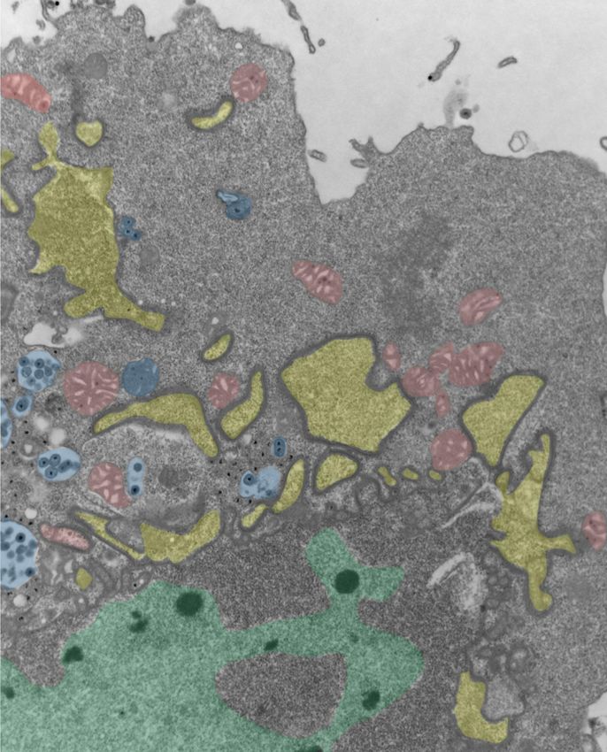
3. Developmental regulation of germline syncytium organization in C. elegans
Jack Bauer1, Mei Zhen3, Jean-Claude Labbé1,2
1
Institut de Recherche en Immunologie et en Cancérologie et 2Département de Pathologie et biologie cellulaire,
Université de Montréal, QC. 3Lunenfeld-Tanenbaum Research Institute, University of Toronto, ON.
Introduction: While a syncytial architecture is common among animal germlines and is required for fertility, the
mechanisms leading to syncytium formation during development are largely unknown. The nematode C. elegans
constitutes a powerful in vivo model to study syncytium formation and organization throughout development. In the
embryo, incomplete division of the germline precursor blastomere P4 gives rise to the two primordial germ cells (PGCs)
that remain stably interconnected by a cytoplasmic bridge enriched in conserved contractility regulators. In the larval and
adult germline, these contractility regulators are found at the stable actomyosin rings that connect germ cells to the central
rachis. We are characterizing the structure of the two PGCs at the L1 larval stage to understand the relationship between
the stable cytoplasmic bridge that forms between the PGCs during embryogenesis and the syncytial germline architecture
of adult animals. Méthodes et résultats: With confocal and electron microscopy we observed the presence of a syncytial
structure in newly-hatched animals. Contractility regulators form actomyosin rings that connect the two PGCs to a
common central cavity (a proto-rachis), to which several membrane lobes are also connected. We monitored the first
division of the PGCs to understand how they remain connected to the proto-rachis after cytokinesis. Our preliminary
results suggest that the pool of contractile regulators in the cytokinetic ring persists and directly contributes to proto-
rachis expansion. Conclusion et pertinence: Investigating the developmental regulation of C. elegans PGCs will provide
a better understanding of the mechanisms required for germline syncytium formation and organization.
4. L'interféron bêta facilite la résolution de l'inflammation pulmonaire induite par E. coli.
Othman A1, Sekheri M1, El Kebir D1, Ariel A2, Filep JG1
1
Département de pathologie et biologie cellulaire et Centre de recherche, HMR
2
Université de Haifa
Introduction: le rôle des interférons de type I dans la défense antibactérienne est peu connu. Récemment, un sous-
ensemble de macrophages a été identifié lors de la résolution de l'inflammation. Ces macrophages présentent un
transcriptome distinct caractérisé par une signature génique associée à l'IFNβ. Ainsi, nous avons voulu savoir si l'IFNβ
pourrait affecter la résolution de l'inflammation induite par la bactérie E-coli.
Méthodes et résultats: l'instillation intratrachéale de E. coli à des souris B57BL/6, provoque une lésion pulmonaire
médiée par les neutrophiles. Cette lésion atteint son pic après 6 h mais elle est complètement résolue dans les 48 h post
injection. Les souris éliminent rapidement les bactéries, suive d`une augmentation des neutrophiles apoptotiques et
d'efferocytose, ceci est associé à des élévations significatives des niveaux d'IFNβ, 24 h après l'injection de E. coli. Le
prétraitement des souris avec un anticorps neutralisant, anti-IFNβ, retarde l`élimination de E. coli, attenue l'apoptose des
neutrophiles et prolonge les lésions pulmonaires. En revanche, le traitement des souris avec un IFNβ exogène au pic de
l'inflammation améliore la clairance bactérienne, redirige les neutrophiles à l'apoptose, améliore l`efferocytose et accélère
la résolution de l'inflammation.
Conclusion et pertinence : nos données impliquent une diaphonie médiée par l'IFNβ entre les macrophages et les
neutrophiles afin de permettre une résolution rapide de l'inflammation bactérienne. Ces résultats identifient un rôle
précédemment peu connu de l'IFNβ dans la défense antibactérienne et suggèrent le potentiel thérapeutique de l'IFNβ dans
l'inflammation chronique et la réparation des blessures. (Supporté par les IRSC MOP-97742 et MOP 102619).33e JOURNÉE SCIENTIFIQUE
Département de pathologie et biologie cellulaire
Mardi 2 avril 2019
5. Spectrométrie de masse clinique de nouvelle génération en immunologie, vaccinologie et immunothérapie
Caron E, Département de pathologie et biologie cellulaire, et Centre de recherche CHU Sainte-Justine, Université de
Montréal.
L'efficacité de l'immunothérapie personnalisée chez les patients atteints de cancer est régie par la qualité et la quantité de
peptides associés aux molécules HLA présentés sur les cellules tumorales, collectivement nommé immunopeptidome, et
la capacité des cellules T à les engager. Par conséquent, une immunothérapie à base de cellules T hautement précise et
efficace n'est pas possible sans 1) une caractérisation moléculaire détaillée de l’immunopeptidome dans les tumeurs de
chacun des patients et 2) une compréhension mécanistique précise de l'engagement des cellules T chez chacun des patients
cancéreux. Pour mieux comprendre de manière globale la relation entre les cellules T et le cancer, leur analyse doit se
faire de manière systématique sur de très grandes cohortes de patients. Au cours des dernières années, j'ai contribué au
développement d'une technologie de spectrométrie de masse de nouvelle génération, appelée SWATH/DIA-MS,
permettant de mesurer quantitativement le protéome et l’immunopeptidome de cellules à partir de grandes cohortes de
patients. Au cours de cette présentation, je montrerai comment cette technologie peut être déployé pour mieux
comprendre la relation entre les cellules T et le cancer, incluant l’identification de nouvelles cibles thérapeutiques pouvant
être utilisées en immunothérapie du cancer. En tant que fondateur du « Human Immuno-Peptidome Project », un projet
d’envergure internationale, je présenterai mon programme de recherche et ma vision à long terme dans le cadre de ce
projet afin d’amener la spectrométrie de masse de nouvelle génération en clinique vers une immunothérapie extrêmement
précise et efficace chez tous les patients atteints du cancer.
6. Corrélation histologie-cytologie de biopsies ganglionnaires/médiastinales par EBUS/EUS
Khullar S, Roméo P, Leduc C, Hadjeres R, Département de pathologie et biologie cellulaire, Centre Hospitalier de
l’Université de Montréal
Introduction: Les biopsies médiastinales visent à déterminer la nature d’une adénopathie suspecte, à stadifier une
néoplasie pulmonaire, et/ou à obtenir du matériel pour analyses immunologiques et génétiques en vue d’un traitement
oncologique personnalisé. L’échographie par voie endobronchique (EBUS) et transoesophagienne (EUS) est une
technique d’acquisition de matériel biopsique qui a l’avantage d’être minimalement invasive, mais qui procure moins de
tissu pour analyse par le pathologiste. Le but principal de cette étude est d’établir le niveau de corrélation diagnostique
entre la cytologie et l’histologie provenant d’une même biopsie par EBUS ou EUS, afin de déterminer la nécessité
d’effectuer ces deux modalités.
Méthodes et résultats: Les biopsies médiastinales obtenues par EBUS ou EUS au CHUM entre mars et mai 2017 pour
lesquelles des lames de cytologie et d’histologie ont été effectuées (n=122) ont été incluses dans l’étude. Pour chaque
modalité, le diagnostic posé, l’adéquation du spécimen (qualité de quantité de matériel), les études moléculaires
demandées, ainsi que la présence d’un bloc cellulaire (cytologie) ont été répertoriés et comparés. L’adéquation des
spécimens est comparable entre l’histologie et la cytologie. La corrélation diagnostique entre ces deux modalités est de
90%. Lorsqu’il y a discordance, l’histologie s’avère plus sensible que la cytologie.
Conclusion et pertinence : Dans notre institution, il semble avantageux de privilégier l’histologie pour les biopsies
médiastinales, notamment afin d’augmenter la précision diagnostique et de préserver du matériel pour des études
supplémentaires à visée thérapeutique.33e JOURNÉE SCIENTIFIQUE
Département de pathologie et biologie cellulaire
Mardi 2 avril 2019
7. Herpes Simplex Virus type-1 Interaction with Human ATP-dependent RNA Helicase DDX3X
Khadivjam B1, Bonneil E2, Thibault P2, Lippé R1, 1Département de Pathologie et biologie cellulaire,et 2Institut de
Recherche en Immunologie et en Cancérologie, Université de Montréal. Herpetic infections are amongst the oldest and
most prevalent infections worldwide. Herpes simplex virus type 1 (HSV-1) latently persists in sensory neurons,
occasionally recurring as a lytic infection which damages the epithelium. Even though HSV-1 causes a mild disease
known as the cold sore in majority of cases, the infection can have catastrophic consequences such as encephalitis and
keratitis in immunocompromised individuals, newborns and rarely in immune competent adults.
Characterization of HSV-1 mature particles by our lab revealed the viral incorporation of approximately 49 cellular
proteins alongside the viral proteins. Next, a high throughput siRNA screen of these host proteins proved that a third of
them are active modulators of viral replication. Among these hits we found the human DDX3X protein, a DEAD-box
ATP-dependent RNA helicase. DDX3X is a nucleocytoplasmic shuttling protein that is implicated in several aspects of
RNA metabolism, translation, IFN induction and apoptosis. Moreover, DDX3X has been already shown to be involved
in the replication of various viruses. Previously, we discovered that HSV-1 yield is sensitive to DDX3X levels, as either
depleting or overexpressing the protein would negatively affect viral gene expression and genome copy numbers.
Interestingly, although DDX3X is involved in the interferon pathway, our findings show that DDX3X acts on this DNA
virus by a novel mechanism. Now, we need to elucidate a detailed mechanism for the effects we observe. Therefore, we
sought to find the interacting partners of DDX3X in the context of infection and distinguish the possible differences
between the viral (incorporated) and cellular pools of this protein.
Although the discovered MS/MS hits require further validation through some functional assays such as IF and co-IP, this
preliminary result might explain the dose-dependent behavior of DDX3X towards the virus. Perhaps, earlier during
infection, virus needs to modulate the IFN stimulatory effects of DDX3X, however, later this protein helps the virus
transport its capsids.
Despite more than 100 years of extensive research on HSV-1, many molecular and cellular aspects of the viral life cycle
remain elusive. Therefore, shedding light on the mechanistic details of HSV-1 infection can help us control this
widespread infection.
8. Rho-dependent control of Citron kinase is required for the contractile ring-to-midbody ring transition during
cytokinesis.
Carim, S.*, Wernike, D.*, El-amine, N.* & Hickson, G.R.X, Centre de Cancérologie Charles Bruneau, Centre
Hospitalier Universitaire Sainte-Justine, Centre de Recherche, Département de Pathologie et Biologie Cellulaire,
Université de Montréal, Montréal.
Introduction:Cytokinesis is the physical separation of two daughter cells following mitosis. Activation of the small
GTPase, Rho mediates the assembly of an actomyosin contractile ring (CR) at the cell equator that drives cell division.
Constriction of the CR gives rise to the midbody ring (MR) that coordinates abscission. Citron kinase or Sticky in
Drosophila, is a putative Rho-GTP binding protein required for the formation of a stable MR, however its regulation and
modes of action, are unclear. This study aims to elucidate how Sticky localizes during cytokinesis and how Sticky
contributes to the formation of a stable MR in Drosophila S2 cells.
Méthodes et résultats:Using live-cell microscopy, we identify two distinct Rho-dependent mechanisms of Sticky
localization to the MR. The first mechanism depends on scaffold protein Anillin, and mapped to the central coiled-
coil(CC) domain of Sticky (miniCC2a). Biochemical assays identified a physical interaction between Sticky-miniCC2a
and the N-terminal domain of Anillin. Furthermore, the localization of Sticky-miniCC2a and Anillin-NTD overlapped
during cytokinesis. The second mechanism of Sticky cortical recruitment occurs through the Rho-GTP binding domain
(RBD), since it was sensitive to a mutation that abolishes interaction with Rho-GTP (L1246N). Both Rho-dependent
inputs were required for robust cortical localization of Sticky. Deletion of Sticky-miniCC2a or perturbation of its RBD,
lead to unstable MR and cytokinesis failure.
Conclusion et pertinence :Sticky is recruited to the CR by two Rho-dependent mechanisms: one via its RBD and one
via Anillin. Multiple Rho-dependent interactions of Sticky are essential for the transition from CR to stable MR, a
prerequisite for successful cytokinesis.33e JOURNÉE SCIENTIFIQUE
Département de pathologie et biologie cellulaire
Mardi 2 avril 2019
9. Retinal vascular mural cell heterogeneity and functional plasticity.
Künzel SE1, Cagnone G2, Eichmann A1, Joyal JS2, Dubrac A2.
1 Department of Internal Medicine, Yale Cardiovascular Research Center, Yale University School of Medicine, New
Haven, CT, 06511, USA.
2 Department of Pathology and Cellular Biology, CHU Sainte-Justine Research Center, Université de Montréal,
Montréal, QC, Canada
Introduction:
The development and maturation of a functional vascular network is critical for tissue growth and depend on the
interaction between the endothelial cells and the pericytes. Pericytes are mural cells that surround capillaries and control
angiogenesis and capillary barrier function. Pericyte dysfunction promotes the onset and progression of several vascular
diseases including neurodegenerative diseases, intracerebral hemorrhage (ICH) and retinopathy. However, due to a
paucity of defining markers, pericyte identification and functional characterization remain ambiguous and data
interpretation problematic.
Méthodes et résultats:
We have recently shown that abnormal, α-SMA-expressing pericytes cover angiogenic sprouts and pathological
neovascular tufts (NVTs) in a mouse model of oxygen-induced retinopathy (OIR), a model that mimics retinopathy of
prematurity (ROP). Genetic lineage tracing demonstrates that pericytes acquire α-SMA expression during NVT
formation. Pericyte depletion decreases NVT formation and impairs revascularization, whereas inactivation of the NCK1
and NCK2 adaptor proteins inhibits pericyte migration, NVT formation, vascular leakage and increases revascularization,
suggesting that pericyte activation promotes the vascular defects observed in the OIR model.
Conclusion et pertinence :
Whereas insight has recently been obtained on the identity of pericytes in different organs, much less is known about the
mechanisms regulating pericyte activation and how activated pericytes contribute to vascular diseases. Then, the main
goal of our research program is to precisely define the identity of pericytes in normal and OIR retinas, investigate new
signaling pathways involved in pericyte activation and study the function of activated pericyte in blood brain barrier
defects and ICH.
10. Smarcd1 subunit of SWI/SNF chromatin remodeling complexes is required for lymphoid specification.
Pierre Priam1, Veneta Krasteva1, Philippe Rousseau1 and Julie A Lessard1
1 Département de pathologie et biologie cellulaire, IRIC, Université de Montréal.
Introduction: Lymphocyte development from hematopoietic stem cells (HSCs) is accompanied by a loss of self-renewal
capacity and a progressive restriction of developmental potential. Although the molecular mechanisms for generation of
mature lymphocytes are beginning to be revealed, a major unanswered question is how the progeny of HSCs initiate
commitment toward lymphoid fate. Increasing evidence indicates that specialized assemblies of ATP-dependent
SWI/SNF chromatin remodeling complexes perform lineage-specific roles in hemopoiesis.
Methods and Results: Our studies show that the Smarcd1 SWI/SNF subunit is specifically required for establishing
lymphoid cell identity and function in the hematopoietic system. Acute deletion of Smarcd1 in adult hematopoiesis causes
lymphopenia with near complete absence of mature B- and T-lymphocytes, whereas the myeloid and erythroid lineages
remain largely unaffected. Lymphoid primed multipotent progenitors are severely reduced, indicating a role in early
lymphoid priming. Genome-wide expression studies revealed that Smarcd1 functionally collaborates with the bHLH E2A
transcription factor to establish a lymphoid-restricted signature. Mechanistically, we show that Smarcd1 directly interacts
with E2A and is required for its recruitment to the promoter of essential regulators of lymphoid specification.
Conclusion: Altogether, these studies identified Smarcd1 as a lineage-specific chromatin remodeler that collaborates
with E2A to specify cell fate and enforce developmental checkpoints in the lymphoid lineage.33e JOURNÉE SCIENTIFIQUE
Département de pathologie et biologie cellulaire
Mardi 2 avril 2019
11. Intégration de biomarqueurs moléculaires à la pratique Clinique
Dr Kurosh Rahimi, Dre Cécile le Page, Dre Diane Provencher, Dre Anne-Marie Mes-Masson.
CR-CHUM, Montréal, Qc, Canada
Introduction :
Le cancer de l’ovaire est le cancer gynécologique le plus meurtrier, et compte environ 2500 nouveaux cas par an au
Canada. L’espérance de vie des patientes varient beaucoup en fonction du sous-type de cancer (histotype, grade, stade de
la maladie). L’identification précise de chaque sous-type est donc primordiale pour le bon diagnostic et la prise en charge
du patient. Au laboratoire, nous développons des modèles biologiques pour identifier, et test, des biomarqueurs afin de
mieux diagnostiquer et prédire la réponse aux thérapies des patientes.
Méthodes et résultats :
Grâce à la banque de tissus gynécologiques du CRCHUM, et à la ressource du consortium Canadien CŒUR, nous avons
construit plusieurs micro-étalage tissulaires (TMA) représentant les différents sous-types de cancer de l’ovaire. Ainsi
nous avons évalué, par immunohistochimie, les marqueurs p53, p16, PR, WT1, PAX8 et Napsin, pour discriminer les
différents sous-types du cancer de l’ovaire. D’autres marqueurs, tel que p16 (CDKN2A) ou la mutation du gène BRCA,
semblent eux aider à prédire le pronostic des patientes.
Conclusion et pertinence :
Les banques de tissus sont des ressources précieuses pour la recherche de biomarqueurs et permettent de construire des
cohortes d’étude pour étudier, et valider, des biomarqueurs biologiques utiles aux cliniciens.
12. The tumor suppressor RASSF1A is an effector of the RGK family of small GTPase proteins
Dhanaraman T1, Singh S1, Killoran R1, Singh A2, Shifman J2 and Smith M J1
1. Département de pathologie et biologie cellulaire, et Institut de recherche en immunologie et en
cancérologie, Université de Montréal. 2. Department of Biological Chemistry, Hebrew University of Jerusalem.
Introduction: RASSF proteins are tumor suppressors that are downregulated by promoter hypermethylation in numerous
cancer cells. There are 10 RASSF homologs (RASSF1-10) in the human proteome, each comprising RAS Association
domain proposed to bind specifically with activated RAS GTPases. RA domains are present in all effector proteins that
interact with RAS, enabling signal transduction to number of downstream processes from proliferation to apoptosis.
Direct binding between RAS and RASSFs has only been reported for RASSF5, and only limited information is available
describing the interactions and functions of other RASSF proteins. In this study, we aim to identify and characterize
RASSF1 interactors. Methods and results: We have begun to characterize RASSF protein interactions with RAS small
GTPases. We find that only RASSF5 interacts with KRAS, with a K d of 1.7 µM. In contradiction to other studies, we
report that only Classical-RASSFs (1-6) bind the MST1 kinase. We propose that RASSF7-10 are actually members of
the ASPP family of GTPase effectors, and show that N-terminal RASSFs interact with p53 regulatory ASPP proteins
(ASPP1/2). Further, we have generated homology models for all RASSFs RA domains based on the crystal structure of
the HRAS-RASSF5 complex. Using our model of the RASSF1 RA domain, we identified key residues that diverge from
those at the RASSF5-RAS interface. These data helped us identify alternative GTPase partners for RASSF1, and we
demonstrate it interacts specifically with the RGK family and RASL small GTPases. We confirmed these interactions
thorough biochemical and co-localization studies. Conclusion:Overall, our study provides a thorough characterization
of the specific interactions mediating RASSF tumour suppressor activity, and suggests that the significant expansion of
this family in vertebrates has evolved to relay signals from diverse RAS superfamily GTPases to the proapoptotic
pathways.33e JOURNÉE SCIENTIFIQUE
Département de pathologie et biologie cellulaire
Mardi 2 avril 2019
13. A PTEN/PLC pathway reduces endosomal PI(4,5)P2 and can compensate for loss of the OCRL phosphatase
Mondin V1, Ben El Kadhi K1, Chagroui J1, Decelle B1, Carréno S 1,2
1-Institute for Research in Immunology and Cancer (IRIC), Université de Montréal, Montreal, QC H3C 3J7, Canada
2-Département de Pathologie et de Biologie Cellulaire, Université de Montréal, Montreal, QC H3C 3J7, Canada
The Oculo-Cerebro-Renal Lowe (OCRL) syndrome is a rare multisystemic disease characterized by anomalies affecting
the eyes, the nervous system and the kidneys. This incurable disease is caused by mutations in the OCRL gene, which
encodes for the phosphatidylinositol (4,5) bisphosphate (PIP 2) phosphatase OCRL1. We previously reported that
depletion of dOCRL, the orthologue of this phosphatase in Drosophila melanogaster promote accumulation of
PtdIns(4,5)P2 on endosomes and cytokinesis defects, leading to multinucleation. By investigating how PtdIns(4,5)P2 is
produced on endosomes of dOCRL-depleted cells, we investigated an important enzyme for direct production of
PtdIns(4,5)P2 in Drosophila S2 cells : the well-known tumor suppressor Phosphatase and tensin homolog deleted on
chromosome 10 (PTEN) that dephosphorylates PtdIns(3,4,5)P3 into PtdIns(4,5)P2.
Here, we make the unexpected discovery that in Drosophila, PTEN reduces PtdIns(4,5)P 2 levels on endosomes,
independently of its phosphatase activity. This new PTEN function requires the enzymatic action of dPLCXD, an atypical
Phospholipase C. Importantly, we discovered that this novel PTEN/dPLCXD pathway can compensate for dOCRL
depletion. Here, we show that PTEN or dPLCXD overexpression prevents the accumulation of PtdIns(4,5)P2 on
endosomes and cytokinesis defects. In addition, we found that chemical activation of this pathway restores normal
cytokinesis in human Lowe syndrome cells and rescues OCRL phenotypes in a zebrafish Lowe syndrome model
(manuscript in revision, JCB).
Our findings identify, a novel PTEN/dPLCXD pathway that controls PtdIns(4,5)P2 levels on endosomes. They also point
to a potential new strategy for the treatment of Lowe syndrome.
14. Chronic endometritis: revision of histological characteristics and fertility outcome
E. Fradette1, A. Bissonnette1, L. Nicholls-Dempsey1, J. Saumet2, N. Patey3,4, D. Dal Soglio3,4
1
Dept. Ob/Gyn, Université de Montréal, 2Dept. Ob/Gyn, Centre hospitalier universitaire Sainte-Justine, Montréal, 3Dept.
de Pathologie, Centre hospitalier universitaire Sainte-Justine, Montréal, 4Superviseures du projet de recherche
Introduction: Chronic endometritis (CE) is defined as a chronic inflammation of the endometrial lining characterized
by unusual infiltration of plasma cells in the endometrial stroma. Studies have suggested the potential negative impact of
CE on uterine receptivity. However, there are no establish standardized guidelines for its diagnosis. Therefore, this pilot
research aimed to investigate the histopathology of CE in women with reproductive issues.Methods and results: This
retrospective study reviewed the charts of 312 patients with a history of recurrent pregnancy loss (RPL), recurrent
implantation failure (RIF) or unexplained infertility between 2013 and 2018. Sixty-eight women diagnosed and treated
with antibiotics for CE and 11 fertile women without CE were included in the study. The endometrial biopsies sampled
before and after treatment were analysed using hematoxylin-eosin-saffron staining and immunohistochemistry staining
for Syndecan-1 (CD138). The following histological features were quantified: nodular lymphocyte infiltrates, plasma
cells by field (x100), plasma cells on the complete section, small and big gatherings of plasma cells (x200), spindle
stromal cells, oedema and fibrosis. Endometrial biopsies of women with RPL, RIF or unexplained infertility were similar
for all the evaluated features. Thus, women were separated according their reproductive outcome after antibiotic
treatment, either having successfully carried a pregnancy (group 1; n =26) or still having fertility issues (group 2; n = 42),
and were compared to the fertile subjects (group 3; n=11). Before treatment, group 1 had significantly more spindle
stromal cells than group 2. Otherwise, both group 1 and 2 showed higher degree of inflammation and more nodular
lymphocyte infiltrates than group 3. After treatment, group 1 demonstrated a significant reduction of nodular lymphocyte
infiltrates only, while group 2 presented a significant diminution of the total number of plasma cells, plasma cells per
field, small and big gatherings of plasma cells, nodular lymphocyte infiltrates and spindle stromal cells.
Conclusion: The impact of CE on fertility as the primary factor of reproductive failure remains ambiguous. Antibiotic
treatments for CE can normalize the endometrial lining, but do not ensure a positive reproductive outcome. This study
suggests that plasma cell density and nodular lymphocyte infiltrates are appropriate elements to consider for the diagnosis
of CE. Moreover, we hypothesize that a moderate degree of inflammation for women diagnosed with CE would be
favorable for the gestational process.number of plasma cells, plasma cells per field, small and big gatherings of plasma
cells, nodular lymphocyte infiltrates and spindle stromal cells.33e JOURNÉE SCIENTIFIQUE
Département de pathologie et biologie cellulaire
Mardi 2 avril 2019
Résumés des
communications
scientifiques
affiches33e JOURNÉE SCIENTIFIQUE
Département de pathologie et biologie cellulaire
Mardi 2 avril 2019
1. Identification des partenaires de gM d’HSV-1 par BioID.
Hugo Boruchowicz, Roger Lippé
Département de pathologie et biologie cellulaire, Département de microbiologie, infectiologie et immunologie,
Université de Montréal
Introduction:
Le virus herpès simplex de type 1 (HSV-1) est un virus à ADN muni d’une enveloppe d’origine cellulaire qui
réplique son génome et assemble de nouvelles particules virales dans le noyau. Parmi les glycoprotéines virales que
le virus contient, la glycoprotéine M (gM), est la première détectée dans les membranes nucléaires. On ne sait
toutefois pas comment gM est ciblé à la membrane nucléaire interne. L’hypothèse est que les protéines destinées à
la membrane nucléaire interne y sont acheminées en interagissant avec d’autres molécules, notamment des protéines
régulant le transport au travers des pores nucléaire et la matrice intranucléaire. Pour clarifier ce processus de ciblage,
mon projet consiste à identifier les partenaires de gM par spectrométrie de masse en utilisant une approche de BioID.
Méthodes et résultats :
Des lignées cellulaires ont été générées dans les HEK293 Flp-In (BirA*Ha, NLSBirA*Ha, UL10BirA*HA). Suite
à l’ajout de biotine et d’une infection au HSV-1 ∆2gM, une purification sur colonne de streptavidin (BioID) est
effectuée. L’identification des éventuels partenaires de gM se fait par spectrométrie de masse (plateforme l’IRIC).
Après analyses des résultats, une validation de la présence et importance des partenaires sera effectuée.
Conclusion et pertinence :
Cette approche permettra d’identifier les molécules interagissant avec gM dont certaines devraient être impliqués
dans le ciblage de cette glycoprotéine virale à la membrane interne. Cette étude bonifiera notre compréhension du
mécanisme qui permet au virus de transiter au travers les membranes nucléaires mais aussi de clarifier comment les
protéines de l’hôte y sont aussi ciblées.
2. Epidermolytic acanthoma: a study of 131 cases
Simon F. Roy MD (1), Feras M. Ghazawi MD (2), PhD, Keith A. Choate MD, PhD (3,4), Jennifer M. McNiff
MD (3,4)
(1)Department of Pathology, University of Montreal, Canada, (2) Department of Dermatology, University of Ottawa, Canada,
(3) Department of Dermatology, Yale School of Medicine, Connecticut, (4)Department of Pathology, Yale School of Medicine,
Connecticut.
BACKGROUND
Epidermolytic acanthoma (EA) is a rare, benign acquired cutaneous keratosis displaying epidermolytic
hyperkeratosis in more than 50% of its surface. Due to the sparsity of comprehensive studies, little is known on the
patient demographics and clinical characteristics of this uncommon entity. We wish to comprehensively characterize
the clinical and demographic features of EA and to differentiate it from its mimickers.
METHODS
We carried out a retrospectively review of 131 cases of EA, recorded clinical and histopathologic features and
performed linear regression of yearly incidence rates to assess for possible under-reporting of this entity.
RESULTS
EA affected both genders equally. We found 9.08 cases per 100,000 biopsy specimens per year and linear regression
analysis demonstrated significant decreasing incidence rates. Analysis of the anatomical site distribution of EA
lesions demonstrated a more frequent genital location in men (39.1% of cases, as compared to 11.3% for women).
Contrary to previous studies, lesions were most frequently single (91.7%) and the mean age of presentation was 57.8
years.
CONCLUSION
The presented largest case series to-date indicates that EA is likely an underdiagnosed entity and establishes the
demographic and clinical features of EA33e JOURNÉE SCIENTIFIQUE
Département de pathologie et biologie cellulaire
Mardi 2 avril 2019
3. Instabilité des microsatellites et protéines MMR : un contrôle de qualité en gynécopathologie
Simon F. Roy, François Gougeon
Introduction: Une des avancées récentes en gynéco-pathologie a été l’identification de défaut des protéines
réparatrices de l’ADN (en anglais le DNA mismatch repair, ou MMR) et le MSI (soit l’instabilité microsatellite)
pour dépister certains syndromes génétiques comme le syndrome de Lynch. Les microsatellites sont des courtes
séquences répétées de nucléotides, parsemées à travers le génome, et susceptibles à des erreurs de réplication du à
des glissements de la polymerase DNA.
Méthodes et résultats: Nous avons recherché par le logiciel Diamic les 250 derniers cas de cancers de l’endomètre
grâce au mot clé « endométrioïde ». Nous voulions établir le pourcentage de cas sans examens
immunohistochimiques demandés, et le pourcentage de cas avec omission du test d’hyperméthylation par PCR. Le
taux d’IHC demandé était élevé (98.4%), mais les omissions se produisaient au niveau du test de méthylation (seuls
86.3% de taux demandé).
Conclusion et pertinence : Nous avons pu détecter 27 patientes qui devront être vues en génétique, afin d’évaluer
pour un syndrome de Lynch. Il demeurerait à investiguer les raisons charnières expliquant notre faible taux de
demande de test de méthylation, qui pourtant devrait être un test réflexe.
4. Multinucleate cell angiohistiocytoma: a clinicopathologic review of 62 cases
Simon F. Roy, Danni Dong, Peggy Myung, Jennifer M. McNiff
BACKGROUND
Multinucleate cell angiohistiocytoma (MCAH) is an uncommon and likely underdiagnosed entity that is thought to
be of vascular and fibrohistiocytic origin.
OBJECTIVE AND METHODS
We retrospectively reviewed all cases diagnosed as MCAH at the Yale Medicine Dermatopathology laboratory
between January 1 st 1990 to September 1st 2018. Sixty-two cases were retained. We performed
immunohistochemistry on the ten most inflamed lesions found and assessed for a possible alteration within the ß-
Catenin/LEF-1 signaling pathway, involved in follicular induction in dermatofibroma. We subsequently established
histologic diagnostic criteria to differentiate MCAH from its mimickers.
RESULTS
MCAH affected both genders equally. The hands or fingers were affected in 51.6% of cases. We found the most
specific histologic criteria to be: 1) presence of odd multinucleated fibroblasts. b) presence of superficial parallel
fibrosis c) presence and thickening of superficial papillary dermal vessels d) absence of perifollicular fibrosis. As
for immunoreactivity, we found positivity to CD138, CD163 and CD117 in the mononuclear inflammatory infiltrate,
yet absence of Beta-Catenin and LEF1 expression indicating that MCAH and dermatofibroma are likely separate
entities.
CONCLUSION
This large case series of MCAH reviews the clinical features of this entity, characterizes immunohistochemically
the mononuclear inflammatory infiltrate and establishes histologic criteria for its diagnosis.33e JOURNÉE SCIENTIFIQUE
Département de pathologie et biologie cellulaire
Mardi 2 avril 2019
5. ERM proteins act downstream of the GPCR TBXA2R to regulate cell motility in breast cancer cells
Leguay K1, Decelle B1, He YY1, Hogue M1, Kobayashi H1, Le Gouill C1, Bouvier M1,2, Carréno S1,3
1
Institut de Recherche en Immunologie et en Cancérologie (IRIC), 2Département de biochimie et médecine
moléculaire, 3Département de pathologie et biologie cellulaire
Introduction: Metastasis is correlated with poor prognosis. For instance, metastasis is responsible for 90% of death
in breast cancer patients. Development of secondary tumors involves invasive cell migration from the primary to the
second site and requires modification of the actin and microtubule cytoskeletons. Proteins of the ERM family (Ezrin,
Radixin and Moesin) bridge these two cytoskeletons with the plasma membrane to control cell morphogenesis. ERM
overexpression and over-activation were shown to promote metastasis development. Here, we show for the first time
that a GPCR activates ERM, and that this activation could be involved in cell migration.
Methods and Results: We developed conformational BRET sensors to follow activation of individual ERM. Using
this tool, we discovered that TBXA2R activates all three ERM in HEK293T cells. We also showed that this
activation is dependent of G12/13 and Gq/11 subunits, RhoA, PKC but not ROCK using CRISPR knockout cell
lines and/or inhibitors. We also found that activation of TBXA2R increases the migration velocity and the
directionality in Hs578T triple negative breast cancer cells.
Conclusion and relevance: In this study, we discovered a new signaling pathway involving GPCR and ERM
proteins. We also showed that activation of another GPCR PAR2 also induces ERM activation suggesting that this
new pathway is conserved between different GPCR. Finally, we propose that TBXA2R could be a new potential
target for anti-metastatic treatment in breast cancer.
6. Corrélation histopathologique entre les biopsies et les traitements à l’anse diathermique (LEEP) du col de
l’utérus, un an d’expérience au CHUM.
Samson, L et Rahimi K, Département de pathologie et biologie cellulaire de l’Université de Montréal et Centre
hospitalier de l’Université de Montréal (CHUM).
Introduction:
La prise en charge des lésions intraépithéliales du col de l’utérus est directement reliée au diagnostic sur biopsie. Or,
il n’existe pas d’étude de rendement interne récent sur la concordance entre la biopsie et le spécimen de LEEP
thérapeutique.
Méthodes et résultats:
Révision de 235 rapports de biopsie cervicale ayant mené à une LEEP et relecture de 24 cas discordants. Parmi ces
derniers, 47% sont attribuables à un biais d’échantillonnage ou à une régression spontanée, 26% à une erreur
d’interprétation histologique et 26% à une erreur d’interprétation liée à l’immunohistochimie p16 pour un taux de
discordance global de 12%.
Conclusion et pertinence :
Le CHUM affiche un taux de discordance total de 12% ce qui est inférieur à ce qui est rapporté dans la littérature
(14 – 26%). Les discordances reliées à l’utilisation de l’immunohistochimie p16 sont attribuables à son utilisation
en dehors des lignes directrices. Ce projet renforce donc l’importance d’utiliser cet outil exclusivement dans des
lésions cervicales ambiguës et non pour confirmer un diagnostic morphologique clair.33e JOURNÉE SCIENTIFIQUE
Département de pathologie et biologie cellulaire
Mardi 2 avril 2019
7. Concordance cytologique-microbiologique des lavages broncho-alvéolaires chez les patients greffés.
Ngo MH et Stephenson P, Département de pathologie et biologie cellulaire, Université de Montréal, et Centre
hospitalier de l’Université de Montréal (CHUM).
Introduction:
Les infiltrats pulmonaires chez les patients greffés pulmonaires représentent souvent des infections. Au CHUM, les
échantillons de lavages broncho-alvéolaires (LBA) chez ces patients sont systématiquement envoyés pour études
cytologique et microbiologique. Le but de cette étude est de vérifier le taux de concordance entre les résultats de ces
deux modalités d’analyse.
Méthodes et résultats:
Étude des résultats de 213 échantillons de LBA chez 99 patients greffés pulmonaires avec stratification pour la
présence de bloc cellulaire, le type d’organisme et les organismes cliniquement significatif. Concordance de 61.6%
lorsque qu’un bloc cellulaire a été effectué par rapport à 47,2% sans bloc. Concordance de 10,4% pour les mycoses,
de 0% pour les mycobactéries. 37% des mycoses ont nécessité des colorations spéciales pour être détectées en
cytologie.
Conclusion et pertinence :
La présence de cas seulement détectables par la microbiologie et d’autres seulement détectables par la cytologie
renforce le fait que ces analyses sont complémentaires dans les LBA. Cette étude a aussi démontré la pertinence de
faire des colorations spéciales en plus du Papanicolaou lorsque possible.
8. Role of host proteins in HSV-1 C-capsid maturation
Mackenzie Thornbury1, Bita Khadivjam1, Nabil El Bilali1, Roger Lippé1
1
Département de Pathologie et Biologie Cellulaire, Faculté de Médicine, Université de Montréal, Montréal,
Québec, H3C 3JT, Canada
Introduction: Herpes simplex type 1 (HSV-1) is a ubiquitous human pathogen that generally causes mild disease
such as cold sores. This enveloped, dsDNA virus has a complex life cycle, one aspect of which is capsid maturation.
Capsid proteins are imported into the nucleus for assembly and DNA packaging. Capsid maturation results in 3
different types of capsids: A-capsids, B-capsids and C-capsids. A- and B- capsids result from defects in DNA
packaging and C-capsids are mature capsids that are preferentially exported from the nucleus to form infectious
virions. The mechanism of preferential export is not well understood.
Méthodes et résultats:
All three capsids types were collected and analyzed by Mass Spectrometry to determine host proteins associated
with each fraction. PCBP-1 was specifically associated with C-capsids. Using RNAi we knocked down PCBP-1 in
HeLa cells and infected them with HSV-1. Extracellular and intracellular virus fractions were collected and tittered.
Extracellular titers decreased by half in PCBP-1 KD cells while intracellular titers did not change.
Conclusion et pertinence :
Our data shows that PCBP-1 may be pro-viral, as PCBP-1 KD decreased extracellular viral titers. As we observed
no difference in intracellular virus, this suggest PCBP-1 has effects on viral egress and release. As PCBP-1 is
involved in pre-mRNA processing I hypothesis it plays a role in translation of host proteins involved in the
trafficking of mature virions from TGN to the plasma membrane. If true, this could help elucidate the intricacies of
HSV-1 egress and release.Vous pouvez aussi lire