Bactériémie en réanimation - C. Dupuis Service de Médecine Intensive et Réanimation CHU Gabriel Montpied
←
→
Transcription du contenu de la page
Si votre navigateur ne rend pas la page correctement, lisez s'il vous plaît le contenu de la page ci-dessous
Bactériémie en réanimation

                     C. Dupuis
    Service de Médecine Intensive et Réanimation
               CHU Gabriel Montpied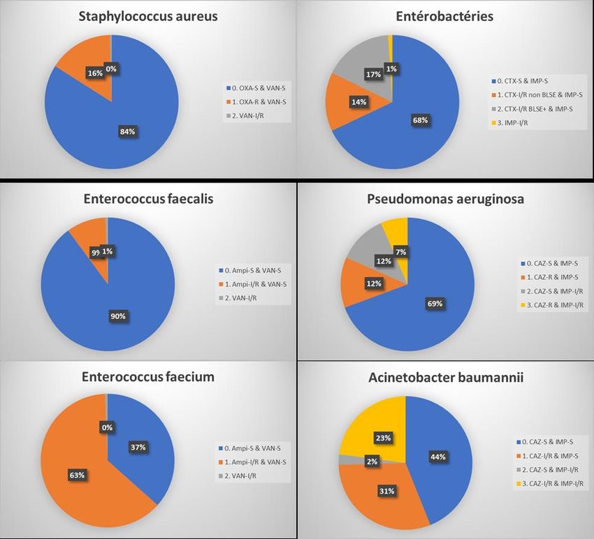
Épidémiologie

• Incidence – sources – microbiologie – résistances – mortalité - COVID

Diagnostic et bilan d’extension

•   Modalité de réalisation des hémocultures
•   Interprétation (bactériémie vrai versus contamination)
•   Les nouveaux outils
•   Bilan d’extension

Les modalités thérapeutiques d’une bactériémie

•   Contrôle de la source
•   Antibiothérapie probabiliste
•   Bi antibiothérapie
•   Monitorage
•   Désescalation
•   Durée
Quelques définitions
• Bactériémie primitive si pas de porte d’entrée
• Bactériémie persistante: si hémocultures toujours positives après trois jours
  d’antibiothérapie bien conduite
• BSI: Bloodstream infection: bactériémie
• Community-acquired BSI (CA – BSI): Bactériémie communautaire identifiée dans les
  48H00 suivant l’admission à l’hôpital chez un patient sans exposition récente au système
  de santé
• Healthcare-associated BSI: Bactériémie associée aux soins identifiée dans les 48H00
  suivant l’admission à l’hôpital chez un patient exposé au système de soins à savoir
  dialysé chronique, vivant en milieu médicalisé, avec une exposition récente aux
  antibiotiques.
• Hospital-acquired BSI: Bactériémie acquise à l’hôpital: identifiée dans les 48H00 après
  l’admission à l’hôpital
• ICU-acquired BSI: Bactériémie identifié dans les 48H00 après l’admission en réanimation
Introduction
   5% des patients en réanimation vont
   présenter une bactériémie

   Entre 30 et 40% des patients admis pour
   sepsis ou choc septique en réanimation
   présente une bactériémie
   Mortalité entre 35 et 50% en réanimation

Timsit, ICM, 2020

Principales sources et germes en cause
                                    • BSI bloodstream infection,
                                    • CAP community-acquired
                                      pneumonia,
                                    • HCAP healthcare-associated
                                      pneumonia,
                                    • UTI urinary tract infection,
                                    • ESBL-PE extended-spectrum
                                      beta-lactamase-producing
                                      Enterobacterales,
                                    • SSTI skin and soft-tissue
                                      infection,
                                    • HAP hospital-acquired
                                      pneumonia,
                                    • VAP ventilator-associated
                                      pneumonia.
Timsit, ICM, 2020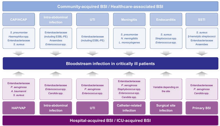
Réa raisin 2014 et Eurobact
    • En 2015:
       • 3.64/100 patients surveillés
                                                N=2 672
       • 3.52 pour 1000 j de séjour en
         réanimation
    • ↗ taux incidence des
      bactériémies entre 2004 et 2015
      de 3.31 à 3.52 pour 1000 j de
      séjour en réanimation
      (+6.3%,pval=0.07)
Dans Eurobact (n=1156):
•    21% infections sur dispositif vasculaire
                                                Souvent origine KT,
•    21% pneumopathie
                                                abdo, respi et non
•    12% infection intra abdominale                 identifiée
•    24% pas de source retrouvée                                      Tabah, ICM, 2012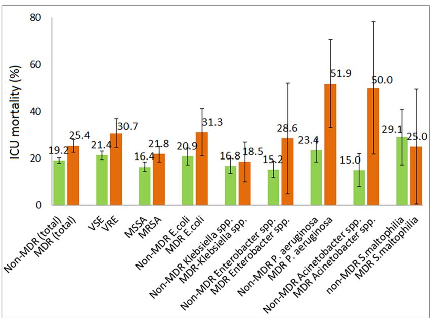
Bloodstream infection
                                                      The increase in BSI rate in the 2020Cov group compared with
                                                      the 2019NonCov group was related to an increase in intra-
                                                      vascular device origin of infection, particularly from peripheral
                                                      catheters, but also from pulmonary origin, whereas bacteraemia of
                                                      digestive origin were less frequent (Table 4).

major effect on the hospital organization, with work
overload, creation of temporary beds in ICUs,
involvement of personnel not usually dedicated to ICUs,
and an initial shortage of personal protective equipment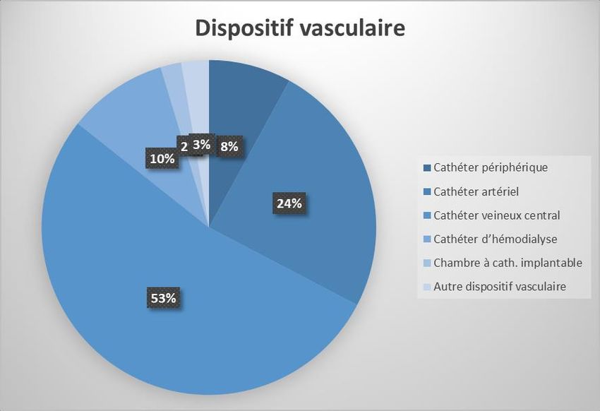
!!!! ↗
   Les germes des CA-BSI en réanimation                                                 Entérobactérie
                                                                                             BLSE

   • Variation en fonction des pays, régions, hôpitaux, source de l’infection, caractéristiques du
     patient
        • Dans 70% des cas : E Coli, S Aureus, K Pneumoniae et S Pneumoniae
        • 5% de pseudomonas en communautaire surtout chez les patients immunodéprimés,
          ou bactériémies liées aux soins, ou origine urinaire ou respiratoire
        • Très peu de souche BMR
             • SARM en plateau
             • Entérobactérie BLSE en augmentation en communautaire, jusqu’à 5% des
               infections urinaires, intra abdominales – voire 20% dans certaines régions

Timsit, ICM, 2020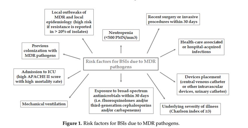
Dispositif invasif   Densité d’incidence pour 1000 cathéter-jours
                                                                             CVC                  0.5 et 2.5 épisodes

    Les HA-BSI en réanimation                                                Cathéter artériel
                                                                             ECMO
                                                                                                  1 épisode
                                                                                                  20 épisodes

• 25% importés/75% acquise en réanimation
• 5 à 7% des patients admis en réanimation présentent une
  ICU-acquired BSI
• Facteurs de risques de ICU-acquired BSI :
    •   Sévérité, durée de séjour prolongée, immunosuppression, pathologie
        hépatique, admission chirurgie, matériel invasif                                            !!!↗ Carba-PE et
                                                                                                    XDR Acinetobacter
• Épidémiologie des MDR varie d’une réanimation à l’autre                                              baumannii
    •   P. aeruginosa and Acinetobacter baumannii surtout dans les pays
        chauds
    •   Sinon surtout ESBL-PE, carba-PE, MDR P. aeruginosa, MDR
        Acinetobacter baumannii, SARM et methicillin-resistant coagulase-
        negative staphylococci
                                                                                    ESBL-PE : Extended-spectrum beta-lactamase-
                                                                                    producing Enterobacteriaceae (ESBL-PE)
                                                                                    MDR: MultiDrug Resistance
                                                                                    SARM: staphylocoque aureus méticilline résistant
 Timsit, ICM, 2020                                                                  Carba-PE: Carbapenemase-producing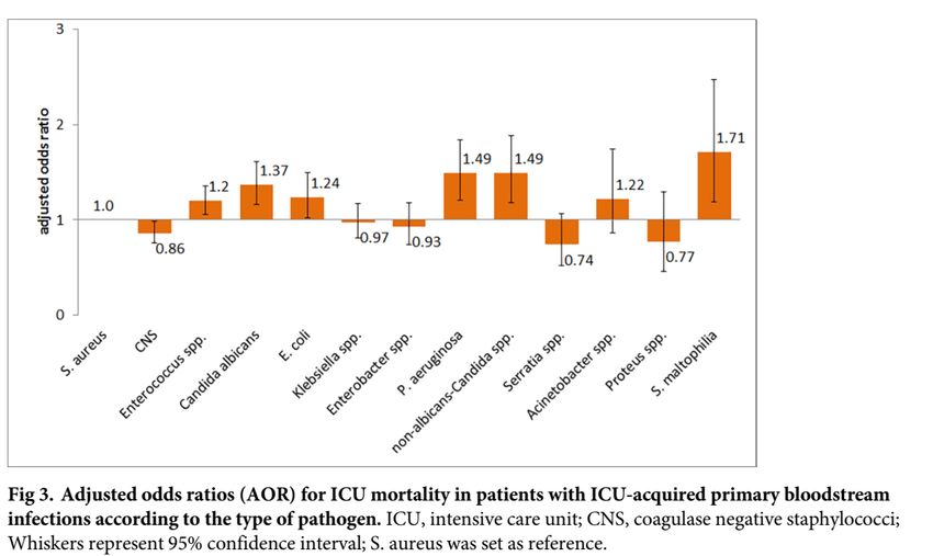
Répartition des micro-organismes
                      Germes                    HA-BSI   ICU-BSI
                      Escherichia coli          10-19%   4-10%
                      Klebsiella spp.           8-9%     4-15%
                      Enterobacter spp.         5-7%     1-8%
                      Pseudomonas aeruginosa    7-10%    2-12%
                      Acinetobacter baumannii   3-7%     2-16%
                      Staphylococcus aureus     16-26%   6-27%
                      CoNS                      10-20%   13-39%
                      Enterococcus spp.         9-11%    8-19%
                      Candida spp.              7-10%    6-15%
                      Autres
Indicateurs de résistance
ESC extended-spectrum cephalosporins, MDR multidrug-resistant
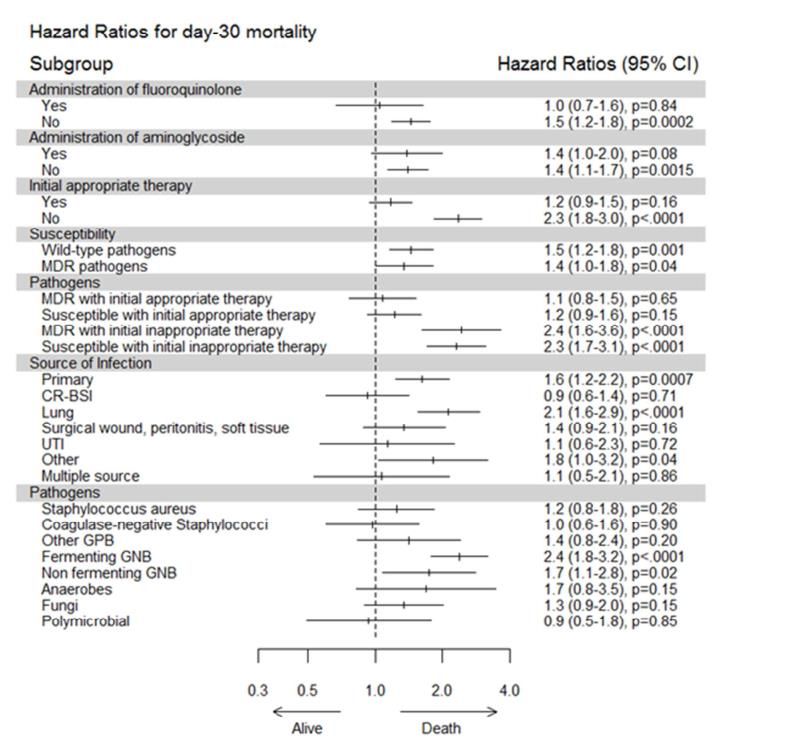
Timsit, ICM, 2020; Réa RaisinN=571 ICU-BSI ↗ risque décès: HR, 1.40; 95% CI, 1.16 to 1.69; p < 0.01. ↗ de 130% quand pas d’adéquation initiale: HR, 2.3; 95% IC, 1.8 à 3.0; ↗ 20% quand adéquation initiale: HR, 1.2; 95% IC, 0.9 à 1.5 Adrie, Journal of Infection, 2016
4,556,360 patients avec 16,978,882 patient jour de 937 ICUs;
                     14,626 BSI dont 12,745 mono-microbiennes

Schwab, Plos one, 2017
Diagnostic et bilan d’extension
Identification - diagnostic
   • Bactériémie = hémocultures positives en dehors des contaminations
   • Inoculation sang dans des flacons d’hémocultures anaérobies et aérobies
     mis en incubation
   • 2 à 3 paires d’hémocultures bien remplis (10 mL) avant antibiothérapie sur
     2 sites différents ou espacés d’au moins 30 minutes
   • En cas de suspicion d’endocardite, les espacer dans le temps
   • Si suspicion infection sur cathéter: prélever paires hémocultures sur
     cathéters et en périphérie
   • Avant toute antibiothérapie sauf purpura fulminans
   • Quand faire les hémocultures:
        •   En cas de fièvre +/- frissons intenses, sueurs, hypotension inexpliquée,
        •   Foyers infectieux multiples ou
        •   Patient neutropénique ou porteur de matériel étranger,
        •   Hypothermie,
        •   En l’absence de fièvre si sujet âgé, immunodéprimé, corticothérapie, traitement
            antipyrétique

Lamy, Frontiers in microb, 2016
Contamination?
• Parmi l’ensemble des hémocultures prélevées dans un hôpital seul 3 à 5% sont positives
• Parmi ces hémocultures, 20 à 56% sont positives à des contaminants
• Bactérie de la flore cutanée: Staphylocoques coagulase négative (SCN), corynebacterium spp.,
Micrococcus spp, Bacillus spp, Propionibacterium acnes …
• Délai de pousse allongé
• Attention particulière si:
• Matériel, quel qu'il soit, notamment endovasculaire
• Contexte clinique: porte entrée cutanée, toxicomanie, neutropénie
• Certaines bactéries: staphylococcus lugdunensis (pouvoir pathogène proche de S. aureus), corynebacterium
jeikeium (infection de matériel chez l’immunodéprimé notamment).
• Ce n’est pas une contamination si:
• Plusieurs hémocultures au même germe
• Hémoculture positive à plusieurs germes possible si foyer digestif, fistule vasculaire, neutropénie
• Une seule hémoculture positive à un germe qui est toujours pathogène: Staphylococcus aureus, streptococcus
pneumoniae, Escherichia Coli, autres entérobactéries, pseudomonas, listeria, pasteurella, candida…
Lamy, Frontiers in microb, 2016Caractériser la bactériémie
• Distinguer:
• La porte d’entrée (plaie cutanée, muqueuse, inoculation, translocation
digestive)
• Le foyer infectieux (pneumonie, pyélonéphrite, colite, méningite…)
• Les localisations secondaires (abcès rénal, spondylodiscite …)
• Les relais endovasculaires (endocardite, thrombophlébite septique, infection
de prothèse endovasculaire …)
• CA-BSI – HCA-BSI – HA-BSI – ICU-acquired BSI
• Et toujours rechercher:
• Les signes de gravités (sepsis, choc septique)
• Le terrain (neutropénie +++) 48H00 pour Antibiogramme
Les méthodes de diagnostic rapide PCR direct sur sang
Sn et Sp médiocre
automates non disponible
pas tous les mécanismes de
résistances
Tests reposant sur des techniques de
résonnance magnétiques : T2Bacteria
Panel, T2Biosystems Meilleur Sn (90%):
rendu en 3.5h à 5 à 8 heures
Tests PCR sur Hémocultures positives
excellente performance
Matrix-assisted laser desorption ionization–
time of flight mass spectrometry (MALDI-TOF)
: slt sur culture + après purification de
l’échantillon bonne performance pour les BGN
( > 90% concordance) mais moins pour CGP
(slt 80%); peux détecter certains mécanisme
de résistance
Plus récemment: next-generation sequencing
Timsit, ICM, 2020 (NGS) methods avenirBilan d’extension
• Hémocultures persistantes
• Faire échographie/doppler des axes vasculaires, imagerie pour les emboles
septiques (angioTDM corps entier) et échocardiographie (surtout si
staphylocoque doré, streptocoque (sauf groupe A), entérocoque, candida),
fond d’oeil
• Hémocultures persistantes à S. Aureus ou entérocoque
• Faire ETO
• Hémocultures persistante et risque EI
• Faire ETO, quel que soit le germe si:
• Hémodialyse, foyers emboliques d’infection, toxicomanie IV, chambre implantable,
dispositif électronique intracardiaque, valve prothétique, ATCD Ei et anomalie
structurelle cardiaque.
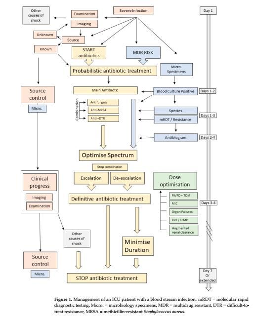
RFE SRLF 2019Prise en charge thérapeutique
Timsit, BMC Infectious Diseases, 2014
Tabah, antibiotics, 2022
Time to antibiotics

Evans, ICM, 2021
Contrôle de la source
• Péritonite, obstacle urinaire, dermo-hypodermite
  nécrosante, pleurésie, péricardite, empyèmes, abcès
  profond…                                                      CONTRÔLE DE LA
                                                               SOURCE dans les 6 à
• Argument pour drainage/débridement                           12 premières heures
   • Diminution de l’inoculum
   • Diminution du risque de récidive
   • Diminution du risque de sélection de mutants résistants
   • Accélère le traitement d’un territoire dans lequel la
     diffusion antibiotique est mauvaise
   • Effet symptomatique antalgique selon les cas
Choix du traitement anti-microbiens
1. Traitement empirique ou documenté
2. Origine de l’infection présumée ou prouvée
3. Suspicion ou présence avérée de résistances
4. Immunosupression
5. Suspicion ou candidémie avérée
• Toujours évaluer la balance bénéfice risque
• ATB large spectre et sélection de résistances
• ATB plus ciblée mais risque d’échec thérapeutique et/ou retard d’instauration d’une ATB efficace
• !!! Stratégie d’épargne des nouvelles beta-lactamines
• Raisonner en fonction de l’écologie locale, du patient (colonisation, exposition ATB, voyage), et
des pratiques de services.
Timsit, ICM, 2020Tabah, antibiotics, 2022
Risques de bactériémies à BMR

                                Tabah, antibiotics, 2022; Di Franco, Life, 2021
Choix du traitement anti-microbiens Timsit, ICM, 2020
Tabah, antibiotics, 2022
TDM « Therapeutique drug monitoring »
1. Dose de charge d’autant plus si inflation hydrosodée
2. Adaptation à la fonction rénale (Insuffisance rénale -
hyperclairance)
3. Connaissance sur la pharmaco-dynamie des antibiotiques:
Temps/CMI , Pic/CMI ou AUC/CMI
• Pour mode administration: continue ou non; TDM +++
• Surtout pour minimiser la toxicité et augmenter la réponse
aux ATB
• TDM disponible seulement pour certaines molécules
• Disponible en routine pour vancomycine, aminosides, de
plus en plus pour les beta lactamines
Timsit, ICM, 2020; Tabah, antibiotics, 2022Mono ou bithérapie pour les ICU-acquired BSI Souvent combinaison beta lactamine et aminoside ou fluoroquinolone Traitement possible en monothérapie pour les BSI à SAMS et à enterobacteries (AmpC hyperproductrice et ESBL) Controverse pour les acineto carba R, le pseudomonas, et les Accélère carba-PE bactéricidie Recommendation bithérapie pour les patients en choc septique, mais pas pour les sepsis sans défaillance hémodynamique Timsit, ICM, 2020; Evans, ICM, 2021
Stratégie de désescalade des antibiotiques
   • Souvent à J2-J3 lors récuperation antibiogramme

   • Réduire le nombre d’antibiotique et le spectre de l’antibiothérapie

   •   ! Quand la source peut être polymicrobienne comme dans les infections abdominales

   •   ! PK/PD.

Timsit, ICM, 2020
Durée de l’antibiothérapie
• Durée suffisante pour prévenir les rechutes et/ou récurrences
• !! durée trop longue expose effets adverses, toxicités, émergences de résistances,
  augmentation prix et des ressources
• J0= Contrôle de la source : traitement chirurgical et/ou négativation des hémocultures
• Clairance de la bactériémie: au moins une culture négative, après J2-J4 de l’infection
• Faire des hémocultures quotidiennes
• Durée de traitement codifiée en fonction du germe, de la source de l’infection, du bilan
  d’extension
• Toujours réévaluer entre J5 et J7 que l’infection soit contrôlé (Fièvre, syndrome inflammatoire,
  PN, CRP, PCT, défaillance d’organe, choc, négativation des hémocultures)
• Répéter les imageries, place de la scintigraphie aux leucocytes marqués, et PET Scanner
                                                                                           Timsit, ICM, 2020
En cas d’aggravation
• Non contrôle de la source de l’infection (abcès, matériel étranger, tissus infecté,
endocardite, sous dosage ATB, acquisition de résistance, nouveau germe)
• Autre infection nosocomiale (KT, PAVM, Urines)
• Refaire bilan infectieux complet
• Timing modification ATB, contrôle de la source en fonction aggravation (choc?)
• En cas de fièvre sans aggravation clinique penser aux fièvres non infectieuses:
allergie, TVP
Timsit, ICM, 2020Durée antibiothérapie
Type d’infection Durée ATB
Bactériémie sans complication à distance à SCN* 3-5 jours
Bactériémie sans complication à distance à BGN* 7 jours
• Liste non exhaustive
Bactériémie sans complication à distance à streptocoque ou 7 jours
entérocoque* *comprend les infections de cathéters après
Bactériémie sans complications à distance à Staph Aureus§ ou 14 jours
ablation du cathéters et sans complication à
candidémie*
distance
Thrombophlébite septique et endocardites 4 à 6 semaines **corps étrangers, métastases septiques, micro-
abcès
Bactériémie et complications à distance** 4 à 6 semaines
§ Staphylococcus lugdunensis idem que
Arthrites 4 à 6 semaines Staphylococcus aureus
Ostéo-arthrites; Spondilodiscites 6 à 12 semaines • Rôle imagerie et évolution clinique-biologique
pour guider durée
Abcès cérébrale 6 à 12 semaines
Bactériémies d’origine urinaire Idem infection
urinaire
Gauzit, SPILFRaccourcissement de la durée de l’antibiothérapie? Pour Contre • Pas de comorbidités • Immunosuppression • Source contrôlée • Pas de contrôle de la source • CMI basse, bactéricidie • BMR ou XDR • ATB adapté dès le début • Faible bactéricidie • Bonne diffusion • Mauvaise diffusion • Pas de matériel étranger • Matériel étranger • Évolution clinique rapide • Évolution lente ou défavorable
Les points clés
• SCN= la bactérie la plus fréquente/ savoir apprécier son rôle pathogène ou
  contaminant
• Contrôle de la source primordiale - drainer/débrider toute collection ou
  atteinte tissulaire
• L’antibiothérapie:
   • IV en optimisant la bactéricidie
   • L’association d’AB à justifier (élargir le spectre , accélérer la bactéricidie)
• La recherche localisations associées et porte d’entrée systématique, mais
  négative dans 15 à 25% des cas
• Durée de traitement
   • En fonction des caractéristiques de la bactériémie, du bilan d’extension, de
     l’évolution
Merci de votre attention
Bassetti M, Righi E, Carnelutti A: Bloodstream infections in the Intensive Care Unit. Virulence 2016; 7:267–
 279

 Di Franco S, Alfieri A, Pace MC, et al.: Blood Stream Infections from MDR Bacteria. Life 2021; 11:575

• Gauzit R. et al. Anti-infectious treatment duration: The SPILF and GPIP French guidelines and
    recommendations (Durées des traitements anti-infectieux. Recommandations françaises SPILF et GPIP).
    Infect. Dis. Now.

 Giacobbe DR, Giani T, Bassetti M, et al.: Rapid microbiological tests for bloodstream infections due to
 multidrug resistant Gram-negative bacteria: therapeutic implications. Clinical Microbiology and Infection
 2020; 26:713–722

 Kimmig A, Hagel S, Weis S, et al.: Management of Staphylococcus aureus Bloodstream Infections. Front Med
 2021; 7:616524

 Réseau REA-Raisin: SURVEILLANCE DES INFECTIONS NOSOCOMIALES EN RÉANIMATION
 ADULTE [Internet]. 2015; Available from: https://www.santepubliquefrance.fr/maladies-et-
 traumatismes/infections-associees-aux-soins-et-resistance-aux-antibiotiques/infections-associees-aux-
 soins/documents/rapport-synthese/surveillance-des-infections-nosocomiales-en-reanimation-adulte.-reseau-
 rea-raisin-france-resultats-2015

 Tabah A, Koulenti D, Laupland K, et al.: Characteristics and determinants of outcome of hospital-acquired
 bloodstream infections in intensive care units: the EUROBACT International Cohort Study. Intensive Care
 Med 2012; 38:1930–1945

 Timsit J-F, Baleine J, Bernard L, et al.: Expert consensus-based clinical practice guidelines management of
 intravascular catheters in the intensive care unit. Ann Intensive Care 2020; 10:118

 Timsit J-F, Laupland KB: Update on bloodstream infections in ICUs: Current Opinion in Critical Care 2012;
 18:479–486

 Timsit J-F, Ruppé E, Barbier F, et al.: Bloodstream infections in critically ill patients: an expert statement.
 Intensive Care Med 2020; 46:266–284

 Timsit J-F, Soubirou J-F, Voiriot G, et al.: Treatment of bloodstream infections in ICUs. BMC Infect Dis 2014;
 14:489
• According to the origin of the infection, either community-acquired (CA-BSI), hospitalacquired (HA- BSI) or intensive care unit (ICU)–acquired (ICU-BSI). 2. Either secondary to a source of infection or primary, when there is no identified source [2]. 3. Complicated or uncomplicated, which was recently defined as a having definite source (among urinary, catheter, intra-abdominal, pneumonia, skin or soft tissues), and effective source control, in a non-immunocompromised patient, and with clinical improvement after 72 h of antimicrobial therapy (at least defervescence and haemodynamic stability) [3]. 4. By clinical severity, which is the absence or presence of organ failures and the need for organ supportive therapy in the ICU.
• Antimicrobial therapy
   • The Importance of Getting It Right from the Start

   • Kumar and colleagues described in 2006 a 12% increase in crude
     mortality for each hour of delay to administer antimicrobials from the onset of hypotension and septic
     shock [10]. The above-mentioned study by Adrie and colleagues shows
     a 30% increase in mortality when no adequate treatment is given in the first 24 h [8]. In
     the evaluation of a multifaceted intervention to decrease sepsis mortality in a group of
     40 German hospitals, Bloos and colleagues report an increase in the risk of death of patients
     with sepsis or septic shock of 2% for each hour of delay of antimicrobial therapy and 1% for
     each hour of delay in source control [11]
     Hranjec and colleagues i
     e conservative period, immediate antibiotic therapy was
     recommended for patients with shock.
     While controversy remains
     and these data present all the biases inherent to observational studies, they highlight how
     important it is that patients with BSIs receive early appropriate antimicrobial therapy
• 2.1.2. Broad-Spectrum Antibiotics and Combination Therapy? When the source is known, antibiotics should be targeted at the most common pathogens for the source as detailed in Table 1. Molecule choice takes into account risk factors for multidrug-resistant (MDR) or specific pathogens for the patient, according to their history and setting as shown in Table 2. For hospital-acquired infections, knowledge of colonisation from previous clinical or surveillance cultures is a valuable tool to optimise this choice [15,16]. Combination therapy can provide very broad empirical coverage for different classes of pathogens by adding anti-MRSA and antifungal agents or molecules targeted at MDR Gram-negative bacteria (GNB). These should be used with parsimony, in patients with significant risk factors, and only as part of the empirical regimen with a plan to subsequently de-escalate all drugs that are not required [17,18].
• 2.1.3. The Importance of Sending Blood Cultures before Starting Antimicrobials
  2.1.4. The Advent of Molecular Rapid Diagnostic Testing
  as matrix-assisted laser desorption/ionisation–time of flight (MALDI-TOF) mass
  spectrometry [23]. Integrated solutions such as the Accelerate Pheno system automate both
  the identification and AST, providing accurate results in 90 min and 7 h, respectively. In
  a multicentre study, comparing with conventional BC processing, it accurately identified
  14 common bacterial pathogens and 2 Candida sp. with sensitivities ranging from 94.6% to
  100% [24]. The performance of AST results for methicillin-resistant Staphylococcus aureus
  (MRSA) and Staphylococcus sp. had an agreement of 97% with conventional processing. For
  GNB, the agreement on a panel of 15 antimicrobials was 94%, making this system suitable
  for prime clinical use [24].
  Colorimetric assays are relatively inexpensive and extremely accurate benchtop solutions to detect
  extended-spectrum beta-lactamase-producing (ESBL-Es) or carbapenemaseproducing
  Enterobacterales (CPEs) [21,25].
REDUIRE SPEDCTRE ATB
• Etended-Spectrum b-Lactamase-Producing Enterobacterales

• The MERINO trial randomised 391 patients with
  a BSIs due to ceftriaxone-resistant Escherichia coli or Klebsiella pneumoniae to piperacillin–
  tazobactam or meropenem [32]. Mortality was 12.3% for piperacillin–tazobactam compared with 3.7% for meropenem,
  rejecting non-inferiority and not supporting the use of
  piperacillin–tazobactam in severe infections due to ESBL-Es.
  Alternatives for cases where a
  carbapenem cannot be used include fluoroquinolones and trimethoprim-sulphamethoxazole.
  Those are especially interesting for BSIs with a urinary source as they concentrate in the
  urine [30]. While ceftolozane–tazobactam and ceftazidime–avibactam (CAZ-AVI) are potential alternatives, their use
  should be restricted as reserve antibiotics for those pathogens
  that cannot be treated otherwise

• we should avoid using piperacillin–tazobactam in patients with severe infections due
  to pathogens with inducible AmpC [36,37]. Cefepime is a good treatment choice as it is a
  weak inducer, and it is relatively stable against AmpC β-lactamases. Caution is warranted
  in pathogens with a MIC ≥ 4 µg/mL for cefepime as they may harbour an ESBL, making
  them prone to treatment failure. All carbapenems are stable and recommended for the
Vous pouvez aussi lire