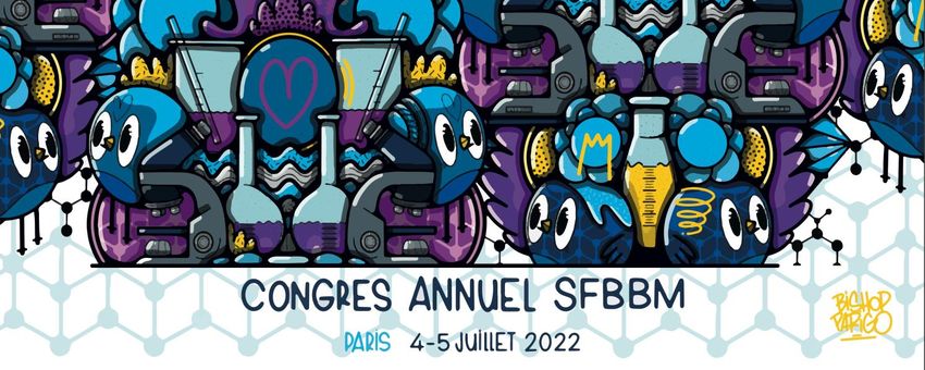
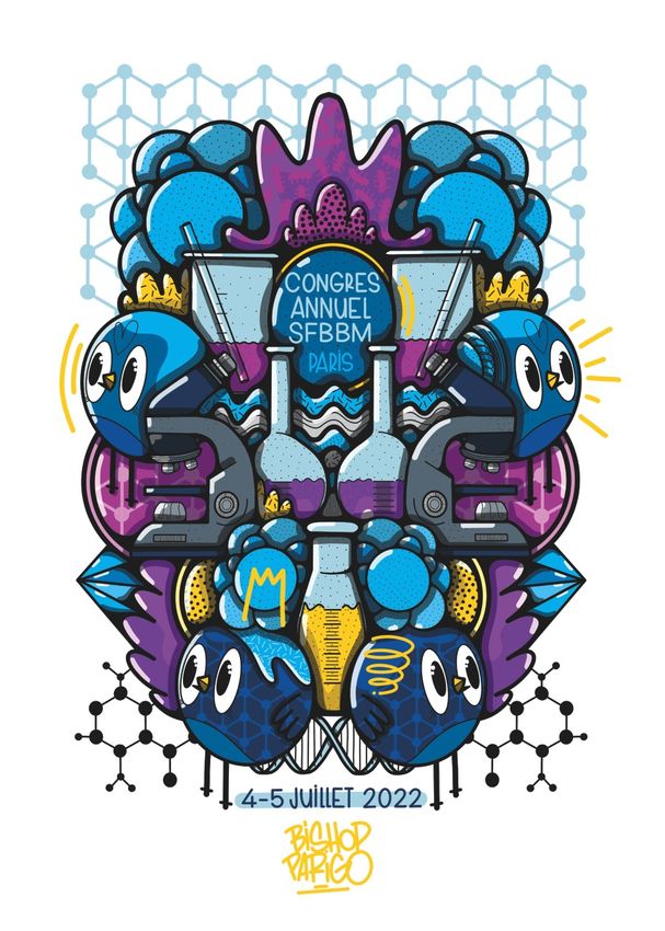
Congrès annuel de la Biologie Moléculaire - Société Française de Biochimie et A la Faculté de Pharmacie de Paris - sfbbm
←
→
Transcription du contenu de la page
Si votre navigateur ne rend pas la page correctement, lisez s'il vous plaît le contenu de la page ci-dessous

Congrès annuel de la
Société Française de Biochimie et
 Biologie Moléculaire

A la Faculté de Pharmacie de Paris
Chers collègues, chers amis,

C’est avec grand plaisir que nous vous souhaitons la bienvenue pour le Congrès Annuel 2022 de la
SFBBM !

Après le succès de l’édition 2021, ce congrès sera à nouveau l’occasion d’offrir à notre Société un
moment d’échanges et de convivialité. Nous aurons le plaisir d’accueillir des conférenciers de
renom, les lauréats des prix de notre société (prix Maurice Nicloux, prix de la fondation Dina
Surdin, prix Biochimie, …) mais aussi des jeunes chercheurs dont les résumés ont été sélectionnés.

Merci à nos prestigieux orateurs invités, à tous les jeunes chercheuses et chercheurs pour leurs
nombreuses contributions, aux sponsors mais aussi au comité local d’organisation et au conseil
scientifique du congrès, dont le dynamisme et l’efficacité ont permis d’établir un programme
passionnant, divers et riche, à l’image de notre chère société.

Organisé pour la deuxième année dans le cadre unique de la Faculté de Pharmacie, notre congrès
bénéficiera à nouveau d’un environnement propice aux échanges et aux débats inspirants dans
une atmosphère décontractée et amicale.

 Au nom de la SFBBM, je vous souhaite à toutes et tous un excellent congrès !

 Martin Picard
 Président de la SFBBM
Présentation de la SFBBM
 Société Française de Biochimie et Biologie Moléculaire

La Société Française de Biochimie et Biologie Moléculaire (SFBBM) est une société savante fondée
par le Professeur Maurice Nicloux en 1914 au Collège de France à Paris. Association loi 1901, la
SFBBM a été reconnue comme établissement d’utilité publique par décret le 27 avril 1933.

Ses missions permettent :
- de rassembler les biochimistes et biologistes moléculaires de toute la France
- d’animer via ses groupes thématiques des actions auprès de sa communauté aussi bien dans la
 recherche que dans l’enseignement
- de représenter sa communauté auprès des instances politiques et scientifiques nationales et
 internationales comme la FEBS

La SFBBM organise des congrès, des réunions scientifiques et des journées sur l’innovation
pédagogique. Elle finance ces actions au travers de prix scientifiques, de bourses et aides financière
pour la participation à des congrès en France comme à l’étranger.

La SFBBM est impliquée dans des publications scientifiques. BIOCHIMIE (revue de la SFBBM) a
succédé en 1971 au Bulletin de la Société de Chimie Biologique, fondé en 1914.

En un demi-siècle, BIOCHIMIE a publié plus de 9000 articles originaux ou de revues, et compte parmi
les journaux scientifiques les mieux connus et les plus respectés dans le domaine. BIOCHIMIE est une
revue internationale publiée en Anglais par Elsevier.

BIOCHIMIE publie des travaux originaux, articles de revue critiques de la littérature, et mini-revues
dans le domaine large de la biologie, englobant biochimie, enzymologie, biologie moléculaire et
cellulaire, régulations métaboliques, génétique, immunologie, microbiologie, biologie structurale,
études « omiques » avec validation fonctionnelle, pharmacotoxicologie et mécanismes moléculaires
des maladies. Son facteur d'impact sur 5 ans est de 4.104.

Le journal publie régulièrement des numéros spéciaux dédiés à des thèmes de recherche d'un intérêt
particulier.
Membres du Comité Local d’Organisation
 Magali Blaud
 magali.blaud@u-paris.fr
 Julien Dairou
 Julien.dairou@u-paris.fr
 Philippe Fossé
 philippe.fosse@ens-paris-saclay.fr
 Julien Henri
 Julien.henri@sorbonne-université.fr
 Zaineb Kammoun
 zaineb.kammoun@parisdescartes.fr
 Hélène Munier-Lehmann
 helene.munier-lehmann@pasteur.fr
 Ariane Pachins
 sfbbm@sfbbm.fr
 Jean-Christophe Paillart
 jc.paillart@ibmc-cnrs.unistra.fr
 Martin Picard
 martin.picard@ibpc.fr
 Julie Soutourina
 Julie.Soutourina@cea.fr
Membres du Comité Scientifique
 Magali Blaud
 magali.blaud@u-paris.fr
 Béatrice Clouet D’Orval
 Beatrice.Clouet-dOrval@ibcg.biotoul.fr
 Bernard Dujon
 bdujon@pasteur.fr
 Laurence Drouard
 laurence.drouard@ibmp-cnrs.unistra.fr
 Christine Ebel
 christine.ebel@ibs.fr
 Bertrand Friguet
 bertrand.friguet@upmc.fr
 Hélène Munier-Lehmann
 helene.munier-lehmann@pasteur.fr
 Fernando Rodrigues-Lima
 Fernando.rodrigues-lima@u-paris.fr
 Emmanuelle Schmitt
 emmanuelle.schmitt@polytechnique.edu
NOS PARTENAIRES

Nous tenons à remercier chaleureusement nos partenaires et sponsors : les éditions Dunod qui
soutiennent depuis de nombreuses années le travail du groupe sur les innovations en pédagogie, les
sociétés Cytiva, Microsynth et Eurofins. Vos contributions à ce congrès nous ont permis de pouvoir
réaliser cet évènement dans les meilleures conditions et d’offrir des prix pour le meilleur poster et la
meilleure présentation orale. Nous tenons à remercier la Faculté de Pharmacie de Paris pour son
accueil et spécialement Samantha Conti (communication et organisation), Mélanie Martin
(installation et organisation) et Stéphane Carraz (TICE).
PROGRAMME LUNDI 4 JUILLET 2022
 8h30 Accueil des participants
Faculté de Pharmacie de Paris – 6 avenue de l’Observatoire/Paris

 9h30 Ouverture du congrès

 10h00 Conférence plénière par Chris Bowler
« Tara Oceans: ecosystems biology at planetary scale »

 11h00 Session 1 : « RNA »
 Conférence par Franck MARTIN
 Cyril Bourgeois : prix Maurice Nicloux 2021
 Jean-Louis MERGNY
 Claire Husser

 12H30 PAUSE DEJEUNER

 13h45 Session 2: « Chemobiology »
 Conférence par Alice LEBRETON
 CD MOHAN : prix de l’article de l’année Biochimie
 Corinne LIONNE

 15H00 SESSION POSTERS ET PAUSE CAFE

 16H00 Présentations par les sponsors
 Mycrosynth, Cytiva, Eurofins et Dunod

 16h30 Session 3: « Cellular proteolysis »
 Conférence par Catherine MOALI
 Yoann SANTIN : prix Dina Surdin
 Paul BIGOT
 Nicolas MATHAS

 18h00 Assemblée Générale de la SFBBM

 18h30-22h COCKTAIL
PROGRAMME MARDI 5 JUILLET 2021
 8h30 Session 4: « Archaea »
 Conférence par Tristan WAGNER
 Solenne ITHURBIDE : prix de l’article de l’année de la SFBBM
 Clément MADRU
 Roxane LESTINI

 10h00 SESSION POSTERS ET PAUSE CAFE

 11h00 Session 5 : « Genetic »
 Conférence par Claire Rougeulle
 Maxime WERY
 Louise le COADOU
 Inge KÜHL

 12H30 PAUSE DEJEUNER

 13h30 Session 6: « Enzymes »
 Conférence par Alain MARTY
 Damien SORIGUE : prix article de l’année SFBBM 2018
 Nicolas JOLY : prix Maurice Nicloux 2021
 Claire CÉRÉ
 Paolo ZECCHIN

 15h15 Session 7: « Education »
 Wilfried GRANGE, Florent BUSI et Caroline CHAUVET

 15h45 Remise des prix et conclusions

 16h30 Session spéciale du Groupe de Travail Enzymes

 WIFI : réseau DESCARTES
 Identifiant de connexion (login) : pharmacie
 Mot de passe : *pharma*

Merci à nos sponsors pour leur soutien dans l’organisation du congrès

Conférence plénière d’ouverture du congrès
 Animée par
 Christine Ebel
 Institut de Biologie Structurale– Grenoble

 «Tara Oceans: ecosystems biology at planetary scale»
 par Chris Bowler
Ecology and Evolutionary Biology Section, Institut de Biologie de l’Ecole normale supérieure (IBENS), Paris, FRANCE

 The ocean is the largest ecosystem on Earth and yet we know very little about it. This is
 particularly true for the plankton that drift within, even though they form the base of marine food
 webs and are key players in Earth’s biogeochemical cycles. Ocean plankton are at least as
 important for the Earth system as the forests on land, but most of them are invisible to the naked
 eye and thus are largely uncharacterized. To increase our understanding of this underexplored
 world, a multidisciplinary consortium, Tara Oceans, was formed around the 36m research
 schooner Tara, which sampled plankton at more than 210 sites and multiple depth layers in all the
 major oceanic regions during expeditions from 2009-2013 (Karsenti et al. Plos Biol., 2011). This
 talk will summarize the foundational resources from the project, which collectively represent the
 largest DNA sequencing effort for the oceans (see Science special issue May 22, 2015 and Cell, Nov
 14, 2019), and analyses that illustrate several aspects of the Tara Oceans’ eco-systems biology
 approach to address microbial contributions to ecological and evolutionary processes. The project
 provides unique resources for several scientific disciplines that are foundational for mapping
 ocean biodiversity of a wide range of organisms that are rarely studied together, exploring their
 interactions, and integrating biology into our physico-chemical understanding of the ocean, as well
 as for identifying new organisms and genes of biotechnological interest. These resources, and the
 scientific innovations emerging to understand them, are furthermore critical towards developing
 baseline ecological context and predictive power needed to track the impact of climate change on
 the ocean.
SESSION 1

 “ RNA ”

• Conférence par Franck Martin
• Cyril Bourgeois : prix Maurice Nicloux 2021
• Jean-Louis Mergny
• Claire Husser

 Animée par
 Laurence Drouard
 Institut de biologie moléculaire des plantes –
 Strasbourg
Conférence invitée session 1

 «Viral and cellular translation during SARS-CoV-2 infection»
 par Franck MARTIN
Université de Strasbourg, Institut de Biologie Moléculaire et Cellulaire, Architecture et Réactivité de l’ARN, CNRS
UPR9002, 2, allée Konrad Roentgen, F‐67084 Strasbourg (France)

 SARS-CoV-2 is a betacoronavirus that has emerged in China in December 2019 and which is the
 causative agent of the Covid-19 pandemic. This enveloped virus contains a large positive-sense
 single-stranded RNA genome. Non-structural proteins are encoded by the genomic RNA and are
 produced in the early steps of infection. In contrast, the structural proteins are produced from
 sub-genomics RNA that are translated in the late phase of the infectious program. Non-structural
 protein 1 (NSP1) is a key molecule that regulates both viral and cellular translation1. In addition,
 NSP1 interferes with multiple steps of in the interferon I pathway and thereby blocks host antiviral
 responses1. Therefore, NSP1 is a drug target of choice for the development of antiviral therapies.
 The 5’UTR part of coronavirus genomes plays key roles in the viral replication cycle and the
 translation of the viral mRNAs. The first 75‐80 nucleotides, also called the leader sequence, are
 identical for the genomic RNA and for the subgenomic RNAs. Recently, it was shown that
 cooperative actions of a 5’UTR segment and the non‐structural protein NSP1 are essential for both
 the inhibition of host mRNAs and for specific translation of viral mRNAs2,3. Sequence analyses of
 both the 5’UTR RNA segment and the NSP1 protein have been done for several coronaviruses with
 special attention to the betacoronaviruses. The conclusions are (i) precise specific molecular
 signatures can be found in both the RNA and the NSP1 protein; (ii) both types of signatures
 strongly correlate between each other. Indeed, definite sequence motifs in the RNA correlate with
 sequence motifs in the protein indicating a co‐evolution between the 5’UTR and NSP1 in
 betacoronaviruses. Experimental mutational data on 5’UTR and NSP1 from SARS‐CoV‐2 using cell‐
 free translation extracts support those conclusions and show that some conserved key residues in
 the N‐terminal half of the NSP1 protein are essential for evasion to the inhibitory effect of NSP1 on
 translation4.
 1 Eriani, G. and Martin, F. (2022) Viral and cellular translation during SARS-CoV-2 infection. FEBS

 Open Bio. DOI: 10.1002/2211-5463.13413.
 2 Miao, Z., Tidu, A., Eriani, G. and Martin, F. (2020) Secondary structure of the SARS-CoV-2 5’UTR.

 RNA Biology. 23, 1-10. DOI: 10.1080/15476286.2020.1814556.
 3 Tidu, A., Janvier, A., Schaeffer, L., Sosnowski, P., Kuhn, L., Hammann, P., Westhof, E., Eriani, G.

 and Martin, F. (2021) The viral protein NSP1 acts as a ribosome gatekeeper for shutting down host
 translation and fostering SARS-CoV-2 translation. RNA. 27, 253-264. DOI: 10.1261/rna.078121.120
 4 Sosnowski, P., Tidu, A., Eriani, G., Westhof, E. and Martin, F. (2022) Correlated sequence

 signatures are present within the genomic 5’UTR RNA and NSP1 protein in coronaviruses. RNA.
 DOI: 10.1261/rna.078972.121
Conférence invitée session 1
 Prix Maurice Nicloux 2021

 «RNA helicase-dependent gene looping impacts messenger RNA
 processing»
 Par Cyrille Bourgeois
Laboratoire de Biologie et Modélisation de la Cellule, Ecole Normale Supérieure de Lyon CNRS UMR5239, INSERM
U1293, Université Lyon 1 46 Allée d'Italie, 69007 Lyon

 DEAD-box RNA helicases DDX5 and DDX17 regulate several aspects of gene expression, especially
 transcription and splicing, through incompletely understood mechanisms. A transcriptome analysis
 of DDX5/DDX17-depleted human cells confirmed the large impact of these RNA helicases on
 splicing and revealed a widespread deregulation of 3’ end processing. In silico analyses and
 experiments in cultured cells showed the binding and functional contribution of the genome
 organizing factor CTCF to chromatin sites near a subset of DDX5/DDX17-dependent exons that are
 characterized by a high GC content and a high density of RNA Polymerase II. We propose that an
 RNA helicase-dependent relationship between CTCF and the dynamics of transcription across DNA
 and/or RNA structured regions contributes to the processing of internal and terminal exons. Our
 work showed that local DDX5/DDX17-dependent chromatin loops spatially connect RNA helicase-
 regulated exons to their cognate promoter, and we provide the first demonstration that de novo
 gene looping modifies alternative splicing and polyadenylation. Overall our findings uncovered the
 impact of DDX5/DDX17-dependent chromatin folding on pre-messenger RNA processing.
«Quadruplexes are everywhere!»
 Jean-Louis Mergnya
 a
 Laboratoire d’Optique et Biosciences, Ecole Polytechnique, CNRS UMR7645 – INSERM U1182,
 Institut Polytechnique de Paris, 91128 Palaiseau cedex, France
 E-mail of corresponding author: jean-louis.mergny@polytechnique.edu

 G-quadruplexes are unusual nucleic acid structures which can find applications in biology,
 medicine, as well as biotech- and nano-technologies 1. We are developping tools to
 understand their folding and polymorphism 2. In parallel, we proposed a new algorithm for
 prediction of G4 propensity of unprecedented accuracy 3. We are now applying this G4-
 Hunter prediction tool to a number of genomes.
 We became interested in quadruplexes quadruplex-prone regions conserved in the
 genome of a number of viruses 4. We recently demonstrated that viruses regularly causing
 persistent infections are enriched in G4 motifs, while viruses causing acute infections are
 significantly depleted in these structures 5. We performed a genome analysis of SARS-
 CoV2 6. One of the viral proteins, Nsp3, contains a SARS-Unique domain (SUD), essential
 for replication, which can bind to G4s. These interactions can be disrupted by mutations
 that prevent the oligonucleotides from folding into G4 structures and, interestingly, by
 molecules called ligands specific for these G4s. Our results pave the way for further studies
 on the role of SUD/G4 interactions during SARS-CoV-2 replication and the use of inhibitors of
 these interactions as potent antiviral compounds. We are performing a global screening of
 molecules capable of inhibiting this interaction and testing their antiviral properties.
 We are also interested in the role of quadruplexes in parasites such as Plasmodium
 falciparum or Trypanosoma brucei 7 and, more recently on parasitic helminths 8, which are
 highly prevalent and infect approximately two billion people worldwide. A nematode,
 Ascaris lumbricoides, was found to be highly enriched in stable quadruplexes. We
 demonstrated that small compounds able to recognize these structures called G-quadruplex
 ligands were able to selectively recognize G4 found in the Schistosoma mansoni genome.
 Two of these compounds demonstrated potent activity both against larval and adult stages
 of this helminth, opening new perspectives for the use of G4 ligands to fight diseases caused
 by these parasites.
 Relevant references:
(1) Mergny & Sen, Chem. Rev. (2019), 119, 6290-6325. Mergny Biochimie (2020), 168, 100-109.
(2) Cheng et al, Nucleic Acids Res. (2018), 46, 9264. Chen et al, Nucleic Acids Res. (2021) 49, 9548;
 Luo et al, Nucleic Acids Res. (2022), accepted.
(3) Bedrat et al, Nucleic Acids Res. (2016), 44: 1746; Brazda et al, Bioinformatics (2019), 35,
 3493 & Bioinformatics (2020), 36, 3246.
(4) Jaubert et al, Sci Adv. (2018) 8: 8120. Abiri et al, Pharmacol Rev. (2021) 73, 897.
(5) Bohálová et al, Biochimie (2021) 186, 13-27
(6) Lavigne et al. Nucleic Acids Res. (2021) 49, 7695
(7) Belmonte-Reche et al, Eur J Med Chem. (2018), 61: 1231; Guillon et al, Chem. Biol. Drug Des.
 (2018), 91: 974 ; Gazanion et al, PLoS Pathogens (2020), 16, e1008917. Belmonte-Reche et
 al, Eur J Med Chem. (2022), 232, 114183.
(8) Cantara et al, Nucleic Acids Research (2022), 50, 2719-2735.
 KEYWORDS: G-quadruplex; Nucleic acid structures; Prediction; RNA ligands
«Development of Fluorogenic RNA-based biosensors by ultra-high
 throughput microfluidic screening»
 Claire Hussera, Michael Ryckelyncka
 a
 Architecture et Réactivité de l’ARN (UPR9002 du CNRS), team "Digital Biology of RNA" Institut
 de Biologie Moléculaire et Cellulaire du CNRS, Université de Strasbourg
 Halogene compounds are implied in many environmental pollutions and are classified
 as Persistent Organic Polluants (POPs) by the Stockholm convention and the World Health
 Organization. On top of that these compounds are highly toxic to both humans and wildlife.
 Their persistence, bioaccumulation, and toxicity lead to the need of the discovery or the
 development of new dehalogenation reaction catalyst.
 Some organisms have been described as able to degrade halogenated compounds leading
 to the idea that enzyme-based biodegradation of halogene coumpounds can be a promising
 solution for contaminants degradation.
 To be able to monitor the degradation of such compounds, one must either be able to
 sense in real time the amount of the compound of interest or be able to sense its degradation
 products. In the case of the research of new enzymes or strains able to degrade fluorinated
 compounds, our choice was to develop a new method of detection of fluoride ions
 Our group is specialized in the development of RNA modules called light-up RNA
 aptamer and in the development of Fluorogenic RNA-based Biosensor (FRB) using μIVC-seq (2) (a
 technology coupling In Vitro Compartmentalization assisted by microfluidics with Next
 generation sequencing and bioinformatic).
 In this talk, I will present how we used our µIVC-seq technology for a rapid and efficient
 identification of Fluoride-FRBs for their capacity to emit fluorescence only in the presence of
 fluoride from a library containing more than 64 000 variants based on the CrcB Fluoride specific
 riboswitch aptamer domain (1). And how this biosensor can be used to detect Fluoroacetate
 dehalogenase activity.

1. Breaker, R. R. New Insight on the Response of Bacteria to Fluoride. Caries Res 2012, 46 (1), 78–81.
2. Autour, A.; Bouhedda, F.; Cubi, R.; Ryckelynck, M. Optimization of Fluorogenic RNA-Based
3. Biosensors Using Droplet-Based Microfluidic Ultrahigh-Throughput Screening. Methods 2019, 161,
 46–53.
4. KEYWORDS: Fluorogenic RNA-based Biosensor, Hight-throughput screening, Fluoride
SESSION 2

 “ Chemobiology ”

• Conférence par Alice Lebreton
• Chakrabhavi Dhananjaya Mohan : prix de l’article de
 l’année Biochimie
• Corinne Lionne

 Animée par
 Hélène Munier-Lehmann
 Institut Pasteur - Paris
Conférence invitée session 2
 «Shedding new light on host-pathogen interactions with
 chemobiological labelling approaches»
 par Alice LEBRETON
 Institut de biologie de l’École normale supérieure (IBENS), CNRS, INSERM, Université PSL, 75005 Paris et
 INRAE, IBENS, 75005 Paris.

 Caroline Peron-Canea,b, Arnaud Gautierc, Nicolas Desprata,b and Alice Lebretona,d
 a
 Institut de biologie de l’École normale supérieure (IBENS), CNRS, INSERM, Université PSL, 75005
 Paris. b Laboratoire de Physique de l’École normale supérieure (LPENS), Université PSL, Université
 Paris Cité, Sorbonne Université, CNRS, 75005 Paris.c Sorbonne Université, École normale
 supérieure, Université PSL, CNRS, LBM, 75005 Paris. d INRAE, IBENS, 75005 Paris.
 E-mail of corresponding author: alice.lebreton@ens.psl.eu
 Objectives: The onset of host-pathogen interactions constitutes a remarkable example of
 dynamic phenomena in biology, where both the host and pathogen respond to each-other. In
 this molecular crosstalk, secretion of protein effectors is key to the virulence of microbial
 pathogens, by allowing them to interact with their host and subvert molecular mechanisms
 to their benefit. However, tracking the dynamics of secreted virulence factors in real time has
 long been impaired by a paucity of appropriate fluorescent tools.
 Methods: We took advantage of the properties of the fluorogenic probe FAST to analyse by
 live microscopy and quantify the secretion of virulence factors from Listeria monocytogenes during
 infection. This versatile tool allowed us to monitor infection dynamics in real time among a
 population of Listeria-infected epithelial cells, as well as to assess the residence time of Listeria
 inside internalisation vacuoles.
 Results: We unveiled a surprizing heterogeneity in residence time of Listeria inside its entry
 vacuoles. By tracking the localisation of the secreted pore-forming toxin listeriolysin O over time,
 we observed that it labelled not only the membrane of entry vacuoles, but also compartments
 that enlarged over time and where the bacteria could multiply as fast as in the cytosol.
 Conclusions: We provide evidence that these compartments result from a process analogous to
 LC3- associated phagocytosis and constitute an alternative replication niche for Listeria in epithelial
 cells.
 Relevant references:
1. Peron-Cane C, Fernandez J-C, Leblanc J, Wingertsmann L, Gautier A, Desprat N, Lebreton A.
 Fluorescent secreted bacterial effectors reveal active intravacuolar proliferation of Listeria
 monocytogenes in epithelial cells. PLoS Pathog (2020) 16(10):e1009001.
2. Chekli Y, Peron-Cane C, Dell’Arciprete D, Allemand J-F, Li C, Ghigo J-M, Gautier A, Lebreton A,
 Desprat N et Beloin C. 2020. Visualizing the dynamics of exported bacterial proteins with the
 chemogenetic fluorescent reporter FAST. Sci Rep. 10(1):15791.
 KEYWORDS: Up to 5
 Listeria monocytogenes; Virulence factors; Bacterial secretion; Pore-forming toxin; Fluorogenic
 labelling.
Conférence invitée session 2
 Prix de l’article de l’année Biochimie

 «Targeting the STAT3 pathway in human cancers by natural
 compounds»
 Par Chakrabhavi Dhananjaya Mohan
Department of Studies in Molecular Biology, University of Mysore, Mysore 570006, India

STAT3 is an oncogenic transcription factor that is overactivated in various types of human cancers.
STAT3 controls the expression of genes associated with oncogenic functions such as cell
proliferation, apoptosis, angiogenesis, and metastasis. We are involved in the discovery of new
small molecule-based (natural and synthetic) inhibitors of the STAT3 signaling pathway in human
malignancies. In one of our studies, we examined the effect of vitexin on the
constitutive/inducible activation of STAT3 signaling in hepatocellular carcinoma (HCC) cells. Vitexin
effectively inhibited sustained activation of JAK1, JAK2, Src, and STAT3 in HCC cells. Interestingly,
treatment with tyrosine phosphatase inhibitor altered the vitexin-induced STAT3 phosphorylation,
and the attenuation of STAT3 by vitexin was found to be driven through the upregulation of
PTPεC. The combinational studies indicated that vitexin can exhibit substantial apoptotic effects
with doxorubicin and sorafenib. It also suppressed the CXCL12-induced cell invasion. Overall, this
study demonstrated that vitexin can act as a potential blocker of the STAT3 signaling cascade and
mitigate the survival as well as invasion of HCC cells.
«Structure-based design of NAD+ analogues targeting bacterial
 NAD kinases, promising targets for new antibiotics»
Rahila Rahimova1, Pauline Nogaret2, Valérie Huteau3, Clarisse Leseigneur4, Muriel Gelin1, David
Clément3, Olivier Dussurget4, Gilles Labesse1, Sylvie Pochet3, Anne Blanc-Potard2, Corinne
Lionne1
1
 Centre de Biologie Structurale (CBS), Université Montpellier, CNRS UMR 5048, INSERM
U1054, 29 rue de Navacelles, F-34090 Montpellier, France.
2
 Laboratory of Pathogen Host Interactions (LPHI), Université Montpellier, CNRS UMR 5235,
Place Eugène Bataillon, CC107, F-34095 Montpellier cedex 5, France
3
 Institut Pasteur, Université de Paris, CNRS UMR3523, Unité de Chimie Biologique Epigénétique,
28 rue du Dr Roux, F-75015 Paris, France.
4
 Institut Pasteur, Université de Paris, Sorbonne Paris Cité, Unité de Recherche Yersinia, 28 rue du
Dr Roux, F-75015 Paris, France

E-mail of corresponding author: lionne@cbs.cnrs.fr

Multi-drug resistance is a major public health problem that requires the urgent development of new
antibiotics and therefore the identification of novel bacterial targets. The activity of nicotinamide
adenine dinucleotide kinase, NADK, is essential in all bacteria tested so far, including many human
pathogens that display antibiotic resistance leading to failure of current treatments. Inhibiting
NADK is therefore a promising and innovative antibacterial strategy since there is currently no drug
on the market targeting this enzyme. Through a drug design approach based on substrate-derived
fragments, we have recently developed NAD+-competitive inhibitors of NADKs, which displayed
in vivo activity against Staphylococcus aureus or Pseudomonas aeruginosa in animal models of
infection [1-3].

Funding and supports: Agence Nationale de la Recherche (ANR-17-CE18-0011-02), Institut
Pasteur, Centre National de la Recherche Scientifique (CNRS), Institut National de la Santé et de la
Recherche Médicale (INSERM), University of Montpellier.

Relevant references:
[1] Clément DA, Leseigneur C, Gelin M, Coelho D, Huteau V, Lionne C, Labesse G, Dussurget O,
Pochet S (2020) New chemical probe targeting bacterial NAD kinase. Molecules. doi:
10.3390/molecules25214893.
[2] Gelin M, Paoletti J, Nahori MA, Huteau V, Leseigneur C, Jouvion G, Dugué L, Clément D,
Pons JL, Assairi L, Pochet S, Labesse G, Dussurget O (2020) From substrate to fragments to
inhibitor active in vivo against Staphylococcus aureus. ACS Infect Dis. doi:
10.1021/acsinfecdis.9b00368.
[3] Rahimova R, Nogaret P, HuteauV, Gelin M, Clément D, Labesse G, Pochet S, Blanc-Potard A,
Lionne C (2022) Structure-based design, synthesis and biological evaluation of a NAD+ analogue
targeting Pseudomonas aeruginosa NAD kinase. In Preparation.

KEYWORDS: Antibiotic resistance, Drug design, Inhibitors, Pseudomonas aeruginosa, X-ray
crystallography
“ Présentation par les sponsors ”
SESSION 3

 “ Cellular proteolysis ”

• Conférence par Catherine Moali
• Yoann Santin : prix Dina Surdin
• Paul Bigot
• Nicolas Mathas

 Animée par
 Bertrand Friguet
 Sorbonne Université – Paris
Conférence invitée session 3

 «A tale of a protease enhancer becoming a protease inhibitor»

 par Catherine Moali
Laboratoire de Biologie Tissulaire et Ingénierie Thérapeutique (LBTI), UMR5305 CNRS-Université de Lyon, France

 It is a well-accepted paradigm in the protease field that every protease should work with a specific
 inhibitor to regulate its activity and avoid the deleterious effects of uncontrolled proteolytic
 activity. BMP-1/tolloid-like proteinases (BTPs) have been described several decades ago and are
 known to cleave more than 50 extracellular substrates. They play important roles in extracellular
 matrix assembly, angiogenesis, growth factor activation and peripheral nervous system
 development but, strikingly, no endogenous inhibitor is presently reported for this enzyme family
 (apart from the broad-spectrum a2-macroglobulin). In contrast, BTPs seem to be regulated by
 several substrate-specific enhancers such as PCPE-1 (procollagen maturation), periostin (LOX
 cleavage), twisted gastrulation (chordin degradation) or WFIKKN1 (myostatin activation).
 In my presentation, I will explain how we identified the first endogenous and specific inhibitor of
 BTPs. This discovery was made completely incidentally and has broad consequences for the
 functions and regulation of BTPs.
Conférence invitée session 3
 Prix Dina Surdin 2021

 « From interbacterial competition to endobiotic predation: Size does
 matter! »

 Par Yoann Santin
De Duve Institute - B-1200 Brussels - Belgium

 Size regulation is an important process that occurs at all scales of life. Thus, the study of the influence
 of size on biological systems is a prerequisite for better understanding their functioning. During my
 Ph.D., I worked on a bacterial macromolecular system, the type VI secretion system (T6SS), which is
 depicted as a nano-crossbow that is used by some bacteria to inject toxic effectors directly into
 competitor cells. Since this system spans the entire cell, we wondered how its length is controlled. By
 using a combination of biochemical assays and live fluorescence microscopy we first determined that
 T6SS length is dictated by the cell width and then, that TagA, a new T6SS partner, acts as a molecular
 latch to ensure proper firing events. Recently, I started a post-doc to work on a predatory bacterium,
 Bdellovibrio bacteriovorus, which grows by filamentation inside its prey. Once the prey content is
 consumed, the filamentous predator cell divides synchronously to produce a variable number of
 progenies. Interestingly, how the prey cell size influences the proliferation of their predators remains
 poorly understood. By monitoring growth of Bdellovibrio in preys that exhibit different cell sizes, we
 demonstrated that the initial volume of the prey influences both the predator growth rate and the
 number of final progenies. Strikingly, although larger preys give rise to higher numbers of newborn
 predators at the single-cell level, global predator proliferation seems to be negatively impacted by
 increased average prey volume. Through these two examples, ranging from molecular to cellular
 scale, I hope to convince you that in biology, size always matters!
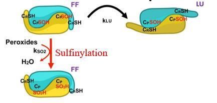
« Exposure to cigarette smoke impairs permeability of lung
 epithelial barrier through proteolytic cleavage of tight junction
 occludin by cathepsin S during COPD »
Paul Bigot a, Simon Chesseron a, Ahlame Saidi a, Damien Sizaret b, Agnès Petit-Courty a,
 Yves Courty a, Fabien Lecaille a, Gilles Lalmanach a
 a
 Université de Tours, Tours, France & INSERM, UMR 1100, Research Centre for Respiratory Diseases (CEPR),
 Team: "Proteolytic Mechanisms in Inflammation", Tours, France
 b
 The University Hospital (CHU Tours), Pathological Anatomy and Cytology, Tours, France
 Corresponding author: paul.bigot-2@etu.univ-tours.fr
 Objectives: Smoking is accountable for more than 80% of chronic obstructive pulmonary
 disease (COPD, 3rd cause of death worldwide), which is characterized by emphysema,
 chronic bronchitis associated to an impaired epithelial permeability. Exposure of the lung to
 cigarette smoke elicits the expression of elastinolytic cathepsin S (CatS) (1). Despite an
 oxidizing environment, the reactivity of the nucleophilic Cys25 within the active site (sharing a
 thiolate (C25)-imidazolium (H159) dyad) remains partially preserved, due to the reversible
 formation of a sulfenic acid, followed by a slower conversion to sulfinic acid (2-3). Recently,
 junctional and/or adhesion molecules were pinpointed as putative CatS targets, suggesting
 that CatS could proteolytically alter epithelial integrity during COPD.
 Methods: Clinical features. Immunochemical analysis of human COPD and non-COPD lung
 biopsies revealed a decreased expression level of tight junction occludin for smokers (vs non-
 smokers). Statistical analysis demonstrated that occludin level correlates negatively with the
 smoking history (number of pack-years), COPD grades as well with proteolytic activity of CatS.
 Results: Molecular mechanisms. Exposure of macrophages to cigarette smoke extract (CSE)
 or nicotine, a major CSE component, triggered expression of secreted and catalytically
 active CatS through the mTOR/TFEB signaling pathway, while incubation of lung epithelial
 cells with CatS is associated with an increased proteolysis of occludin, a decreased trans-
 epithelial electrical resistance and an amplified epithelial permeability. In a model of co-
 cultured macrophages/epithelial cells, an increase of epithelial permeability was observed
 following exposure to CSE. Conversely, both pharmacological inhibition of CatS as well its
 transient transcriptional inhibition by siRNAs restored the basal permeability of lung epithelial
 cells.
 Conclusions: Altogether with its deleterious elastinolytic activity favoring emphysema, the
 uncontrolled enzymatic activity of CatS displays detrimental effects on the integrity of lung
 epithelial barriers, therefore strengthening the therapeutic relevance of targeting CatS in
 COPD (4-5).
 References :
 (1) Cigarette smoke induces overexpression of active cathepsin S in lungs from current smokers with or without COPD. P-M. Andrault, A. C.
 Schamberger, T. Chazeirat, D. Sizaret, J. Renault, C. A. Staab-Weijnitz, E. Hennen, A. Petit, M. Wartenberg, A. Saidi, T. Baranek, S. Guyetant,
 Y. Courty, O. Eickelberg, G. Lalmanach & F. Lecaille (2019) Am. J. Physiol. Lung Cell. Mol.
 Physiol. 317, L625–L638
 (2) Regulation of the proteolytic activity of cysteine cathepsins by oxidants. G. Lalmanach, A. Saidi, P. Bigot, T. Chazeirat, F. Lecaille & M.
 Wartenberg (2020) Int. J. Mol. Sci. 21 :1944 (doi 10.3390/ijms21061944).
 (3) Oxidation of cathepsin S by major chemicals of cigarette smoke. M. Wartenberg, P-M. Andrault, A. Saidi, P. Bigot, L. Nadal-
 Desbarats, F. Lecaille & G. Lalmanach (2020) Free Radic. Biol. Med. 150, 53-65
 (4) Cysteine cathepsins and cystatins: from ancillary tasks to prominent status in lung diseases. G. Lalmanach, A. Saidi, S.
 Marchand-Adam, F. Lecaille & M. Kasabova (2015) Biol. Chem. 396, 111-130
 (5) Protean proteases: at the cutting edge in lung diseases? C. Taggart, M.A. Mall, G. Lalmanach, D. Cataldo, A. Ludwig, S.
 Janciauskiene, S. Meiners, C. Overall, C. Schultz, B. Turk, K.S. Borensztajn (2017) Eur. Resp. J. 49: 1501200
 [https://doi.org/10.1183/13993003.01200-2015]
 Keywords: cysteine protease, chronic obstructive pulmonary disease (COPD), epithelial
 barrier, inflammation, tight junction protein.
«A sensitive HPLC assay with fluorescence detection for the DJ-1
 activity (park7), a parkinson-associated deglycase»
Nicolas MATHAS1, Emmanuelle BRAUD1, Erwan GALARDON1, Catherine LAURENT1, Lucie
LARIGOT2, Daniel MANSUY1, Mélanie ETHEVE-QUELQUEJEU1, Marie-Agnès SARI1, Beatrice LE
GRAND2, Etienne BLANC2, Xavier COUMOUL2 and Julien DAIROU1

1 Université de Paris cité, CNRS, Laboratoire de Chimie et de Biochimie Pharmacologiques et
Toxicologiques, F-75006 Paris, France. 45 rue des Saints-Pères, 75006 Paris, France

2 INSERM UMR-S 1124, Université Paris Cité, Toxicologie Pharmacologie et Signalisation
cellulaire, 45 rue des Saints-Pères, 75006 Paris, France

E-mail of corresponding author: nicolas.mathas@parisdescartes.fr

Abstract: Glycation is an inevitable nonenzymatic covalent reaction between nucleophilic groups
in proteins and nucleotides and endogenous reducing sugars or dicarbonyls (methylglyoxal) that
results in protein and/or nucleotides covalent modification. After which a series of dehydrations,
oxidations and rearrangements leads to amyriad of products called advanced glycation end
products (AGEs). Previously, we showed that DJ-1 (or Park7) is a protein deglycase that repairs
methylglyoxal-glycated biomolecules by acting on early glycation intermediates and releases
repaired biomolecule and lactate. Moreover, the DJ-1 gene, also known as PARK7, is associated
with recessive and sporadic forms of Parkinson’s disease so over the last years, the function of DJ-
1 has appeared as an important topic for the understanding on the etiology of Parkinson
disease. In this context, availability of sensitive and quantitative enzyme assays is of prime
importance to understand the role of DJ-1 and to develop specific effectors. Here, we describe a
new method to measure DJ-1 activity based on the separation and quantification of glycated and
unglycated fluorescent nucleotide by High Pressure Liquid Chromatography (HPLC). Kinetic and
mechanistic analyses using recombinant DJ-1 confirmed the reliability of this approach. In
addition, this assay was further validated using cellular lysats. Our results indicate that this novel
DJ-1a assay is easy, sensitive, and specific.

KEYWORDS: Up to 5
DJ1, Methylglyoxal, Guanosine Fluorescein, enzyme assay, Electrophile stress.
SESSION 4

 “ Archaea ”

• Conférence par Tristan Wagner
• Solenne Ithurbide : prix de l’article de l’année de la SFBBM
• Clément Madru
• Roxane Lestini

 Animée par
 Emmanuelle Schmitt
 Ecole Polytechnique - Palaiseau
Conférence invitée session 4
 «What are the molecular tricks of methanogenic archaea to save energy?»
 par Tristan Wagner
Max Planck Institute for Marine Microbiology, Celsiusstraβe 1, 28359, Bremen, Germany.
 Nevena Maslaća, Marion Jespersena, Marie-Caroline Müllera, Olivier Lemairea, Seigo Shimab, Sylvain
 Engilbergec, and Tristan Wagnera.
 a
 Max Planck Institute for Marine Microbiology, Celsiusstraβe 1, 28359, Bremen, Germany. twagner@mpi-
 bremen.de
 b
 Max Planck Institute for Terrestrial Microbiology, Marburg, Germany.
 c
 European Synchrotron Radiation Facility (ESRF), Grenoble, France.
 Objectives. Methanogenic archaea drive the final step in anaerobic organic compound mineralization, dictating
 the carbon flow of Earth’s diverse anoxic ecosystems in the absence of inorganic electron acceptors. In other
 words, the methanogens had, have, and will have a predominant impact on our planet due to their ability to
 generate methane (CH4) from different carbon sources. While the molecular process of CH4 generation has been
 studied during the past decades, one question prevails: How do hydrogenotrophic methanogens (reduce CO2 to
 CH4 with H2) grow so efficiently with an energy metabolism providing 60 times less ATP than O 2 respiration? With
 such a low energy yield, it would be impossible for this organism to fix CO 2 via known conventional strategies.
 This presentation will illustrate how these archaea avoid ATP-consuming steps and circumvent non-favorable
 reactions by tunneling effect strategies and regulatory networks [2,3,4,5].
 Methods. Methanothermococcus thermolithotrophicus is a marine methanogen able to duplicate as fast as
 Escherichia coli in a fermenter gassed with H2 and CO2. However, in comparison to E. coli, M.
 thermolithotrophicus grows in a minimum mineral medium with stunning chemolithoautotrophic capabilities. To
 investigate the metabolic pathway of this archaeon, our laboratory developed a pipeline to extract, purify,
 characterize and crystallize the proteins from the methanogen under an anaerobic atmosphere. Enzymology and
 biophysical experiments confirm protein functions, while X-ray crystallography provides structural information.
 Results. All the reactions involved in methanogenesis are ATP-independent [1], even the CO2-fixation step. In
 this process, CO2 is firstly reduced to formate and then covalently fixed on a carrier. The formate accumulation
 in the enzyme core drives its fixation on the carrier molecule [2]. The electrons required for CO 2-reduction are
 provided by another complex, which “energizes” electrons obtained from H2 via another coupled reaction called
 electron-bifurcation [3]. Biosynthetic pathways also rely on these tunneling effects. For instance, the unfavorable
 first step in archaeal lipid biosynthesis is triggered by the next step, which is highly exergonic [4]. Transcriptome
 analyses combined with the structural/biochemical elucidation of the whole sulfur and nitrogen assimilation
 pathways were accomplished. This unprecedented success highlighted new catalytic reactions, allosteric
 regulation nodes, and an overall view of the stress response to starvation.
 Conclusions. Hydrogenotrophic methanogens fuel their catabolic and anabolic pathways with H2, the simplest
 molecule in the universe. While hydrogenases harness the reducing power of H2, energy converters energize the
 captured electrons and distribute them through the metabolism via carriers. A myriad of processes using directly
 or indirectly these electrons are dispatched through the central metabolism to save cellular energy [5]. Among
 them are new catalysts, bypass of ATP-consuming reactions, enzymatic coupling, and regulation via allosteric
 effectors. These results were obtained by native exploration, an unbiased approach that must be applied to
 unveil the unsuspected.
 Relevant references.
 [1] Shima S, Huang G, Wagner T, Ermler (2020) U. Annu Rev Microbiol. [2] Wagner T, Ermler U and, Shima S.
 (2016). Science
 [3] Wagner T, Koch J, Ermler U and, Shima S. (2017). Science
 [4] Vögeli B, Engilberge S, Girard E, Riobé F, Maury O, Erb TJ, Shima S, Wagner T. (2018 ) PNAS [5] Jespersen M
 and Wagner T (2022) Nat. Chemical Biology. Under review
 Keywords. Methanogenic archaea, Anaerobic metabolism, Biochemistry and structural biology, Enzymatic
 complex, Energy-saving strategies, Allosteric regulation.
Conférence invitée session 4
 Prix de l’article de l’année de la SFBBM

«Cell division in the archaeon Haloferax volcanii relies on two FtsZ proteins with
 distinct functions in division ring assembly and constriction»

 Par Solenne Ithurbide
 University of Freiburg, Institute of Biology II, Molecular Biology of Archaea lab.

 In bacteria, the tubulin homologue FtsZ assembles a cytokinetic ring, also called the Z ring, and plays a key
 role in the machinery that constricts to divide the cells. Many archaea encode two FtsZ proteins from
 distinct families, FtsZ1 and FtsZ2, with previously unclear functions but early observations by
 immunolabelling of one FtsZ as a band at mid cell predicted of a bacterial-like FtsZ division mechanisms.
 Our recent studies, in the archaeal model organisms Haloferax volcanii, showed fundamental differences of
 FtsZ-based archaeal division compared to Bacteria. Indeed, the 2 FtsZs were revealed to have fundamental
 but different roles in archaeal cell division. The two FtsZs, FtsZ1 and FtsZ2 colocalize to form the dynamic
 division ring. However, FtsZ1 can assemble rings independently of FtsZ2, and stabilizes FtsZ2 in the ring,
 whereas FtsZ2 functions primarily in the constriction mechanism. FtsZ1 also influenced cell shape,
 suggesting it forms a hub-like platform at midcell for the assembly of shape-related systems too. Many
 archaea also encode for homologues of MinD family proteins which are playing an important role into the
 mid-cell placement of the Z ring. However, we showed that none of the MinD homologues is involved in the
 Z ring placement in H. volcanii, highlighting further the differences between FtsZ based cell division
 between Archaea and Bacteria. Interestingly, both FtsZ1 and FtsZ2 are widespread in archaea with a single
 S-layer envelope, but the minority of archaea with a pseudomurein wall and division septum only have one
 FtsZ1. This suggest that an early duplication of FtsZ and then a lost of one FtsZ , concomitant with the
 pseudomurein apparition in Archaea have led to the development of a 1 FtsZ based cell division.
«Molecular specificities of the Replication Protein A in the third
 domain of life»
 Clément Madrua, Markel Martinez-Carranzaa, Maelenn Chevreuilb, Bertrand Raynalb,
 Sebastien Laurentc, Didier Flamentc, Ahmed Haouzd, Mart Krupovice, Marc Delaruea, Pierre
 Legrandf and Ludovic Saugueta.

a
 Unit of Architecture and Dynamics of Biological Macromolecules, CNRS UMR 3528, Institut
Pasteur, 75015 Paris, France.
b
 Plateforme de Biophysique Moléculaire, CNRS UMR 3528, Institut Pasteur, 75015 Paris, France
c
 Laboratoire de Microbiologie des Environnements Extrêmes, CNRS, UMR 6197, Ifremer, Université
de Brest, F-29280 Plouzané, France.
d
 Plateforme de cristallographie, CNRS UMR 3528, Institut Pasteur, 75015 Paris, France.
e
 Archaeal Virology Unit, Institut Pasteur, Université de Paris, 75015 Paris, France. f Synchrotron
SOLEIL, L'Orme des Merisiers, Gif-sur-Yvette, France. clement.madru@pasteur.fr;
ludovic.sauguet@pasteur.fr

Replication Protein A (RPA) is the major single stranded DNA-binding protein in both eukaryotes
and archaea with essential roles in DNA replication, recombination and repair. RPA binds to
exposed ssDNA to protect it from nucleases, participates in a myriad of nucleic acid transactions
and coordinates the recruitment of other important players. RPA is a heterotrimer and coats long
stretches of single-stranded DNA (ssDNA). We present the first structure of an archaeal RPA in
both its apo form and bound to ssDNA. It includes a trimeric core that is conserved with
eukaryotes and additional domains that are specific to archaea. In its apo form, the RPA adopts a
tetrameric assembly that dissociates upon binding to DNA. By using an integrative approach that
combines X-ray crystallography, cryo-electron microscopy protein-protein and protein-nucleic
acids interaction measurements, we investigated the role of each individual domain of RPA and
present a structural model of how the archaeal RPA assembles on long ssDNA.

KEYWORDS: DNA replication; Archaea; ssDNA binding protein
« Understanding replication dynamics and chromosomal
 organisation in the archaea Haloferax volcanii»
 C. Cockram2, D. Noury1, A. Thierry2, R. Koszul2, O. Nicolas1 and R. Lestini1
 1
 Laboratoire d’Optique et Biosciences, Ecole Polytechnique, CNRS UMR7645 – INSERM U1182, Institut Polytechnique
 de Paris, 91128 Palaiseau cedex, France.
 2
 Institut Pasteur, Unité Régulation Spatiale des Génomes, CNRS, UMR 3525, F-75015 Paris, France.
 DNA replication is essential to all proliferating cells. However, little is known concerning this
 important process in archaea. Because the archaeal DNA replication machinery is largely
 homologous to that of eukaryotes, expanding our knowledge of replication to archaea will
 deepen our understanding of replication dynamics in all domains of life.
 Archaeal main chromosome is circular, a bacterial-like feature, but often contains multiple
 active replication origins, a eukaryotic-like feature. This is notably the case of the main
 chromosome of the archaea Haloferax volcanii which possesses four replication origins. H. volcanii
 is a halophilic archaea easily grown in laboratory (120 minutes generation time at 45°C on rich
 media) for which powerful tools have been developed. This includes genetic tools and the use of
 protein labeling by the green fluorescent protein GFP to study their localization in living cells,
 which are essential to tackle the in vivo study of fundamental processes such as DNA replication.
 Using wide-field and 3D-SIM super-resolution live cell imaging, we have provided unexpected
 insight
 into the intracellular dynamics of DNA replication in H. volcanii cells (1,2). Considering the 4
 replication origins of the main chromosome and its high copy number (18 copies of the genome
 on average), an unexpected low number of replication foci was observed. These results prompted
 us to investigate the 3D organization of the chromosome in H. volcanii. Using HiC method with a
 resolution of up to 1 kb, we have revealed a chromosomal organisation composed of self-
 interacting domains and chromatin loops (3). This organisation is regulated by both transcription
 and the archaeal SMC protein. We could also show that replication is not strongly implicated in
 the chromosomal structuring in H. volcanii. Thus, it is unlikely that a chromosomal organisation
 bringing replication forks close to one another accounts for the relatively low number of
 replication foci observed. Yet, this method also relies on a population of cells, and, as a result,
 reflects an average of the population which may mask cell to cell variation. And oligoploidy is in
 itself another limitation, as interchromosomal contacts cannot be distinguished from
 intrachromosomal contacts. So it could be that replication affects chromosome organisation but
 that this effect has been averaged out by the heterogeneity of cells in the population. This why
 we are developing super-resolution imaging to map the spatial organization of chromosomal
 copies at the cell-to-cell level (4).
(1) Lestini, R., Laptenok, S.P., Kuhn, J., Hink, M.A., Schanne-Klein, M.C., Liebl, U. and Myllykallio, H. (2013) Intracellular
 dynamics of archaeal FANCM homologue Hef in response to halted DNA replication. Nucleic Acids Research, 41,
 10358-10370.
(2) Delpech F., Collien Y., Mahou P., Beaurepaire E., Myllykallio M., Lestini R. (2018) Snapshots of archaeal DNA
 replication and repair in living cells using super-resolution imaging, Nucleic Acids Research, 46(20), 10757–10770
(3) Cockram, C., Thierry, A., Gorlas, A., Lestini, R., and Koszul, R. (2021). Euryarchaeal genomes are folded into SMC-
 dependent loops and domains, but lack transcription-mediated compartmentalization. Mol Cell 81, 459-472 e410.
(4) Lestini, R., Collien, Y., Olivier, D., Olivier, N. and Myllykallio, H. (In press) BrdU Incorporation and Labeling of
 Nascent DNA to Investigate Archaeal Replication Using Super-Resolution Imaging. Methods Molecular Biology,
 Vol. 2522, Sébastien Ferreira-Cerca (Eds): Archaea.
SESSION 5

 “ Genetic ”

• Conférence par Claire Rougeulle
• Maxime Wery
• Louise Le Coadou
• Inge Kühl

 Animée par
 Bernard Dujon
 Institut Pasteur - Paris
Conférence invitée session 5

 «X chromosome inactivation, at the interface between development,
 chromatin and the noncoding genome»

 par Claire Rougeulle
Université Paris Cité, CNRS, Epigenetics and Cell Fate, F-75013 Paris, France

 X chromosome inactivation (XCI) in mammals is an essential epigenetic process which

 compensates for X chromosome imbalance between sexes. XCI is established early during female

 development, at peri-implantation stages, and is triggered by the accumulation of the long

 noncoding RNA XIST. This process has been mainly studied in the mouse where embryonic stem

 cells (ESCs) have been instrumental to characterize the actors of the process, and to unravel the

 kinetics of the molecular events leading to the transcriptional silencing of one of the two X

 chromosomes. However, it is now known that X-inactivation initiates through remarkable diverse

 strategies in different species. We are using primate ESCs as a model system for early primate

 development, to characterize the early stages of X chromosome inactivation and to identify

 regulators of the process in primates. We are in particular exploring the extent to which long

 noncoding RNA contribute to the variation in XCI strategies between species.
«Translation is a key determinant controlling the fate of
 cytoplasmic long non- coding RNAs»

 Sara Andjus1,4, Ugo Szachnowski1,4, Nicolas Vogt1, Isabelle Hatin2, Chris Papadopoulos3,
 Anne Lopes3, Olivier Namy2, Maxime Wery1,5 & Antonin Morillon1,5
 1
 ncRNA, epigenetic and genome fluidity, Institut Curie, PSL University, Sorbonne Université, CNRS
 UMR3244, 26 rue d’Ulm, F-75248 Paris Cedex 05, France
2
 Genomics, Structure and Translation, Institute for Integrative Biology of the Cell (I2BC), CEA, CNRS,
 Université Paris-Sud, Université Paris-Saclay, 91198 Gif-sur-Yvette cedex, France
3
 Molecular Bio-informatics, Institute for Integrative Biology of the Cell (I2BC), CEA, CNRS, Université
 Paris-Sud, Université Paris-Saclay, 91198 Gif-sur-Yvette cedex, France
4
 equal contributions
5
 corresponding authors: maxime.wery@curie.fr; antonin.morillon@curie.fr
 The hidden face of genomes produces thousands of long non-coding (lnc)RNAs.
 Although they were initially presumed to lack coding potential, recent works revealed that
 lncRNAs can be translated into micropeptides, which might represent proteins “in progress”
 throughout evolutionary constraints, or have actual intra/extra-cellular roles, or be the source
 of neoantigens driving the immune response. However, despite the interest they arouse, the cis-
 and trans-acting mechanisms controlling the synthesis of these peptides remain poorly
 characterized. In yeast, we previously showed that the cytoplasmic Xrn1-sensitive lncRNAs are
 degraded via the translation-dependent Nonsense Mediated mRNA Decay (NMD) pathway,
 suggesting that translation determines their degradation. Here, we show that most cytoplasmic
 lncRNAs are actively translated, modulating their cellular abundance and providing an
 opportunity for the cell to produce novel peptides. We found that NMD-sensitive lncRNAs
 accumulate in wild-type (WT) cells treated with translation elongation inhibitors. Our data
 indicate that translation also affects lncRNAs decay independently of NMD, by interfering
 directly with their decapping. Ribo-Seq analyses confirmed ribosomes binding to a substantial
 fraction of lncRNAs and identified actively translated small (sm)ORFs in their 5’-proximal region.
 Mechanistic analyses revealed that the NMD sensitivity of lncRNAs depends on the length of the
 3’ untranslated region, following the smORFs. Finally, we show that translation of an NMD-
 sensitive lncRNA reporter gives rise to a detectable peptide in WT cells. Our work highlights the
 role of translation in the metabolism of lncRNAs. We propose that the translation of lncRNAs
 could contribute to expose genetic novelty to the natural selection, while NMD would restrict
 their expression.
 KEYWORDS: lncRNA/Xrn1/NMD/translation
«Enzymatic and structural characterization of the recurrent
 S1624C oncogenic mutation of the histone lysine
 methyltransferase SETD2»
 Louise LE COADOUa, Jérémy BERTHELETb, Ariel MECHALYc, Christina MICHAILa, Linh-Chi BUIa,
 Ahmed HAOUZc, Jean-Marie DUPRETa, Fernando RODRIGUES LIMAa
 a b
 Université Paris Cité, Unité de Biologie Fonctionnelle et Adaptative (BFA), UMR 8251, CNRS, Paris, France;
 c
 Université Paris Cité, Centre Epigénétique et Destin Cellulaire (CEDC), UMR 7216, CNRS, Paris; Institut Pasteur,
 Plate-forme de Cristallographie-C2RT, CNRS UMR 3528, Paris, France
 E-mail of corresponding author: louise.le-coadou@etu.u-paris.fr

Objectives: The histone methyltransferase SETD2 is responsible for the trimethylation of lysine 36 on
histone H3 (H3K36me3) [1]. This epigenetic mark is crucial for transcriptional regulation, splicing and DNA
damage repair [2]. Many studies have shown that SETD2 is a tumor suppressor and a cancer diver gene,
frequently mutated in cancer [2],[3]. In particular, the S1624C recurrent oncogenic mutation has been
identified in patients with T-cell lymphoma [4], adenocarcinoma or adrenal cortical carcinoma [5],[6]. So
far, this mutation has not been characterized. Interestingly, it is located in the SET catalytic domain of
SETD2 and may thus affect the enzymatic properties of SETD2. The objective of this project is to
characterize the structural and functional impact of this mutation on SETD2.
Methods: Different biochemical, enzymatic and structural approaches were used such as: methylation
assays (western blot, radioactivity and HPLC), crystallography, thermal shift assay (TSA), cell transfection
and cellular enzymology.
Results: Methylation assays (HPLC, western blot/radioactivity) showed that the lysine methyltransferase
activity of SETD2 S1624C mutant is drastically reduced (residual activity around 10%). Strong decrease
of H3K36me3 mark is also observed on histones extracted from cells transfected with S1634C mutant.
On the other hand, a crystallographic structure of the S1624C mutant in complex with the cofactor
SAM (S-adenosylmethionine) and a histone H3 substrate peptide was obtained and showed that the
mutation does not impair co-factor and substrate binding. Local and very slight deformations can be
observed compared to the structure of SETD2 WT. Further results indicate that the replacement of the
Ser residue by a bulkier cysteine reduces the stability of the mutant thus increasing its tendency to
form aggregates. In addition, increased zinc release from the active-site zinc fingers of the enzyme is
observed for the S1624C form thus further contributing to the formation of aggregates. Formation of
reversible disulfide-dependent aggregates is also increased for the S1624C mutant thus supporting that the
replacement of the Ser residue by a Cys increases redox sensitivity of the mutant form compared to WT
SETD2.
Conclusions: Our study provides the molecular basis for the altered lysine methyltransferase activity of the
oncogenic SETD2 S1624C mutant form. This mutation leads to a poorly active enzyme that retains the
ability to bind its SAM cofactor and a H3 peptide substrate. The bulkier Cys compared to Ser leads to
increased instability of the enzyme and aggregation. Moreover, increased disulfide- dependent
aggregation of the mutant is observed. Ongoing studies (mass spectrometry and molecular dynamics)
will provide further understanding of the molecular impact of the SETD2 S1624C mutation.
Keywords: Histone methyltransferase, SETD2, Mutant, Cancer, H3K36 trimethylation
Relevant references:
[1] E. J. Wagner et P. B. Carpenter, Nat. Rev. Mol. Cell Biol., 2012 [2] D. Husmann et O. Gozani, Nat. Struct. Mol. Biol., 2019
[3] X. Zhu et al., Nat. Genet., 2014
[4] A. Roberti et al., Nat. Commun., 2016
[5] A. Zehir et al., Nat. Med., 2017
[6] S. A. Forbes et al., Curr. Protoc. Hum. Genet., 2008
Vous pouvez aussi lire