PET scan Medical practice variations - For a Healthy Belgium
←
→
Transcription du contenu de la page
Si votre navigateur ne rend pas la page correctement, lisez s'il vous plaît le contenu de la page ci-dessous
Nuclear medicine - PET scan

               Medical practice variations

                                    PET scan

   Analysis of the distribution and evolution of medical practice in Belgium,
                in terms of volume and expenditure per insured
    (analysis and trends by region, province and district), for the year 2017

NIHDI – Healthcare Service – Directorate for Research, Development and Quality promotion
                                 Appropriate care unit
                      Pascal Meeus, Virginie Dalcq, Anneleen Van Geystelen
                          Contact: appropriatecare@riziv-inami.fgov.be

                             Date of report: 29 March 2019

                                                                                                           1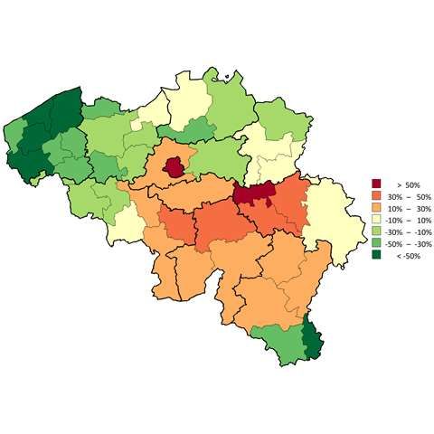
Nuclear medicine - PET scan

CONTENTS

CONTENTS ................................................................................................................................................................................................................................. 2

1.        INTRODUCTION ................................................................................................................................................................................................................. 3

2.        SPECIFIC METHOD OF ANALYSIS ........................................................................................................................................................................................ 4

     A.       NIHDI NOMENCLATURE CODES SELECTED FOR ANALYSIS....................................................................................................................................................................... 4
     B.       PAST HISTORY OF NOMENCLATURE CODES ......................................................................................................................................................................................... 5
     C.       SOURCE OF DATA .......................................................................................................................................................................................................................... 6
     D.       SPECIFIC SELECTION CRITERIA .......................................................................................................................................................................................................... 7
     E.       STANDARDISATION........................................................................................................................................................................................................................ 7

3.        RESULTS ............................................................................................................................................................................................................................ 8

     A.       NATIONAL STANDARDISED RATE OF USE............................................................................................................................................................................................. 8
     B.       BREAKDOWN OF NOMENCLATURE CODES PROVIDED, BY VOLUME ........................................................................................................................................................... 9
     C.       SPECIALISATION OF HEALTHCARE PROVIDERS .................................................................................................................................................................................... 10
     D.       SPECIALISATION OF PRESCRIBERS.................................................................................................................................................................................................... 11
     E.       STANDARDISED RATE OF USE BY GENDER AND AGE GROUP................................................................................................................................................................... 12
     F.       STANDARDISED RATE OF USE: HOSPITAL AND OUT-PATIENT CARE .......................................................................................................................................................... 16
     G.       STANDARDISED RATE OF USE BY REIMBURSEMENT RATE ...................................................................................................................................................................... 18
     H.       TRENDS IN STANDARDISED RATES OF USE ......................................................................................................................................................................................... 20
     I.       GEOGRAPHICAL VARIATIONS IN STANDARDISED RATES OF USE .............................................................................................................................................................. 23
     J.       STANDARDISED HEALTHCARE EXPENDITURE BORNE BY THE INSURANCE .................................................................................................................................................. 28

4.        KEY DATA SUMMARY ...................................................................................................................................................................................................... 31

                                                                                                                                                                                                                                                            2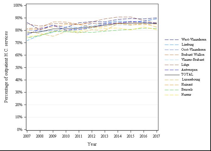
Nuclear medicine - PET scan

    1. INTRODUCTION

The Appropriate Care Unit was set up within the NIHDI’s Directorate for Research, Development and Quality under NIHDI’s Admin-
istration Contract for 2016-20181. Article 35 of this contract refers to ‘the setting up of an Appropriate Care Unit, aiming specifically
to promote an integrated approach to the rational use of resources’. The Appropriate Care Unit has been up and running since the
second quarter of 2017.

The tasks of the Unit were set out formally in the ‘2016-2017 Healthcare monitoring Action plan’, published by NIHDI on 18 July
20162. This plan lists around thirty measures designed to make healthcare provision more efficient, by encouraging appropriate
practice and tackling unnecessary or inappropriate care.

The plan states that one of the tasks of the Appropriate Care Unit is to analyse the ‘appropriateness of care’, in order to identify un-
explained variations in consumption patterns, identified after standardisation. Such variations can potentially point to non-optimal
use of resources.

‘Variations in medical practice’ documents report on the analyses carried out in this framework. Each report focuses on a particular
topic.

In this document, we present the figures and graphs relating to analyses 3 of practice in the area of PET scan, and give the explana-
tions necessary to understand these.

We have deliberately chosen not to attempt to interpret the figures, preferring to present the results to experts who are in a better
position to do so. This document has nevertheless been made available to the public in order to provide objective, open input to
discussions on this issue.

1
  (Institut national d'assurance maladie-invalidité, 2016)
2 (Institut national d'assurance maladie-invalidité, 2016)
3 Readers interested in the methodology used in these quantitative analyses should consult the document entitled ‘Variations in practice – Methodology’.

                                                                                                                                                           3
Nuclear medicine - PET scan
2. SPECIFIC METHOD OF ANALYSIS
A. NIHDI nomenclature codes selected for analysis
The NIHDI nomenclature codes selected for the analysis are listed below:
Outpatient Inpatient Used for rates ? Used for expenses ? Inclusive Exclusive Label Creation Deletion Group N Value
Exa men s ci ntigra phi que fonctionnel du cœur
comportant deux exa mens tomogra phi ques
s ucces s i fs a vec tra i tement pa r ordi na teur
442595 442606 yes yes 01-11-1998 N46 N435
comprena nt a u moi ns deux pl a ns non pa ra l l èl es
de recons truction, a vec protocol e et documents
i conogra phi ques
Tomogra phi e à émi s s i on de pos i tons pa r
détection en coïnci dence a vec protocol e et
documents , pour l 'ens embl e de l 'exa men, s i , da ns
442676 442680 yes yes l e ca s d’une i ntervention chi rurgi ca l e prévue pour 01-01-2016 N46 N250
une i ns uffi s a nce corona ri enne compl ètement
documentée récemment, un doute s ubs i s te encore
qua nt à l a vi a bi l i té du myoca rde concerné
Tomogra phi e à émi s s i on de pos i tons pa r
détection en coïnci dence a vec protocol e et
documents , pour l 'ens embl e de l 'exa men, s i l a
442691 442702 yes yes théra pi e s ous forme d’une i ntervention 01-01-2016 N46 N250
chi rurgi ca l e es t i nfl uencée de ma ni ère déci s i ve ,
pour l a l oca l i s a tion d'un foyer épi l eptogène d’une
épi l eps i e réfra ctai re
Tomogra phi e à émi s s i on de pos i tons pa r
détection en coïnci dence a vec protocol e et
442713 442724 yes yes 01-01-2016 N46 N250
documents , pour l 'ens embl e de l 'exa men, pour
des i ndi ca tions i nfectieus e ou i nfl a mma toi re
Tomogra phi e à émi s s i on de pos i tons pa r
détection en coïnci dence a vec protocol e et
442735 442746 yes yes 01-01-2016 N46 N250
documents , pour l 'ens embl e de l 'exa men, pour
des i ndi ca tions neurodégénéra tives
Exa men tomogra phi que à émi s s i on de pos i tons
pa r détection en coïnci dence , a vec protocol e et
documents , pour d’a utres i ndi ca tions que cel l es
442750 442761 yes yes 01-01-2016 N46 N250
mentionnées a ux pres tations 442971-442982,
442676-442680, 442691-442702, 442713-442724 ou
442735-442746
Tomogra phi e à émi s s i on de pos i tons pa r
détection en coïnci dence a vec protocol e et
442971 442982 yes yes 01-01-1991 N46 N250
documents , pour l 'ens embl e de l 'exa men, pour
des i ndi ca tions oncol ogi ques
This table shows the NIHDI nomenclature codes selected for this analysis, stating whether or not they were included in the analyses of services and expenditure, and
giving, for each one, a description, dates of creation and deletion, where appropriate, their N group (in the NIHDI nomenclature) and their value. 4Nuclear medicine - PET scan
B. Past history of nomenclature codes
Outpatient Inpatient Date Label Group N Value
Test scintigraphique fonctionnel comportant deux examens tomographiques successifs avec traitement par
ordinateur comprenant au moins deux plans non parallèles de reconstruction, avec protocole et documents
442595 442606 01-11-1998 N46 N435
iconographiques, non cumulable avec les prestations 442411 - 442422, 442455 - 442466, 442610 - 442621 et 442632 -
442643 pour l'examen d'une même fonction effectué au moyen d'un même produit marqué
Examen scintigraphique fonctionnel du cœur comportant deux examens tomographiques successifs avec
442595 442606 01-01-2016 traitement par ordinateur comprenant au moins deux plans non parallèles de reconstruction, avec protocole et N46 N435
documents iconographiques
Tomographie à émission de positons par détection en coïncidence avec protocole et documents, pour l'ensemble
442676 442680 01-01-2016 de l'examen, si, dans le cas d’une intervention chirurgicale prévue pour une insuffisance coronarienne N46 N250
complètement documentée récemment, un doute subsiste encore quant à la viabilité du myocarde concerné
Tomographie à émission de positons par détection en coïncidence avec protocole et documents, pour l'ensemble
442691 442702 01-01-2016 de l'examen, si la thérapie sous forme d’une intervention chirurgicale est influencée de manière décisive, pour la N46 N250
localisation d'un foyer épileptogène d’une épilepsie réfractaire
Tomographie à émission de positons par détection en coïncidence avec protocole et documents, pour l'ensemble
442713 442724 01-01-2016 N46 N250
de l'examen, pour des indications infectieuse ou inflammatoire
Tomographie à émission de positons par détection en coïncidence avec protocole et documents, pour l'ensemble
442735 442746 01-01-2016 N46 N250
de l'examen, pour des indications neurodégénératives
Examen tomographique à émission de positons par détection en coïncidence, avec protocole et documents, pour
442750 442761 01-01-2016 d’autres indications que celles mentionnées aux prestations 442971-442982, 442676-442680, 442691-442702, N46 N250
442713-442724 ou 442735-442746
442971 442982 01-01-1991 Tomographie à positons avec protocole et documents, pour l'ensemble de l'examen. N46 N1300
Tomographie à positrons par détection en coïncidence avec protocole et documents, pour l'ensemble de
442971 442982 01-07-1999 N46 N1150
l'examen
Tomographie à émission de positons par détection en coïncidence avec protocole et documents, pour l'ensemble
442971 442982 01-01-2016 N46 N250
de l'examen, pour des indications oncologiques
This table shows the history of the NIHDI nomenclature codes selected for analysis, as well as their past value and N group (in the NIHDI no-
menclature). If no changes have taken place over time, only the current information is shown.
5Nuclear medicine - PET scan
C. Source of data
The data used in the analyses have been taken from the following databases:
for the utilisation rate and amount of expenses of insured persons (who meet
Document N the selection criteria) whose age, sex, preferential regime and residence are
known 2007-2017
for the utilisation rate and amount of expenses of insured persons (who meet
Document P
the selection criteria) by type of medical specialities in 2015-2017
'N Documents' are monthly data sent by the sickness funds to NIHDI, within three months. These data show the number of services provided, dates and
the fees involved. Every six months, these data are compiled by the insurers, which also add data on patients: age, gender, social category and district of
residence. N Documents, however, cannot be used to analyse the combinations of services received by individual patients.
'P Documents' are six-monthly data sent by the sickness funds to NIHDI, within four months. These data show the services provided, the service-
provider, the prescriber, the place of provision of service, and the hospital where patients were treated. P Documents can be used to monitor medical
consumption and pricing, but not (yet) to analyse services per patient.
6Nuclear medicine - PET scan
D. Specific selection criteria
Several filters may have been applied to the data, so that only one section of the population is considered in analyses. If so, the fil-
ters used are shown in the table below:
FILTERS APPLIED TO DATA
Gender Women and men
Age All
E. Standardisation
The data are standardised before analysis per year, based on age, sex and preferential regime per arrondissement, province and re-
gion.
Standardisation renders populations comparable in relation to one or several criteria. If a difference is observed between these populations, we can
therefore assume that it is not due to the criteria covered by the standardisation process.
7Nuclear medicine - PET scan
3. RESULTS
A. National standardised rate of use
TOTAL
Average number of interventions per yea 78.671
Standardised rate of use
722
per 100,000 insured persons
8Nuclear medicine - PET scan
B. Breakdown of nomenclature codes provided, by volume
9Nuclear medicine - PET scan
C. Specialisation of healthcare providers
Specialisation of the provider Total providers Concerned providers % Providers Median of H.C. services Number of H.C. services % Total H.C. services Expenses % Expenses
Nuclear medicine 214 130 61% 422,00 78.359,00 99% 13.395.920,55 99%
Other specialities 1843 12 1% 13,00 494,00 1% 98.135,02 1%
Total 2057 142 7% 377,00 78.853,00 100% 13.494.055,57 100%
This table shows the following non-standardised data, by medical specialities (average figures for the period 2017) :
- The number of service-providers per specialisation who have recorded at least one service falling within the N group(s) (of the NIHDI nomen-
clature) covered by this analysis;
- The number of service-providers recording services under the nomenclature codes selected for this analysis;
- The service-providers for these codes as a percentage of the total number of service-providers recording provision of at least one service fall-
ing within the N group(s) (of the NIHDI nomenclature) covered by this analysis;
- The median number of services per service-provider (recording provision under these codes);
- The number of services provided;
- The service percentage, i.e. the number of services recorded for this specialisation as a percentage of total services provided;
- Expenditure;
- The expenditure percentage, i.e. the expenditure on this specialisation as a percentage of total expenditure.
10Nuclear medicine - PET scan
D. Specialisation of prescribers
Specialisation of the prescriber Total prescribers Concerned prescribers % Prescribers Median of prescriptions Number of prescriptions % Prescriptions Expenses % Expenses
Medical oncology 292 260 89% 43,00 14.150,00 18% 2.403.822,72 18%
Pneumology 621 502 81% 16,00 11.487,00 15% 1.961.754,64 15%
Gastroenterology 782 576 74% 6,00 7.823,00 10% 1.330.448,69 10%
Haematology 166 147 89% 40,00 7.039,00 9% 1.197.063,65 9%
Specialists in training 6684 1211 18% 2,00 5.656,00 7% 976.544,55 7%
Unknown 3 2 67% 2.203,00 4.406,00 6% 759.062,27 6%
Internal medicine 1418 498 35% 3,00 3.711,00 5% 639.714,68 5%
Neurology 637 380 60% 4,00 3.492,00 4% 597.078,75 4%
Radiotherapy 226 162 72% 13,00 3.328,00 4% 565.251,63 4%
Urology 488 292 60% 6,00 2.885,00 4% 487.449,69 4%
General surgery 1770 485 27% 2,00 2.838,00 4% 481.843,84 4%
Gynaecology and midwifery 1744 416 24% 2,00 2.173,00 3% 369.164,96 3%
Otorhinolaryngology 757 240 32% 3,00 1.932,00 2% 329.647,51 2%
Rheumatology 276 151 55% 5,00 1.212,00 2% 206.414,20 2%
Geriatrics 345 201 58% 3,00 1.152,00 1% 200.906,62 1%
Other specialities 38919 1535 4% 2,00 5.569,00 7% 987.887,17 7%
Total 55128 7058 13% 3,00 78.853,00 100% 13.494.055,57 100%
This table shows, in order, the following non-standardised data per specialities (average figures for the period 2017) :
- The number of prescribers who have prescribed at least one service falling within the N group(s) (of the NIHDI nomenclature) covered by this analysis;
- The number of prescribers prescribing the nomenclature codes selected for this analysis;
- The prescribers prescribing these codes as a percentage of the number of prescribers prescribing at least one service falling within the N group(s) (of the
NIHDI nomenclature) covered by this analysis;
- The median number of services per prescriber (prescribing these codes);
- The number of services prescribed;
- The percentage of services prescribed, i.e. the number of prescriptions issued for this specialisation as a percentage of total services prescribed;
- Expenditure;
- The expenditure percentage, i.e. expenditure on this specialisation as a percentage of total expenditure.
11Nuclear medicine - PET scan
E. Standardised rate of use by gender and age group
TOTAL
Average number of interventions per year 78.671
Median age (years) 65
Mean age (years) 63,38
Max/Min Ratio of the median age
1,06
(by district)
Percentage of women 46,88%
Max/Min Ratio:
The max/min ratio measures the dispersion of values. It is calculated as the ratio of the maximum value found
for the variable, in all districts, to the minimum value. If this minimum value is equal to zero, the max/min ratio
cannot be calculated, and should be given as ‘NA’ (‘not applicable’).
12Nuclear medicine - PET scan
This figure is made up of bar
charts for each gender. The
coefficient of variation, shown by
the red line, measures the rela-
tive dispersion of the standard-
ised rates of use observed for
each district, by age group and
gender (standard deviation
divided by the mean). This line is
shown in bold for age groups
where the variation coefficient
can be validly interpreted (i.e.
for age groups in which there
are sufficient insured persons
per district to allow for a proper
comparison).
The left-hand vertical axis of the
graph plots the standardised
rate of use, and the right-hand
axis plots the variation coeffi-
cient. The horizontal axis shows
the division by age group. The
horizontal dotted lines show the
total values of the standardised
rates of use (in blue) and of the
variation coefficient (in red).
Standardised rate of use per 100,000 insured persons, and variation coefficient for the districts, by age
group and gender, for the year 2017
13Nuclear medicine - PET scan
Comparison of the standardised rates of use for women and men (per 100,000) in 2017
14Nuclear medicine - PET scan
This histogram shows standard-
ised rates of use by province and
by gender. The grey bars show
the rates for men, while the
green bars show the rates for
women, for each province. The
grey and green broken lines
show the total standardised
rates of use, again grey for men,
green for women.
Standardised rate of use per 100,000 insured persons, by gender and by province for the year 2017
15Nuclear medicine - PET scan
F. Standardised rate of use: hospital and out-patient care
TOTAL
Average number of interventions per year 78.671
Percentage of out-patient care 85,42%
Max/min ratio of out-patient care percentage
1,19
(by district)
This graph shows the per-
centage of out-patient ser-
vices (including hospital day
admissions), i.e. the number
of out-patient services pro-
vided as a percentage of total
services (out-patient + hospi-
tal stays). As well as one bar
per region, there is a bar for
the whole Belgian population.
This overall ratio is also
shown by a dotted line.
Percentage of out-patient care, total and by region
16Nuclear medicine - PET scan
Change over time in the percentage of out-patient care, by province
17Nuclear medicine - PET scan
G. Standardised rate of use by reimbursement rate
TOTAL
Average number of interventions per year 78.671
Percentage provided under the preferential reimbursement rate 25,35%
Standardised rate of use with preferential reimbursement rate
987
(per 100,000)
Standardised rate of use without preferential reimbursement rate
662
(per 100,000)
Ratio Preferential rate /General rate 1,49
18Nuclear medicine - PET scan
This graph shows the stand-
ardised rate of use with (in
red) and without (in grey) the
preferential reimbursement
rate, by region and in total.
The red and grey dotted lines
show the overall standardised
rates of use, with and without
the preferential reimburse-
ment rate, respectively.
Standardised rate of use by reimbursement rate and by region
19Nuclear medicine - PET scan
H. Trends in standardised rates of use
TOTAL
Average number of interventions per year 78.671
Trend (2007-2017) 3,43%
Trend (2015-2017) -1,70%
This graph shows a coloured
curve for each region and a
black curve for the whole
Belgian population. The x-axis
shows the years, and the y-axis
shows the standardised rate of
use per 100,000 insured per-
sons.
Trends in the standardised rate of use per 100,000 insured persons, by region
20Nuclear medicine - PET scan
This graph shows a coloured
line for each district and a
black line for the whole Bel-
gian population. The x-axis
shows the years, and the y-
axis shows the standardised
rate of use per 100,000 in-
sured persons.
To better highlight changes
over time, the rates shown
are rolling averages of the
rates for the three years pre-
ceding the year in question
(including the year itself).
The graph only shows the 5
districts with the highest
average rates, and the 5 dis-
tricts with the lowest average
rates over the last 3 years
studied.
Trends in the standardised rate of use per 100,000 insured persons, by district
21Nuclear medicine - PET scan
Rate This table shows the standardised rates of use (or of
Annual increase
of use intervention) for the last year analysed (2017), but
2017 also the average rates of increase, by province, by
2007- 2015- Statistical region and in total, for the longer period (2007-
(per 105
2017 2017 significance 2017) and the shorter period (2015-2017).
insured)
West Flanders 377,39 1,75% -7,60% *** The statistical significance has been calculated to
show whether the change in data over the last
East Flanders 538,62 2,96% -0,77% three years of the period analysed differs from the
Antwerp 652,36 7,08% 2,77% change over the whole period, by province and by
region. Linear regressions have therefore also been
Limburg 709,77 4,49% 3,00% ** used. This method calculates a regression line by
721,77
Provinces
Flemish Brabant 0,05% -3,11% period studied (2007-2017 and 2015-2017). The
slope of this line is the regression coefficient. The
Brussels 1168,95 -2,63% -11,51% statistical significance reflects the size of the differ-
Walloon Brabant 911,85 4,85% 1,18% ** ence between the regression coefficients in the
various periods analysed, i.e. whether or not trends
Hainaut 826,34 5,52% 11,12% have changed.
Liège 943,94 15,03% -5,91% ***
The asterisks show the degree of statistical signifi-
Namur 923,33 4,92% 2,18% cance of the data observed, with the following
Luxembourg 632,03 4,14% -3,49% *** values: * Value from p ≤ 0.05 / ** Value from p ≤
Flanders 590,5 3,36% -0,60% 0.01 / *** Value from p ≤ 0.001. If there is no aster-
Regions
isk, the trend observed is not statistically signifi-
Brussels 1168,95 -2,63% -11,51% cant.
Wallonia 872,4 7,54% 1,60%
‘NA’ is shown where the nomenclature codes se-
TOTAL 722,36 3,43% -1,70% lected for the analysis have not been used for the
whole of the three-year period.
Trends in the rates of use, by province and region
22Nuclear medicine - PET scan
I. Geographical variations in standardised rates of use
TOTAL
Average number of interventions per year 78.671
Coefficient of Variation (2017) 36,65
Max/Min Ratio* of the standardised rates of use
1,98
(by region)
Max/Min Ratio* of the standardised rates of use
5,15
(by district)
Coefficient of Variation (2015-2017) 35,27
Coefficient of Variation (2007-2009) 51,15
Statistically significant difference? (p-value) Yes (0,042)
* An ‘NA’ result indicates a ratio which cannot be calculated, i.e. the minimum value = zero (cf. E. Standardised rate of use by gender and age
group)
23Nuclear medicine - PET scan
A dot plot is a distribution chart
which is useful for highlighting
grouped data, gaps in distribu-
tion and outlying values. Here,
each dot represents the rate of
use of a district, for its whole
population or broken down by
gender.
The rates are rounded to the
nearest unit, ten, hundred,
etc., depending on the value of
the maximum rate, in order to
better group the values.
The graph also shows, as ‘box-
es’, the 25th, 50th and 75th per-
centiles of the non-rounded
standardised rates of use for all
patients. The bottom line of
the box represents the 25th
percentile, while the upper line
represents the 75th percentile.
The line inside the box repre-
sents the 50th percentile.
‘Dot plot’ showing standardised rates of use by district, by gender
24Nuclear medicine - PET scan
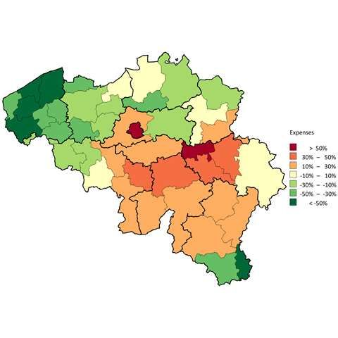
On this map of Belgium, the
boundaries of the districts are
shown by thin lines, while the
provincial borders are shown by
thick lines. The districts are col-
oured using a colour scale based
on the level of rate of use in the
district compared to the Belgian
average. This comparison is ex-
pressed as a percentage: e.g. 0%
if the district rate is equal to the
overall rate, 20% if the rate is
20% above the total rate, and -
20% if the rate is 20% below the
overall rate. The percentages are
calculated using the mean stand-
ardised rate of use of the last
three years, and are displayed in
bands of 20%. The following
colour coding applies:
Colour Category
More than 50%
Between 30% and 50%
Between 10% and 30%
Between - 10% and 10%
Between -30% and -10%
Between -50% and - 30%
Less than -50%
No use made
Map showing distribution of standardised rates of use, by district
25Nuclear medicine - PET scan
On this map of Belgium, the
boundaries of the districts are
shown by fine lines, while the
provincial borders are shown by
thick lines. The districts are col-
oured using a colour scale based
on the level of expenditure in the
district compared to Belgian
average expenditure. This com-
parison is expressed as a per-
centage: e.g. 0% if expenditure in
the district is equal to the overall
expenditure, 20% if it is 20%
higher, and -20% if it is 20% low-
er. The percentages are calculat-
ed using the mean standardised
expenditure for the last three
years and are displayed in bands
of 20%. The following colour
coding applies:
Colour Category
More than 50%
Between 30% and 50%
Between 10% and 30%
Between - 10% and 10%
Between -30% and -10%
Between -50% and - 30%
Map showing distribution of standardised expenditure, by district Less than -50%
No expenditure
26Nuclear medicine - PET scan
In this graph, the standardised rate of
use in a district is placed according to
the size of its population. As well as
the dots for districts, the confidence
intervals (expected variation in the
standardised rate of use when the only
source of variation is random) are also
shown on the graph (horizontal per-
centile lines). These are independent
of the size of the districts. The thicker
horizontal line shows the national
standardised rate of use. The outlier
districts are indicated by values above
P90 and below P10.
As long as the practice analysed only
occurs once per year per insured, this
graph can also be interpreted as a
funnel plot. The confidence levels here
are typically shaped like a funnel: for
small population-sizes, the expected
variation is larger than for more popu-
lous districts. The curves shown by
broken lines represent the 95% and
99.7% confidence intervals. The dis-
tricts situated beyond the upper and
lower limits of the 99.7% confidence
levels are defined as ‘outliers’
‘Funnel plot’ showing the distribution of standardised rates of use by district,
by number of insured persons
27Nuclear medicine - PET scan
J. Standardised healthcare expenditure borne by the insurance
TOTAL
Average number of interventions per year 78.671
Average annual expenditure (€) 13.470.705
Average cost per intervention (€) 171,23
Average annual expenditure per insured (€) 1,24
Max/Min Ratio* of expenditure per insured
1,98
(by region)
Max/Min Ratio* of expenditure per insured
5,14
(by district)
* An ‘NA’ result indicates a ratio which cannot be calculated, i.e. the minimum value = zero (cf. E. Standardised rate of use by gender and age
group)
28Nuclear medicine - PET scan
Standardised expenditure
(per insured)
West Flanders 0,64 €
East Flanders 0,92 €
Antwerp 1,11 €
Limburg 1,24 €
Provinces
Flemish Brabant 1,23 €
Brussels 2€
Walloon Brabant 1,57 €
Hainaut 1,41 €
Liège 1,61 €
Namur 1,58 €
Luxembourg 1,08 €
Flanders 1,01 €
Regions
Brussels 2€
Wallonia 1,49 €
TOTAL 1,24 €
Regional and provincial distribution of standardised expenditure (2017)
29Nuclear medicine - PET scan
Nomenclature 2007 2008 2009 2010 2011 2012 2013 2014 2015 2016 2017 Average annual growth rate
442595-442606 275,96 280,05 291,14 295,31 297,16 300,95 303,78 308,17 308,80 308,80 309,88 1,17%
442676-442680 0,00 0,00 0,00 0,00 0,00 0,00 0,00 0,00 0,00 169,46 170,72 0,74%
442691-442702 0,00 0,00 0,00 0,00 0,00 0,00 0,00 0,00 0,00 173,05 173,21 0,09%
442713-442724 0,00 0,00 0,00 0,00 0,00 0,00 0,00 0,00 0,00 171,38 172,21 0,48%
442735-442746 0,00 0,00 0,00 0,00 0,00 0,00 0,00 0,00 0,00 170,32 170,89 0,34%
442750-442761 0,00 0,00 0,00 0,00 0,00 0,00 0,00 0,00 0,00 170,03 170,35 0,19%
442971-442982 160,20 162,66 169,27 171,74 172,89 174,47 176,52 179,40 179,66 170,06 170,50 0,63%
Change over time in expenditure, by service and by nomenclature code
30Nuclear medicine - PET scan
4. KEY DATA SUMMARY
TOTAL
Main healthcare providers
Nuclear medicine 99,00%
Main prescribers
Medical oncology 18,00%
Standardised rate of use
Average number of interventions per year 78.671
Standardised rate of use per 100,000 insured persons 722,36
Median age (years) 65
Max/min ratio* of the median age (by district) 1,06
Percentage of women 46,88%
Percentage of out-patient care 85,42%
Ratio Preferential rate/General rate 1,49
Trends
Trend (2007-2017) 3,43%
Trend (2015-2017) -1,70%
Statistically significant difference? (p-value) No (0,402)
Geographical variations
Coefficient of Variation (2015-2017) 35,27
Coefficient of Variation (2007-2009) 51,15
Statistically significant difference? (p-value) Yes (0,042)
Max/min Ratio* of number of interventions
1,98
per 100,000 insured persons (by region)
Max/min Ratio* of number of interventions
5,15
per 100,000 insured persons (by district)
Standardised expenditure
Average annual expenditure (€) 13.470.705
Average annual expenditure per insured (€) 1,24
Max/Min Ratio* of expenditure per insured (by region) 1,98
Max/Min Ratio* of expenditure per insured (by district) 5,14
Average cost of interventions (€) 171,23
* An ‘NA’ result indicates a ratio which cannot be calculated, i.e. the minimum value = zero
(cf. E. Standardised rate of use by gender and age group)
31Vous pouvez aussi lire


