Lipides Nutrition & Santé - 23 Mai 2018 Faculté de Médecine, Amphithéâtre Limagne Clermont-Ferrand - INSA Lyon
←
→
Transcription du contenu de la page
Si votre navigateur ne rend pas la page correctement, lisez s'il vous plaît le contenu de la page ci-dessous
18ème journée scientifique
du GIS IMBL

Lipides Nutrition & Santé
23 Mai 2018
Faculté de Médecine, Amphithéâtre Limagne
Clermont-Ferrand
Programme
08h30 – 09h30 Accueil des participants

9h30 – 9h45         Introduction de la journée

9h45 – 10h15        Influence des acides gras alimentaires sur le métabolisme des protéines et
                    des acides aminés dans le contexte du syndrome métabolique.
                    Dominique Hermier (INRA, Agro ParisTech, Paris).

10h15 – 10h45 Structure et fonction des lipides alimentaires.
                Carole Vaysse (Iterg, Bordeaux).

10h45 – 11h15 Pause café

11h15 – 12h15 Flash présentation des Posters

12h15 – 14h00 Buffet et discussion autour des posters

14h00 – 14h30 Implication de l’acide lysophosphatidique dans le diabète et les pathologies
                associées. Jean Sébastien Saulnier-Blache (INSERM, Toulouse).

14h30 – 15h00 Rôles physiopathologiques des oxystérols dans la prostate.
                Jean Marc Lobaccaro (UCA, Clermont-Ferrand).

15h00 – 15h20 Pause

15h20 – 16h30 Communications orales sélectionnées : 3 communications de doctorants (5-10’)
                    & 1 communication collaboration IMBL (20’)

                   -Amandine Rocher (Les isoprostanoïdes, métabolites à double fonction)

 -Thibault Léger (Effets de l’acide eicosapentaénoïque et/ou d’un extrait de thé vert sur la fonction cardiaque
                                   dans un modèle murin de diabète de type 2)

   -Houda Nacir (Bioavailability in human of DHA from either TAG or the structed phospholipid 1-acetyl,2-
                          docosahexaenoyl-glycerophosphocholine (AceDoPC®))

 Marine Milard (Milk Polar lipids in a high-fat diet can prevent body weight cain, improve gut barrier marker
                                      and increase bifidobacteria in mice)

16h30 –             Discussion générale et conclusion de la journée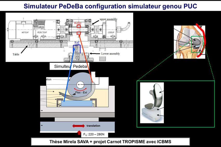
18ème Journée de l’IMBL (23 mai 2018)
 Résumés des Conférences et Posters
Com’ Flash 1 :
Les isoprostanoïdes, métabolites à double fonction
Amandine Rocher1, Claire Vigor1, Guillaume Reversat1, Camille Oger1, Alexandre Guy1, Valérie Bultel-Poncé1,
Jean-Marie Galano1, Joseph Vercauteren1, Thierry Durand1
1
Institut des Biomolécules Max Mousseron, IBMM, UMR 5247, Université de Montpellier, CNRS, ENSCM,
Faculté de Pharmacie - 15 Av. Charles Flahault - BP 14 491 - 34093 Montpellier Cedex
5, France
Le stress oxydant résulte d’un déséquilibre entre la balance pro-oxydante et anti-oxydante (polyphénols,
systèmes enzymatiques) de l’organisme. Plusieurs facteurs endogènes ou exogènes peuvent conduire à
une surproduction d’espèces oxygénées réactives (EOR) attaquant majoritairement l’ADN, les protéines
et les lipides. La peroxydation des acides gras polyinsaturés (AGPIs) par voie enzymatique conduit
notamment à la formation de prostaglandines, résolvines, protectines et par voie non enzymatique aux
isoprostanoïdes. Cinq AGPIs, l’acide α-linolénique (ALA, C18:3 ω3), l’acide arachidonique (AA, C20:4
ω6), l’acide adrénique (AdA, C22 :4 6), l’acide eicosapentaénoïque (EPA, C20:5 ω3) et l’acide
docosahexaénoïque (DHA, C22:6 ω3) conduisent respectivement aux phytoprostanes, isoprostanes,
dihomo-isoprostanes, et neuroprostanes. Cette peroxydation des AGPIs donne ainsi accès à plus de 300
métabolites (la 15-F2t-Isoprostane (15-F2t-IsoP) étant l’analyte le plus étudié dans la littérature)1. Outre
les travaux de synthèse totale de plusieurs de ces métabolites développés au sein de notre laboratoire
depuis de nombreuses années, nous avons initié plus récemment un projet de dosage de ces
isoprostanoïdes par microLC-MS/MS. Présents à l’état de traces dans les échantillons biologiques,
l’extraction et la concentration de ces métabolites d’intérêt, nécessitant plus ou moins d’étapes, a été
optimisée dans différentes matrices (humaine, animale et végétale). L’intérêt diagnostic de ces composés
a déjà été reconnu notamment dans certaines pathologies comme par exemples les maladies
neurodégénératives, cardiovasculaires, rénales, hépatiques et sont aussi impliqués dans certains facteurs
de risques de maladies cardiovasculaires. A titre d’exemple nous illustrerons leur rôle diagnostique avec
le syndrome de Rett2. De par leurs propriétés biologiques, ces métabolites pourraient également
présenter un intérêt en thérapeutique et notamment la 4-F4t-NeuroP par son activité anti-arythmique3.
Nous avons récemment diversifié nos recherches, en nous focalisant plus uniquement sur les fluides
biologiques (plasma, urines, tissus…) mais aussi, en s’ouvrant à l’exploration de matrices autres, telles
que les huiles de poisson ou les algues. Ces derniers résultats nous ont permis de mettre en lumière des
profils intéressants en ces isoprostanoïdes et révéler ainsi le potentiel de ces dernières en nutrition/santé.
1
Jean-Marie Galano et al., “Isoprostanes, Neuroprostanes and Phytoprostanes: An Overview of 25years of
Research in Chemistry and Biology,” Progress in Lipid Research 68, no. Supplement C (October 1, 2017): 83–
108, https://doi.org/10.1016/j.plipres.2017.09.004.
2
Claudio De Felice et al., “F2-Dihomo-Isoprostanes as Potential Early Biomarkers of Lipid Oxidative Damage in
Rett Syndrome,” Journal of Lipid Research 52, no. 12 (December 2011): 2287–97,
https://doi.org/10.1194/jlr.P017798.
3
J. Roy et al., “Non-Enzymatic Oxidized Metabolite of DHA, 4(RS)-4-F4t-Neuroprostane Protects the Heart
against Reperfusion Injury.,” Free Radical Biology & Medicine 102 (January 2017): 229–39,
https://doi.org/10.1016/j.freeradbiomed.2016.12.005.
Com’ Flash 2 :

  Effets de l’acide eicosapentaénoïque et/ou d’un extrait de thé vert sur la
      fonction cardiaque dans un modèle murin de diabète de type 2.

Thibault Leger1, Beibei He1, Jean-Paul Rigaudière1, Kasra Azarnoush2, Luc Demaison1.
1
  Université Clermont Auvergne, INRA, UNH, Clermont-Ferrand, France.
2
  Service de chirurgie cardiaque, Hôpital Gabriel Montpied, Clermont-Ferrand, France.

Introduction et but de l’étude : L’insuffisance cardiaque représente 65% de la mortalité dans
le diabète de type 2. Les effets cardioprotecteurs de l’acide eicosapentaénoïque (EPA) et du thé
vert (TV) ont été constatés en cas d’atteintes cardiovasculaires. L’objectif de cette étude était
d’évaluer leur impact sur la fonction cardiaque dans le diabète de type 2 (DT2).

Matériel et méthodes : Pour cela, 50 rats mâles Wistar étaient répartis en groupes identiques en
nombre selon le régime suivi pendant 9 semaines. Le groupe contrôle a été nourri avec des
croquettes normolipidiques (3%). Les autres animaux ont suivi une alimentation riche en
graisses saturés (HF, 35,2% de lipides) et ont été divisés en 4 groupes : i) HF ; ii) HF + EPA
(1,2% du régime) ; iii) HF + extrait de TV (0,5% du régime) ; iv) HF + EPA + extrait de TV
(respectivement 1,2% et 0,5% du régime). Après 21 jours, ces derniers ont subi une injection
de streptozotocine (STZ, 33 mg/kg) dans le but de déclencher un DT2 en association avec le
régime HF. A la fin de la période de régime, la fonction cardiaque a été évaluée ex vivo par la
méthode du cœur isolé de Langendorff. Immédiatement après, les mitochondries cardiaques ont
été extraites afin d’étudier l’oxydation phosphorylante mitochondriale.

Résultats : Les groupes injectés avec la STZ ont présenté une chute de l’insulinémie (-54%, p
< 0,05) et une hausse ≥ 3 g/L de la glycémie (p < 0,001). Une réduction de la survie des animaux
diabétiques a été constatée, en particulier chez les animaux du groupe HF + EPA (p < 0,05).
Après la période de régime, le groupe HF a montré une forte tendance à la baisse de la
contractilité cardiaque (-38%, NS) et une chute significative de la réactivité coronaire (-77%, p
< 0,001) par rapport au groupe contrôle. L’ajout d’extrait de TV mais aussi d’EPA + extrait de
TV dans les régimes HF ont permis de limiter ces altérations par rapport aux rats HF
(respectivement : +35% NS et +30%, NS pour la contractilité cardiaque ; +55%, NS et +150%,
p < 0,001 pour la réactivité coronaire). L’oxydation phosphorylante mitochondriale a été réduite
par le DT2 pour les substrats des complexes I et II (-39% et -27%, p < 0,05), sans différence
significative entre les différents régimes HF.

Conclusion : Les effets délétères du DT2 sur la fonction cardiaque et l’impact létal de l’EPA
sur les rats diabétiques sont partiellement limités grâce à l’extrait de TV. Des analyses
complémentaires sont nécessaires pour évaluer l’étiologie de ces observations.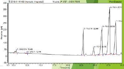
Com’ Flash 3 :
     Bioavailability in human of DHA from either TAG or the structed phospholipid 1-
              acetyl,2-docosahexaenoyl-glycerophosphocholine (AceDoPC®)

Picq Ma, Hachem Ma, Nacir Ha, Belkouche Ma, Bernoud-Hubac Na, Pruneta-Deloche Va,
Windust Ab Mellier Lc, Sauvinet Vc, Feugier Nc, Lambert-Porcheron Sc, Laville Ma,c, Lagarde
Ma

a: Univ-Lyon, Inserm UMR 1060, Inra UMR 1397, INSA-Lyon, Villeurbanne, France

b: National Research Council Canada, Ottawa, Ontario, Canada

c: Hospices Civils de Lyon, Oullins, France

       AceDoPC® is a structured phospholipid that targets the brain with DHA (Hachem et al.
2016) and is neuroprotective in the experimental ischemic stroke (Chauveau et al., 2011).
AceDoPC® is a stabilized form of the physiological 2-DHA-LysoPC with an acetyl group at
the sn-1 position of the glycerolipid preventing the migration of DHA from the sn-2 to sn-1
position.

The bioavailability of 13C-labeled DHA after oral intake of one dose (50 mg DHA) in either
AceDoPC® (preparedfrom U-13C-DHA provided by Anthony Windust in Canada), or 13C-
labeled-DHA in trigylcerides (TG) (prepared by Nestlé France from Crypthecodinium cohnii)
was evaluated. Both lipid forms of DHA were compared in a double-blind study in three human
healthy volunteers (60-70 years-old), with a washout period of 4 months between the second
intake.

Blood samples were takenbefore the intake of DHA sample and right after 1, 3, 6, 24, 72 and
144 hours. Blood red cells were separated from plasma. Total phospholipids from plasma, and
choline and ethanolamine phospholipids from red cells were separated by TLC. After lipid
transmethylation, DHA methylester from FAMEs was analyzed by gas chromatography-
combustion-mass spectrometry to assess the 13C enrichment δ0/00 at each time.
13
 C-DHA enrichment in plasma phospholipids was higher after AceDoPC® compared with TG-
DHA, peaking after 24h in both cases. In red cells, 13C-DHA enrichment in choline
phospholipids from both sources of DHA was also transient, with a maximum after 72h,
whereas the 13C-DHA enrichment in ethanolamine phospholipids was higher from AceDoPC®
compared to TG-DHA, and continued to increase at 144h, especially from AceDoPC®.

Overall, these results indicate that the bioavailability of DHA from AceDoPC® is higher than
from TG-DHA, with a sustained accumulation in red cell ethanolamine phospholipids, which
might mimick the brain accretion (Liu et al. 2014).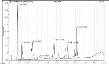
Poster 2 :
    IR spectroscopy analysis of pancreatic lipase-related protein 2 interaction
                               with phospholipids
          Eduardo Mateos-Diaz1, Priscila Sutto-Ortiz1,2, Moulay Sahaka1 and Frédéric Carriere1
1
 CNRS-Aix Marseille Université, UMR7281 Bioénergétique et Ingénierie des Protéines, Marseille,
France
2
 Centro de Investigación y Asistencia en Tecnología y Diseño del Estado de Jalisco A.C. (CIATEJ),
Zapopan, Jalisco, México

Aim/hypothesis- Pancreatic lipase-related protein type 2 (PLRP2) hydrolyzes many lipid substrates in
vitro, but displays however some selectivity depending on the supramolecular structure of substrate and
the presence of surfactants. It displays for instance a phospholipase A1 activity on phospholipids present
in mixed micelles but in liposomes or multilamellar vesicles. We assumed that this selectivity results
from a discriminative interaction of PLRP2 with micelles and not phospholipid bilayers.
Research design and Methods- We used transmission infrared (IR) spectroscopy to study the
interactions between phospholipids (DPPC), surfactants and guinea pig PLRP2 under conditions close
to those of the GI tract. To study the adsorption step independently from hydrolysis, a PLRP2 inactive
variant (S152G) was produced in the yeast Pichia pastoris. Various phospholipid dispersions were
prepared with different aggregation states: multilamellar (MLV) and large unilamellar (LUV) vesicles
and mixed micelles with surfactants. DPPC hydrolysis by PLRP2 was also monitored by IR
spectroscopy.
Results- PLRP2 was found to hydrolyze DPPC present in mixed DPPC-bile salt (NaTDC or NaGDC)
micelles but was inactive on DPPC vesicles and DPPC-Triton X100 micelles. IR analysis of PLRP2
S152G interaction with the DPPC-bile salt system showed a decrease in the conformational disorder
and mobility of the acyl chains, a dehydratation of the interface, and changes in the orientation and H-
bonding of DPPC polar head-groups. These effects were not observed with MLV, LUV and DPPC-
Triton X100 micelles, thus indicating a specific recognition of DPPC in mixed phospholipid-bile salt
micelles, in agreement with phospholipase activity measurements. DPPC hydrolysis by PLRP2 was also
monitored by recording time-course variations in the carbonyl stretching region of the IR spectra. These
variations were correlated to changes in the concentrations of DPPC, lysophospholipids and free fatty
acids. Each lipolysis product was thus quantified in the course of the reaction and IR spectroscopy
allowed the estimation of enzyme activity.
Conclusions- IR spectroscopy is a powerful approach to study PLRP2 interactions with various
phospholipid aggregates, as well as to quantify phospholipolysis with quantification of residual
phospholipids and all lipolysis products.
References
[1] Eduardo Mateos-Diaz, Jean-Claude Bakala N’Goma, Deborah Byrne, Sylvie Robert, Frédéric Carrière and Hélène Gaussier.
IR spectroscopy analysis of pancreatic lipase-related protein 2 interaction with phospholipids: 1. Discriminative recognition of
mixed micelles versus liposomes. Chem Phys Lipids (2018) 211:52-65;
[2] Eduardo Mateos-Diaz, Priscila Sutto-Ortiz, Moulay Sahaka, Deborah Byrne, Hélène Gaussier and Frédéric Carrière. IR
spectroscopy analysis of pancreatic lipase-related protein 2 interaction with phospholipids: 2. Discriminative recognition of
various micellar systems and characterization of PLRP2-DPPC-bile salt complexes. Chem Phys Lipids (2018) 211:66-76;
[3] Eduardo Mateos-Diaz, Priscila Sutto-Ortiz, Moulay Sahaka, Deborah Byrne, Hélène Gaussier and Frédéric Carrière. IR
spectroscopy analysis of pancreatic lipase-related protein 2 interaction with phospholipids: 3. Monitoring DPPC lipolysis in
mixed micelles. Chem Phys Lipids (2018) 211:77–85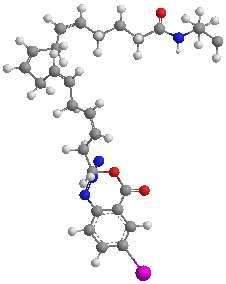
Poster 5 :
 Live determination of triacylglycerol quantity and lipid droplets size in the microalgae
                      P. tricornutum using NMR-based methods.

Antoine Jaussaud[1], Josselin Lupette[2], Marina Gromova[1], Armel Guillermo[1], Pierre-Alain
Bayle[1], Eric Maréchal[2], Michel Bardet [1]
[1]
      Laboratoire de Résonance Magnétique, INAC, CEA Grenoble, France
[2]
      Laboratoire de Physiologie Cellulaire Végétale, BIG, CEA Grenoble, France

         Aim/hypothesis: Because of the foreseen exhaustion of fossil fuel reserves and their
negative effects on climate, sustainable energy production without massive CO2 release is a
critical issue that needs to be addressed [1] [2]. In this context, microalgae, such as Phaeodactylum
tricornutum, have emerged as a potential resource for biofuel and green chemistry alternative.
Coping with environmental stresses, like nitrogen starvation, microalgae are able to produce
and store neutral lipids, mostly triacylglycerol (TAGs) [3]. These neutral lipids accumulate
inside lipid droplets (LDs). Current research conducted in the biofuel industry as well as in the
agro-food industry requires LDs monitoring methods, ideally complying with live analyses.
         Research design, Methods and Results: Different methods to determine LD size are
already used but are time consuming. We investigated the possibility to use a rapid in vivo
method, based on molecular diffusion, allowing the measurement of LD size, and based on
Pulse Field Gradient Nuclear Magnetic Resonance (PFG NMR) [4]. Microalgae were subjected
to nitrogen starvation for seven day and analyzed daily. Two different types of experiments
were performed with NMR based methods. Firstly, in vivo radius determination of LDs inside
microalgae was accomplished with PFG NMR. Secondly, the amount of TAGs accumulated
inside LDs from microalgae was estimated with 1H NMR. PFG NMR measurements from
microalgae LDs were compared to microscopy measurements, and similar results were
obtained, acknowledging the PFG NMR method as suitable to determine LDs size in
microalgae.
         Conclusions: This work presents a non-invasive, rapid, efficient, and sample-
preparation-free method that allows in vivo measurement of LDs in microalgae, such as P.
tricornutum. Potential applications are discussed.

Bibliography

[1] B. Kamm, P. Gruber et M. Kamm, «Biorefinery industrial processes and products,» Status and future
    direction, vol. 1&2, 2010.
[2] T. Tanaka, Y. Maeda, A. Veluchamy, M. Tanaka, H. Abida, E. Maréchal, C. Bowler, M. Muto, Y. Sunaga,
    M. Tanaka, T. Yoshino, T. Taniguchi, Y. Fukuda, M. Nemoto, M. Matsumoto, P. Wong, S. Aburatani et W.
    Fujibuchi, «Oil accumulation by the oleaginous diatom Fistulifera solaris as revealed by the genome and
    transcriptome,» The Plant Cell, vol. 27, pp. 162-176, 2015.
[3] H. Abida, L. Dolch, C. Meï, V. Villanova, M. Conte, M. Block, G. Finazzi, O. Bastien, L. Tirichine, C.
    Bowler, F. Rébeillé, D. Petroutsos, J. Jouhet et E. Maréchal, «Membrane glycerolipid remodeling triggered
    by nitrogen and phosphorus starvation in Phaeodactylum tricornutum,» Plant Physiology, vol. 167, pp. 118-
    136, 2015.
[4] J. Tanner et E. Stejskal, «Restricted self-diffusion of protons in colloidal systems by pulsed gradient spin-
    echo method,» J ChemPhys, vol. 49, pp. 1768-1777, 1968.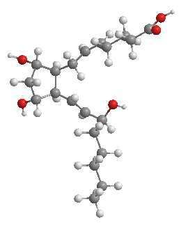
Poster 6 :

    Muscle loss associated changes of oxylipin signatures during biological aging: an
                      exploratory study from the PROOF cohort.
Céline Dalle1, Annika I. Ostermann2,3, Thade Konrad3, Cécile Coudy-Gandilhon1, Alice
Decourt1, Jean-Claude Barthélémy4, Frédéric Roche4, Léonard Féasson5, André Mazur1, Daniel
Béchet1, Nils Helge Schebb2,3, and Cécile Gladine1*

1
 Université Clermont Auvergne, INRA, UNH, Unité de Nutrition Humaine, CRNH Auvergne,
F-63000 Clermont-Ferrand, France.
2
Institute for Food Toxicology, University of Veterinary Medicine Hannover, Bischofsholer
Damm 15, 30173 Hannover, Germany
3
Chair of Food Chemistry, Faculty of Mathematics and Natural Sciences, University of
Wuppertal, Gaußstraße 20, Wuppertal, Germany
4
 Service de Physiologie Clinique et de l'Exercice, CHU Nord, Faculté de Médecine Jacques
Lisfranc, PRES de Lyon, Université Jean Monnet, F-42055 Saint-Etienne, France.
5
 Laboratoire Interuniversitaire de Biologie de la Motricité-EA 7424, Univ Lyon, UJM-Saint-
Etienne, Unité de Myologie, Centre Référent Maladies Neuromusculaires Rares Rhône-Alpes,
CHU Saint-Etienne, Saint-Étienne, France
*Corresponding author : Dr Cécile Gladine, Université Clermont Auvergne, INRA, UNH,
Unité de Nutrition Humaine, CRNH Auvergne, F-63000 Clermont-Ferrand, France,
cecile.gladine@inra.fr

Characterizations of the multiple mechanisms determining biological aging are required to
better understand the etiology and identify early biomarkers of sarcopenia. Oxylipins are a large
family of signaling lipids involved in the regulation of various biological processes that become
dysregulated during aging.
To investigate whether comprehensive oxylipin profiling could provide an integrated and fine
characterisation of the early phases of sarcopenia, we performed a quantitative targeted
metabolomics of oxylipins in plasma of 81-year old subjects from the PROOF cohort with
decreased (n=12), stable (n=16) or increased appendicular muscle mass (n=14).
Multivariate and univariate analyses identified significant and concordant changes of oxylipin
profiles according to the muscle status. Of note, 90% of the most discriminant oxylipins were
derived from EPA and DHA and were increased in the sarcopenic subjects. The oxylipins
signatures of sarcopenic subjects revealed subtle activation of inflammatory resolution
pathways, coagulation processes and oxidative stress and the inhibition of angiogenesis. Heat
maps highlighted relationships between oxylipins and the cardiometabolic health parameters
which were mainly lost in sarcopenic subjects.
This exploratory study supports that targeted metabolomics of oxylipins could provide relevant
and subtle characterization of early disturbances associated with muscle-loss during aging.
Poster 8 :
Self-Organization of Bacterial Acyl Steroid Glycosides:
Investigation of -CAG and BbGL-1 and Analogues
Zonglong Yang,1 Milène Nitenberg,1 Laurent Soulère,1 Mohammed Ahmar,1 John W.
Goodby,2 Stephen J. Cowling, 2 Agnes P. Girard-Egrot 1 and Yves Queneau 1
1
Université de Lyon, Institut de Chimie et de Biochimie Moléculaires et Supramoléculaires
(ICBMS), UMR 5246 CNRS, Université Lyon 1, INSA Lyon, CPE Lyon, Bâtiment Lederer,
F-69622 Villeurbanne Cedex, France.
2
Department of Chemistry, The University of York, York, UK.
Aim/hypothesis- α-CAG and BbGL-1, two acyl steroid glycosides (ASGs) found in the
membrane of pathogenic bacteria H. pylori and B. burgdorferi,1,2 show similar -gluco and -
galacto structures. Their self-organization behavior has been investigated with respect to their
thermotropic properties and as components of biomimetic membrane Langmuir monolayers.
Research design and Methods- The -gluco α-CAG and the -galacto BbGL-1 have been
compared with analogous ASGs varying in chain lengths (C6 to C18 and oleic) and anomeric
configuration. A series of 32 acyl cholesteryl glycosides has thus been synthesized allowing to
stress the main structural parameters governing their behavior. Structure-property relationships
have been established.
Results- A physicochemical insight into their thermotropic liquid crystal behavior and their
ability to promote lipid domains formation in model membrane monolayers is given.3,4 While
the chain length is the most remarkable parameter, significant differences in the way lipids
redistribute and form domains within monolayers resulted from variations in the anomeric
configuration and sugar nature.
Conclusions- These observations corroborate, on the physico-chemical viewpoint, the
biological investigations showing lipidic exchange during infection by H. pylori (in which
CAG becomes a C18 analogue) in relation with its increased pathogenicity.5
References:
1 Y. Hirai, M. Haque, T. Yoshida, K. Yokota, T. Yasuda, K. Oguma, Y. Hirai, M. Haque, T. Yoshida, K. Yokota, T. Yasuda
and K. Oguma, J. Bacteriol., 1995, 177, 5327–5333.
2 G. Ben-Menachem, J. Kubler-Kielb, B. Coxon, A. Yergey and R. Schneerson, Proc. Natl. Acad. Sci., 2003, 100, 7913-
7918.
3 Z. Yang, R. Xu, F. Ali-Rachedi, S. Chambert, N. M. Xavier, L. Soulère, M. Ahmar, G. Mackenzie, E. J. Davis, J. W.
Goodby, S. J. Cowling and Y. Queneau, Liq. Cryst., 2017, 44, 2089-2107.
4 Z. Yang, L. Soulère, M. Ahmar, J. W. Goodby, S. J. Cowling, A. P. Girard-Egrot and Y. Queneau, manuscript in
preparation.
5 H.-M. Jan, Y.-C. Chen, Y.-Y. Shih, Y.-C. Huang, Z. Tu, A. B. Ingle, S.-W. Liu, M.-S. Wu, J. Gervay-Hague, K.-K. T.
Mong, Y.-R. Chen and C.-H. Lin, Chem. Sci., 2016, 7, 6208-6216.Poster 10 :
Lipid classes content and composition in plasma of dairy goats and cows fed similar
diets supplemented or not with lipids
Hélène Fougère1, Laurence Bernard1, Yves Chilliard1, Pablo G. Toral2, Vincent Dietz1 and
Carole Delavaud1
1
Université Clermont Auvergne, INRA, VetAgroSup, UMR Herbivores, F-63122 Saint-Genès-
Champanelle, France
2
Instituto de Ganaderia de Montaña (CSIC-ULE), 24346 Grulleros, León, Spain
Aim/hypothesis: The objectives of this work were i) to develop a method of separation and
quantification of simple lipid classes by HP-TLC (High Performance-Thin Layer
Chromatography), and ii) to determine plasma lipid content and composition in dairy cows and
goats fed diets that induced milk fat depression (MFD) in cows, but not, or to a lesser extent,
in goats.
Research design and Methods: Twelve cows and 14 goats, at a similar lactation stage, were
assigned to a 3x3 latin square design, and successively fed a basal diet without addition of lipids
(Ctrl), or supplemented with sunflower oil plus wheat starch (SOS, 9% DM), or fish oil (FO,
3.6% DM). Plasma lipids were extracted from individual blood collected before morning
feeding at the end of each nutritional 26-d period. Lipid classes were separated by HP-TLC
(Camag, Switzerland). After derivatization and heating, the plates were analyzed by
densitometry (VideoScan, Camag, Switzerland). In order to quantify the major lipid classes, 5
standard curves with increasing quantities of pure C17:0-phospholipid (PL), Cholesterol
(Chol), C23:0-fatty acid (NEFA), cis9-C18:1-triglyceride (TG) and C15:0-cholesteryl ester
(CE), were deposited on each plate with individual samples, and concentrations were expressed
in µg/µL of lipid extract. Data were analysed with proc MIXED of SAS (SAS Institute Inc.,
Cary, NC), and effects of diet, species, and their interaction were determined.
Results: Whatever the species, and compared to Ctrl, FO increased plasma CE (+31%,
PPoster 12 :
Photoenzymatic conversion of lipids into hydrocarbons
Cyril Aselmeyer1,2, Frédéric Carriere1 and Frédéric Beisson2
1
CNRS-Aix Marseille Université, UMR7281 Bioénergétique et Ingénierie des Protéines, Marseille,
France
2
CEA Cadarache, UMR7265 Laboratoire de Bioénergétique et Biotechnologie des Bactéries et
Microalgues
Aim/hypothesis-
The production of bio-sourced hydrocarbons for biofuels and green chemistry is a major
biotechnological challenge. The LB3M laboratory in Cadarache has discovered a microalgal
enzyme, the Fatty Acid Photodecarboxylase (FAP), which produces hydrocarbons from free
fatty acids (FFA). FAP only uses light as energy source and could be useful to convert oils into
hydrocarbons. Our aim is to find out what is the best way to provide the fatty acid substrate to
the FAP, and fully characterize the behaviour of this enzyme at different lipid-water interfaces.
Research design and Methods-
In order to characterize the interfacial behaviour of the newly discovered FAP, devices such as
the drop tensiometer or the Wilhelmy plate will be used (allowing to record the adsorption
and/or the activity of interfacial proteins). Those tools will also be used to follow the
modification of the interfaces as free fatty acids are turned into hydrocarbons, until the possible
collapse of the lipid aggregates and their reorganization. Molecular biology cloning techniques
will be of common use for the creation of different variants of the enzyme. Produced
hydrocarbons will be analysed and quantified via techniques such as GC-MS/FID analysis or
also TLC. Modelisation softwares will also be used to get a better understanding of the
behaviour of the protein vis-à-vis the substrates and different interfaces.
Results-
Results show that the FAP has a strongest photodecarboxylation activity on long chain fatty
acids, with a maximum affinity on palmitic acid. Preliminary investigations on its affinity for
lipid bilayers suggest a potential to bind phosphatidylcholine membranes (under the form of
liposomes) in slightly acidic conditions. Moreover, other preliminary results tend to suggest an
influence of the self-organization of free fatty acids on the ability of the FAP to decarboxylate
shorter/longer chain substrates, underlining the necessity to control the physical state of free
fatty acids.
Conclusions-
FAP is the third light driven enzyme to be identified and the first one known to act in lipid
metabolism. It has thus a strong biotechnological potential. A better comprehension
physiological role of FAP, as well as its use for bio-production of alkanes and alkenes will
necessitate more detailed studies on its behaviour at lipid-water interfaces.Poster 13 :
Bis(monoacylglycero)phosphate : a possible biomarker of drug-induced
phospholipidosis in human urines
Maxence Rabia1,2, Baptiste Fourmaux1,3, Céline Luquain-Costaz1, Philippe Moulin1, Isabelle Delton1 and
Françoise Hullin-Matsuda1,
1
INSERM UMR1060-INSA, Laboratoire CarMeN, 69621 Villeurbanne cedex, France
2
Université Lyon 1, 69622, Villeurbanne cedex, France.
3
Plateforme de lipidomique fonctionnelle, Laboratoire CarMeN/IMBL-INSA de Lyon, 69621
Villeurbanne cedex, France
Aim/Background: Phospholipidosis is a genetic or drug-induced disease characterized by an
accumulation of phospholipids in the endolysosomal organelles of various tissues. Amiodarone, an
iodinated antiarrhythmic drug belonging to the cationic amphiphilic drug family, is inducing
phospholipidosis during long-term treatment, especially in lung, thyroid and liver.
Bis(monoacylglycero)phosphate (BMP), also known as lysobisphosphatidic acid (LBPA), has been
recently described as a possible biomarker of lysosomal storage disorders and drug-induced
phospholipidosis [1]. BMP is a unique phospholipid specifically enriched in the endolysosomal
compartments [2] and a structural isomer of phosphatidylglycerol (PG) [3]. The aim of our study is to
confirm the presence of BMP in urines of amiodarone-treated patients in order to evaluate its use as a
biomarker of drug-induced phospholipidosis in clinical follow-up.
Methods: The study is carried out on the urines of patients treated with amiodarone with or without
dysthyroidism, healthy subjects, and patients with non-alcoholic steatohepatitis (NASH). Lipids are
extracted according to the Bligh and Dyer method in acidic conditions [4]. To analyse the different
molecular species of BMP, we developed a reversed-phase chromatography method coupled to ESI
tandem mass spectrometry (RPLC-MS/MS) using a triple quadrupole AB Sciex 4500 QTRAP working
in positive ion mode. Thanks to this method, we can analyse samples containing pmol amounts of BMP.
Results: Our study show a significant increase of the urinary BMP content of patients treated with
amiodarone with or without dysthyroidism, compared to healthy patients. Interestingly, there is a
selective increase of unsaturated species. Furthermore, there is no differences between urinary BMP
content and composition of healthy patients and NASH patients.
Conclusion: Our study indicates that urinary BMP can be used as a biomarker of endolysosomal
perturbations induced by amiodarone in human patients. We are now looking for the vehicles of BMP
in the urines.
References
[1] Meikle, P.J et al. (2008). Biochem J 411(1): 71-78.
[2] Hullin-Matsuda F and al. (2014). Semin Cell Dev Biol 31: 48-56.
[3] Tan H.H et al. (2012). Angew Chem Int Ed Engl 51(2), 533-535.
[4] Bligh and Dyer (1959). Canadian Journal of Biochemistry and Physiology 37 (8), 911-917Poster 14 :
Endogenous omega 3 fatty acids prevent gut microbiota dysbiosis, colon
mucus layer thickness alteration and endoplasmic reticulum stress induced
by high fat diet
Escoula Q1,2,3, Bellenger S1,2,3, Geissler A5, Lagrost L2,3,4, Narce M1,2,3 and
Bellenger J1,2,3.
1
University of Bourgogne Franche-Comté, UFR Sciences de la Vie, de la Terre et de
l’Environnement, 6 Boulevard Gabriel, 21000 Dijon, France.
2
INSERM, Lipides Nutrition Cancer UMR1231, 21000 Dijon, France.
3
LipSTIC LabEx, Fondation de Coopération Scientifique Bourgogne-Franche Comté, 21000
Dijon, France
4
University of Bourgogne Franche-Comté, UFR Sciences de Santé, 7 Boulevard Jeanne d’Arc,
21078 Dijon Cedex, France
5
CellImaP - UFR des Sciences de Santé - 7 Boulevard Jeanne d'Arc - 21079 Dijon, France
Aim/hypothesis
An obesogenic high fat high sucrose (HFHS) diet leads to gut microbiota dysbiosis,
intestinal barrier alteration, metabolic endotoxemia and chronic low grade inflammation.
Thickness alteration of the intestinal mucus layer, -mainly constituted of Muc2 glycoprotein
produced by goblet cells- has been reported after HFHS diet, and linked to an induction of
inflammation and endoplasmic reticulum (ER) stress. Omega3 fatty acid tissue enrichment
exhibit anti-inflammatory capacities and is able to prevent intestinal barrier dysfunction and
low grade inflammation induced by HFHS diet.
Thus, the main objective of this study was to investigate whether omega3 fatty acid
tissue enrichment could prevent gut microbiota dysbiosis and colon mucus layer alteration
induced by an obesogenic HFHS diet.
Research design and Methods
For that, fat-1 transgenic mice -able to convert omega6 into omega3-, and WT mice
were fed a HFHS or a control diet during 12 weeks. Fecal microbiota analysis was performed,
colon mucus layer thickness was measured after BA/PAS staining and inflammation and ER
stress markers were quantified by RT-QPCR.
Results
Our results showed that endogenous omega 3 fatty acids prevent microbiota dysbiosis
induced by HFHS. Moreover, compared to WT mice, Muc2 expression remained unchanged
and colon mucus layer thickness was preserved in fat-1 mice fed HFHS. Furthermore, the
expression of inflammation markers (Tnf-α and Il1-β) and ER stress (Xbp1 and Grp78) were
not modified in fat-1 mice fed HFHS, whereas they were increased in WT mice.
Conclusions
Such protective effects represent additional arguments to use omega3 fatty acids as a
therapeutic strategy to prevent obesity and related intestinal alterations. The relationship
between omega3 tissue enrichment, microbiota « remodeling » and colon mucus layer
prevention in fat-1 mice on DIO conditions still needs to be highlighted.
In this context, metabolic pathways driving such effects are being investigated and
transfer of cecal microbiota from fat-1 to WT and/or germ-free mice fed a HFHS diet will be
performed.Poster 15 : Soybean polar lipids differently impact adipose tissue inflammation and the endotoxin transporters LBP and sCD14 in flaxseed vs palm oil-rich diets Lecomte M.1, Couëdelo L.2, Meugnier E.3, Loizon E.3, Plaisancié P.1, Géloën A.1, Joffre F.2, Vaysse C.2, Michalski MC.1, Laugerette F.1 1 Univ-Lyon, CarMeN laboratory, INRA UMR1397, INSERM U1060, Université Claude Bernard Lyon 1, INSA-Lyon, IMBL, F-69620 Villeurbanne, France, 2ITERG-ENMS, Université de Bordeaux, rue Léo Saignat, 33076 Bordeaux cedex, France, 3INRA UMR1397, CarMeN laboratory, Univ-Lyon, INSERM U1060, Université Claude Bernard Lyon 1, F-69921 Oullins, France Obesity and type 2 diabetes are characterized by a subclinical inflammatory state. Endotoxins are recognized as an important factor implicated in the development of this inflammation. It has been demonstrated that oil composition of high-fat diets can modulate this phenomenon, however less is known about the impact of soybean polar lipids (SPL), used as emulsifier in numerous food products. We investigated in mice the effect of SPL incorporation into high-fat diets differing in terms of oil composition on inflammation and endotoxemia.C57BL6 mice were divided into 4 groups and fed for 8 weeks with a high fat diet based on palm oil (P) or, (ii) on flaxseed oil (F), or both high-fat diets containing 1.2% of SPL (P-SPL and F-SPL). Plasma endotoxemia was determined using the LAL assay and concentrations of MCP-1, sCD14 and LBP were assayed by ELISA kits. Gene expression of inflammatory proteins were analyzed by qPCR in white adipose tissue (WAT), liver and duodenum. In P-SPL and F-SPL groups, gene expression of MCP-1 and LBP in WAT increased compared with P and F groups. However, only F-SPL diet induced an increased gene expression of pro-inflammatory cytokines (TNF-α, IL-6) and markers of macrophage-infiltration (CD68, CD11c and F4/80) and a higher sCD14 plasma concentration. In the duodenum and liver, LBP expression was lower in both SPL groups, contrary to the WAT where LBP was higher in both SPL groups. In plasma, LBP concentration had the same profile that LBP expression in the liver. Moreover, SRB1 expression, a putative transporter involved in LPS passage across intestinal cells, was higher in the F-SPL group. We have demonstrated that LPS transporters LBP and sCD14 and adipose tissue inflammation can be modulated by SPL in high fat diets differing in oil composition. Corresponding author: fabienne.laugerette@univ-lyon1.fr
Poster 16 :
  Etude de l’interaction entre le surfactant pulmonaire et les particules aéroportées : réaction
                                 inflammatoire et stress oxydatif

Auteurs : Messaoui A*, Vincent M**, Catinon M**, Guichardant M***, Trunfio-Sfarghiu A-M*

*Univ Lyon, INSA, Mechanics of Contacts and Structures Laboratory LaMCoS, UMR 5259, F-69621
Lyon, France
** Société MINAPATH DEVELLOPEMENT, F-69100, Villeurbanne, France
*** Univ-Lyon, CarMeN laboratory, (Inserm UMR 1060, Inra UMR 1397), Université Claude
Bernard Lyon 1, INSA-Lyon, IMBL, F-69100, Villeurbanne, France
                  Le surfactant pulmonaire est la première barrière physiologique du poumon
profond contre les particules aéroportées présentes dans l’air ambiant. Composé principalement
de phospholipides saturés, le surfactant pulmonaire réduit la tension superficielle air/liquide au
niveau des alvéoles pulmonaires et leur confère des propriétés biomécaniques qui assurent un
excellent processus respiratoire. D’autre part les alvéoles pulmonaires sont infiltrées par des
macrophages alvéolaires (cellules immunitaires mobiles) qui déclenchent une réaction
immunologique efficace lors de l’intrusion d’un corps exogène quelle que soit sa nature (Elena
L-P, Jesús P-G., 2014).
Cette réponse immunitaire active différentes réactions biochimiques qui conduisent à la
production d’espèces réactives dérivées de l’oxygène auquel s’ajoute éventuellement des
éléments oxydants provenant des particules elles-mêmes. Le déséquilibre entre les espèces
oxydantes et anti-oxydantes ainsi générées, peut-être à l’origine de diverses pathologies
respiratoires. Ces espèces oxydantes entraînent la peroxydation des acides gras présents dans la
monocouche de surfactant pulmonaire et déstabilisent la structure tridimensionnelle des
phospholipides qui peut in fine aboutir à la déstructuration totale de la barrière biomécanique
et induire une insuffisance respiratoire (Baeza, et al., 2007) (David, 2014) (Baron, 2003).
Il a été démontré que des interactions physico-chimiques peuvent se produire entre les
différentes particules et la couche de surfactant pulmonaire et favoriser ainsi plus ou moins leur
piégeage dans ce liquide alvéolaire. La modification des propriétés biomécaniques des alvéoles
réduit la capacité pulmonaire. (Munteanu, 2014) (Munteanu, 2015). La taille, la morphologie,
la charge ainsi que la nature chimique des microparticules présentes dans l’air ambiant,
pourraient aussi être responsables des pathologies plus ou moins connues (Pachauri et al.,
2013) (Zuo et al., 2008)
Le surfactant biomimétique sera défini à partir des analyses lipidomiques, biomécaniques (cuve
de Langmuir) et physicochimiques (MEB-EDX) des prélèvements de LBA issu de 19 sujets
humains sains (projet Silicosis Minapath). La validation du surfactant biomimétique sera faite
par une exposition aux particules issues du freinage automobile.

Ce travail a donc pour but : d’élaborer un surfactant biomimétique pour analyser efficacement
et systématiquement des particules afin de déterminer d’une part leur effet biomécanique sur
la modification de la capacité pulmonaire et d’autre part leurs effets toxiques en mesurant à la
fois des marqueurs pro-inflammatoires tel que le leucotriène B4 et de la peroxydation des
lipides comme les hydroxyalcénals : (4-hydroxynonenal (4-HNE) et 4-hydroxyhexénal (4-
HHE)).
Poster 17 :
Etude du pouvoir acétylant potentiel de l’AceDoPC® dans un modèle de cellules
neuronales
Rémi Pellegrinato, Houda Nacir, Angélina Cariou, Michel Lagarde, Pascale Plaisancié,
Nathalie Bernoud-Hubac
Univ-Lyon, Inserm UMR 1060, Inra UMR 1397, INSA-Lyon, Villeurbanne, France
Aim/hypothesis:
L’acide docosahexaénoïque (DHA, 22:6n-3) est un acide gras polyinsaturé oméga-3 essential
pour le développement cérébral, la capacité d’apprentissage et la mémoire. Cet acide gras
oméga-3 est par ailleurs déficitaire dans le cerveau de sujets souffrant de maladies
neurodégénératives. Le laboratoire CarMeN a synthétisé une molécule appelée 1-acétyl,2-
docosahexaénoyl-glycérophosphocholine (AceDoPC®, breveté) qui est une forme stabilisée de
la 2-docosahexaénoyl-lysoPC (lysoPC-DHA) et un transporteur privilégié du DHA au cerveau
(1,2). L’AceDoPC® se caractérise aussi par son action neuroprotectrice dans l’accident
vasculaire cérébral (3) et possède des activités anti-inflammatoires (brevet WO2017006047).
Diverses études indiquent que des modifications épigénétiques (dont les modifications
d’acétylation des histones) pourraient être impliquées dans l'étiologie des maladies
neurodégénératives. L’AceDoPC® comporte un groupement acétyle. L’objectif de cette étude
est d’évaluer si l’AceDoPC® posséde des propriétés acétylantes sur les histones H3 et H4 de
cellules neuronales de rat, les B50.
Research design and Methods:
Les cellules B50 ont été incubées avec différentes concentrations d’AceDoPC® (10-9, 10-8 et
10-7M). Les niveaux d’acétylation globaux et de lysines spécifiques des histones H3 et H4 ont
été estimés grâce à l’immunodétection par chimiluminescence.
Results/Conclusions:
Les résultats montrent une modification de l’acétylation des histones en particulier pour une
concentration d’AceDoPC à 10-9 M (modification des taux d’acétylation globaux et sur des
lysines spécifiques). L’étude de l’effet du lysoPC-DHA et de l’activité des enzymes histone
acétyltransférase et désacétylase permettront par la suite de préciser ces résultats.
References
[1] Hachem M, Géloën A, Lo Van A, Fourmaux B, Fenart L, Gosselet F, Da Silva P, Breton G, Lagarde M, Picq
M, Bernoud-Hubac N. Efficient Docosahexaenoic Acid Uptake by the Brain from a Structured Phospholipid. Mol
Neurobiol. 2016;53:3205-15.
[2] A. Lo Van, N. Sakayori, M. Hachem, M. Belkouch, M. Picq, M. Lagarde, N. Osumi, and N. Bernoud-Hubac.
Mechanisms of DHA transport to the brain and potential therapy to neurodegenerative diseases. Biochimie.
2016;130:163-7.
[3] Chauveau F, Cho TH, Perez M, Guichardant M, Riou A, Aguettaz P, Picq M, Lagarde M, Berthezène Y,
Nighoghossian N, Wiart M. Brain-targeting form of docosahexaenoic acid for experimental stroke treatment: MRI
evaluation and anti-oxidant impact. Curr. Neurovascular Reseach 8, 95–102 (2011).Poster 18 :
Les fibres alimentaires modifient l’impact de la suralimentation
sur le métabolisme hépathique
Ahmed Ben MOHAMED1, Didier RÉMOND1, Christophe CHAMBON2, Thierry SAYD2,
Michel HEBRAUD2, Guy DELLA-VALLE3, Frédéric CAPEL1, Benoit COHADE1, Daniel
BÉCHET1, Cécile COUDY-GANDILHON1, Jeremie DAVID1, Dominique DARDEVET1, Joel
DORE4, Sergio POLAKOF 1, Isabelle SAVARY-AUZELOUX1
1
Université Clermont Auvergne, INRA, UMR 1019 Unité de Nutrition Humaine, F-63000
Clermont-Ferrand, France.
2
INRA, UR370 Qualité des Produits Animaux, F-63122 Saint Genès Champanelle, France.
3
INRA, UR 1268 Biopolymères Interactions Assemblages, F-44316 Nantes, France.
4
INRA, UMR 1319 Microbiologie de l'Alimentation au Service de la Santé Humaine, F-
78352 Jouy-en-Josas, France.
Aim/hypothesis- : Notre étude vise à déterminer si et comment, en situation de surnutrition, les
fibres alimentaires peuvent limiter ou stopper l’apparition des dérégulations métaboliques
associées à l’ingestion d’un régime obésogène et notamment à limiter l’infiltration lipidique
dans le foie.
Research design and Methods : Vingt mini-porcs femelles ont été nourris avec un régime contrôle
(C, n=6) (énergie ingérée correspondant aux besoins d’entretien), un régime en surnutrition
supplémenté en pain enrichi (O+F, n=7) ou non (O, n=7) en fibres (pectine, inuline, amidon
résistant). Des prélèvements sanguins ont été réalisés à jeun avant et après 14 et 56 jours de
régime O ou O+F, ainsi que le prélèvement d’une section de foie à l’euthanasie. Des analyses
de paramètres plasmatiques et hépatiques (protéomique, transcriptomique et histologique : red
oil) ont été effectuées. Les effets régimes/cinétiques ont été évaluées par ANOVA un facteur
(mesures dans le foie) ou deux facteurs à mesures répétées (mesures en cinétique), signification
PPoster 19 :
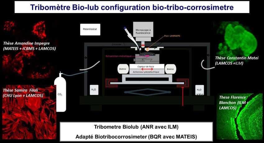
Biomimetic phospholipid vesicles lubricants: Synthesis and multiphysics
characterizations. Application in The Joint Diseases
Darragi Raies N15.,Massardier V1.,Zouari A1.,Maniti O2.,Piednoir A3.,Girard-Egrot A2.,Guichardant M2.,
Landoulsi A5.,Tinland B4,Trunfio-Sfarghiu A.M1
1Univ Lyon, INSA, Mechanics of Contacts and Structures Laboratory LaMCoS, UMR 5259, F-69621 Lyon, France
2Univ Lyon, CNRS, Molecular and Supramolecular Chemistry and Biochemistry Institute ICBMS UMR 5246, F-69622 Lyon,
France
3Univ Lyon, CNRS, Institut Lumière Matière IML UMR 5306, F-69622 Lyon, France
4Univ Aix-Marseille, CNRS, Marseille Interdisciplinary Center of nanosciences CINaM UMR 7325, 13288 Marseille, France
5Univ Carthage, Laboratory of Biochemistry and Molecular Biology LBBM, FSB, Tunisia
Introduction: In mechanical rubbing contacts, a wear phenomenon is invariably observed even
in the presence of lubricant. Its economic impact is tremendous in the industry, and particularly
in the medical world, quoting osteoarthritis as the most common joint pathology in the world
where the used solution is prostheses implantation. However, biolubricant plays a crucial role
in the joint life. Thus, recent studies show that the synovial fluid involves microvesicles filled
with a glycoprotein gel and surrounded by stacked lipid bilayers, which give its excellent
lubricating properties. On this point, this project aims to reproduce and stabilize the synovial
fluid structure in order to understand molecular (lipidomic) origin on the lubricating
performances and to improve therapeutic approaches.
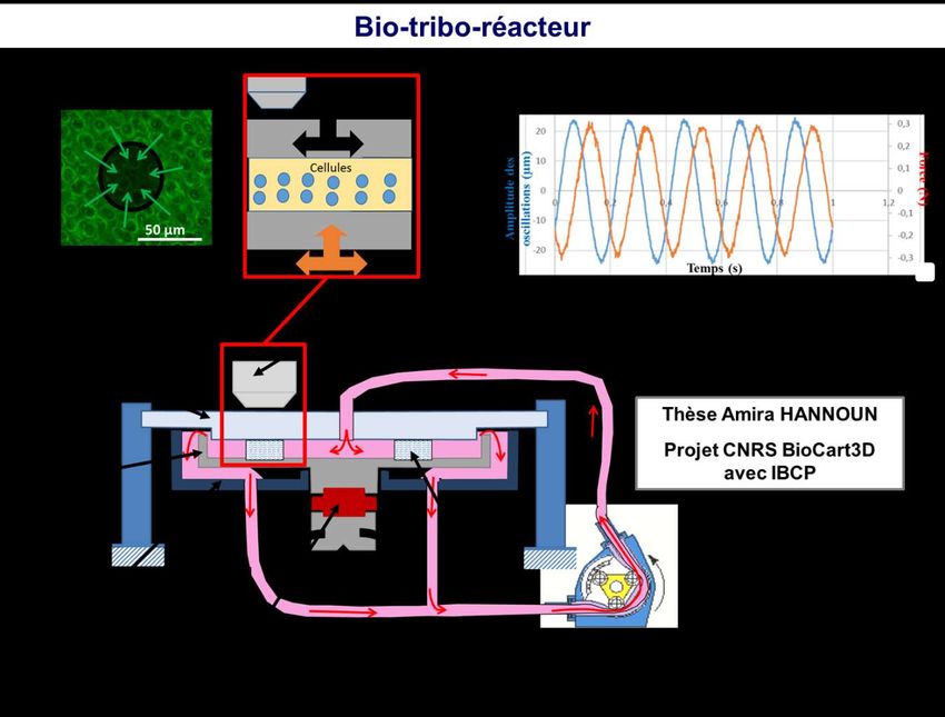
Methodology: Firstly, biochemical, structural and mechanical analysis of the synovial fluid
lipid vesicles of dog samples (surgical waste during surgery on the cruciate ligament) were
performed in order to determine their composition with the aim to produce a biomimetic
structure. Secondly, biomimetic vesicles were prepared by respecting ‘Gel in’ SF Protocol
described by Sava et al.[1]. Those vesicles were fixed on a glass using biotin-streptavidin
coupling. In addition, Confocal Fluorescence Microscopy (Zeiss LSM 700) and Transmission
Electron Microscopy (JEOL 2100F) were used to characterize these multilamellar vesicular
structures. Additionally, the mechanical behaviour of biomimetic phospholipid (PL)
monolayers was compared to that of DPPC (‘’gel” phase at 37°C) and POPC (‘’fluid’’ phase
at 37°C) by measuring isothermal Langmuir compression. Moreover, the biomechanical
properties of the biomimetic PL vesicles were measured using Atomic Force Microscopy
(MFP-3D, Asylum Research) with indentation by a spherical indenter (5µm diameter and
cantilever spring constants 0.2 N/m).
Results and Conclusions: From lipidomic assay results, we propose a synovial fluid
biomimetic composition made up of 20% SpH, 8% POPC, 8% SOPC, 16% DPPC, 16% DSPC,
16% PLPC, 16% SLPC. The Langmuir monolayers results show that this biomimetic
composition of phospholipids increases vesicle rigidity comparing it to that of DPPC. Using
this biomimetic composition, micrometric size vesicles (1 to 10µm) with a multilamellar
membrane were obtained similar to those observed directly in the collection of healthy synovial
fluid and that obtained using DPPC. This Biomimetic vesicular structure is significantly
different from POPC. Whereas, DPPC vesicles occupy a few micrometer sizes (1-5µm) with a
double lipid barrier, POPC vesicles exhibit an unstable diameter of only a few hundred
nanometers with a strong propensity to aggregate. The nano-mechanical and tribological results
reveal that the biomimetic vesicular structure significantly increases the vesicular stiffness and
the lubricant viscosity, which may explain the biolubricant performances of these structures in
vivo.
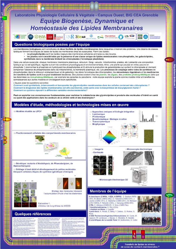
[1] Sava M.M. et al. Comput Methods Biomech Biomed Eng 2013, 16 (1): 216-218.Posters d’équipes
Institut des Biomolécules Max Mousseron
                        UMR 5247 CNRS-UM-ENSCM
                     Equipe Synthèse de Lipides Bioactifs
                               Dr Thierry Durand

                   La synthèse totale
                   * Elaboration de protocoles de synthèses multi-étapes
                   * Mise en oeuvre :
                      cyclisation radicalaire, couplages organométalliques,
                      dédoublement enzymatique
                      préparation de sels de phosphonium et de β-céto-
                      phosphonates,
                      introduction des chaînes latérales par couplage de
                      type Wittig / HWE, métathèse
                      hydrogénation catalytique,
                      deutériation
                      réduction énantiosélective, réduction organocatalysée
                      acylation sélective de polyphénols alkylés
                      cyclisation de Borhan
                      réarrangement de Payne

                         Molécules Synthétisées :
Iso-, Neuro-, Phytoprostanes, Iso-, Neuro-, Phytofurans, Iso-, Neurocétals, diH-
        PUFAs, FAHFAs, deuterated-DHA, Lipophénols, Lipopeptides

                         L’analyse
                         RMN liquide :
                         AMX 300 Bruker (multi noyaux H, C, F, P)
                         Uni et bidimensionnel,
                         Etude en température,
                         AVANCE II 500 MHz (sonde reverse, passeur auto)

                         Chromatographie liquide moyenne pression Combiflash
                         Chromatographie liquide-liquide (CPC 250 ml et 5 l)
                         Extracteur par fluide supercritique WATERS (2 l)
                         CLHP Perkin Elmer Series 200, colonne chirale
                         U-HPLC Accela (Thermo-finnigan) couplée UV3D et LCQ)
                         CPG Clarus 400 Perkin Elmer, Thermo-Finnigan (+DSQ II)
                         Spectrométrie de masse : LCQ-Advantage (ESI MS/MS,
                         Thermo-Finnigan), microLC-MS ABSciex 5500 Qtrap
Atelier Analyse de Lipides
Unité de Nutrition Humaine
Atelier hébergé par l’équipe ASMS
Axe thématique Qualité des Lipides Alimentaires, Lipotoxicité et Pathologies Chroniques
Activité scientifique: Syndrome métabolique, Sarcopénie, Maladies chroniques (Cancer, Polyarthrite
Rhumatoïde), Obésité. L’atelier est aussi impliqué dans diverses collaborations avec d’autres
laboratoires sur des thèmes très variés
Objectifs
Limiter l’impact des situations évoquées ci-dessus sur l’installation de la lipotoxicité musculaire, les
désordres physiologiques et métaboliques des muscles, l’accrétion de masse adipeuse, de la mobilité
et de l’autonomie des individus concernés.
Stratégies
Elaboration d’approches nutritionnelles basées sur la qualité des apports lipidiques et protéiques.
Protocoles d’interventions (activités physiques, combinaisons pharmaco-nutritionnelles)
Matrices Matériel
CPG1 : Thermo Trace Finnigan
Analysées
Colonne Agilent 100 mètres 0,25mm x 0,25µm
Tissus, Organes Profil Acides gras totaux
Cellules, milieu de Profil acides gras dans Phospholipides
culture
CPG2 : Thermo Trace 1300 –
Plasma, érythrocyte
Colonne 2B-SMS 5 mètres 0,25mm x 0,25µm
Matrices
Dosage des DAGs et TAGs
alimentaires
Drosophiles HPLC : Thermo détecteur Corona
2 pompes 600 bar
Analyse des phospholipides, des céramides
L’équipe ASMS dispose aussi d’un
Automate Konelab 20 : Bilan lipidique sanguin
Application au dosage des
principales classes de Profil DAG, TAG,
phospholipides Muscle Souris
Profil Ag
phospholipides
Plasma Rat
Utilisations pour des travaux appliqués à la nutrition
Modulation de la qualité des lipides alimentaires (régulation du ratio, AGS/AGMI, w6/w3…)
Validation de l’incorporation d’AGPI ajoutés dans l’alimentation dans les tissus et membranes
biologiques
Identification de « biomarqueurs » de la consommation de produits particuliers: produits laitiers
(C15, C17, CLA…), produits enrichis en AGPI
Impact d’une intervention (alimentation, exercice) d’un état physiopathologique sur le niveau de
saturation des membranes ou des lipides de réserve (TAG), des dérivés lipotoxiques (DAG)
Contacts : Frédéric Capel (frederic.capel@inra.fr) – Jean Paul
Rigaudière (jean-paul.rigaudiere@inra.fr) – Sarah de St Vincent
(sarah.de_saint_vincent@uca.fr)Vous pouvez aussi lire