Version française - lille inflammation research international center
←
→
Transcription du contenu de la page
Si votre navigateur ne rend pas la page correctement, lisez s'il vous plaît le contenu de la page ci-dessous
Version française

Effet d’une alimentation hyper-protéique sur le renforcement de la barrière
intestinale dans la maladie aiguë du greffon contre l'hôte (GvHDa)

Résumé du sujet de thèse
L’allogreffe de cellules souches hématopoïétiques (allo-CSH) après conditionnement myéloablatif (CMA,
consistant en une chimiothérapie/radiothérapie intense) constitue le traitement de référence des
hémopathies malignes. Chez ces patients, la GvHDa constitue la première cause de mortalité. L’étude de
la GvHDa chez la souris a révélé l’importance de l’atteinte de l’épithélium digestif et de la translocation
consécutives au CMA dans l’apparition de la GvHDa. Chez l’homme, nous avons montré que l’existence
d’une citrulline plasmatique basse avant le CMA reflète la présence d’une atteinte intestinale infra-clinique
qui constitue en post-greffe un facteur de risque indépendant de GvHDa. Chez les patients allogreffés la
sous-alimentation et le jeûne augmentent également le risque de GvHDa. A l’opposé, nous avons montré
que l’incidence et la sévérité de la GvHDa diminue chez les adultes comme chez les enfants qui reçoivent
une nutrition entérale (NE) précoce après leur allo-CSH, probablement grâce à l’action trophique de cette
nutrition sur l’intestin. La littérature confirme qu’en particulier, l’administration de protéines (et des acides-
aminés qu’elles contiennent) par voie entérale jouent un rôle déterminant dans le maintien de la trophicité
de l’intestin grêle. Chez la souris, une alimentation pauvre en protéines entraîne également une atrophie
de l’épithélium grêlique qui potentialise la translocation bactérienne. Inversement, une supplémentation
en acides-aminés (ou une réalimentation entérale) supprime l’atrophie intestinale provoquée par la
restriction alimentaire.
L’objectif est de démontrer qu’une alimentation hyperprotéique permet de limiter l'apparition de la GvHDa
chez les souris allogreffées de moelle.

Descriptif du sujet
1) Le sujet de recherche choisi et son contexte scientifique et économique :
L’allogreffe de cellules souches hématopoïétiques (allo-CSH) constitue le traitement curatif de référence
de la majorité des hémopathies de pronostic défavorable. Dans la semaine qui précède l’allo-CSH on
réalise généralement une chimiothérapie ou une radiothérapie corporelle totale intense qui provoque une
aplasie profonde chez les patients. Ce conditionnement myéloablatif (CMA) permet aux cellules du greffon
de restaurer un tissu hématopoïétique fonctionnel. Dans les 3 mois qui suivent la greffe, 50 % des patients
développent une maladie aiguë du greffon contre l'hôte (GvHDa). Cette pathologie traduit l’alloréactivité
des lymphocytes T (LcT) du donneur vis-à-vis des antigènes de l’hôte (receveur) qui sont présentés par
les cellules présentatrices d’antigènes (CPA) du receveur, puis celles du donneur. Le risque de GvHDa
dépend de multiples facteurs tels que la compatibilité donneur/receveur et l’intensité du conditionnement.
La GvHDa touche habituellement le tube digestif (diarrhées et douleurs abdominales) et/ou le foie
(hépatite cytolytique) et/ou la peau (éruption maculo-papuleuse). Elle constitue la première cause de
morbidité et de mortalité précoce (jusqu’à 95 % de décès pour une GvHDa de grade IV).
L’étude de souris allogreffées de moelle après un conditionnement par irradiation corporelle totale a révélé
l'importance des lésions intestinales et de la translocation bactérienne dans l'initiation et l'amplification de
la GvHDa. En traversant la muqueuse intestinale ainsi endommagée, les produits microbiens ou PAMPs
(pour Pathogen-Associated Molecular Patterns, tels que le LPS), stimulent la sécrétion de cytokines pro-
inflammatoires (TNF-a et IL-1) par les macrophages et la présentation des antigènes de l’hôte par les CPA
aux LcT du donneur qui vont alors exprimer leur alloréactivité 1.

2) L’état du sujet dans le laboratoire d’accueil :
Chez l’homme, l’effondrement de la citrulline plasmatique provoqué par le CMA suggère effectivement
l’effet délétère du conditionnement sur l’épithélium intestinal 2. Depuis, nous avons démontré que
l’existence d’une citrulline plasmatique basse avant la réalisation du conditionnement reflète la présence
d’une atteinte infra-clinique de la barrière intestinale qui constitue, en post-greffe, un facteur de risque
indépendant de GvHDa, en particulier gastro-intestinale 3, 4.

                                David SEGUY - U995 - Lille – France - 2019
Chez ces patients allogreffés, la sous-alimentation et le jeûne liés à l’anorexie, aux troubles digestifs et
aux restrictions alimentaires sont également des facteurs qui augmentent la fréquence et la sévérité de la
GvHDa 2. Inversement, la réalisation d’une décontamination intestinale grâce à une association
d’antibiotiques semble capable de diminuer l’incidence comme la sévérité de la GvHDa . Il en est de même
de la nutrition entérale (NE) précoce, comme nous avons pu le montrer chez les adultes 5, 6 et chez les
enfants 7, 8. Nous pensons que cet effet de la NE est lié à son action bénéfique sur la trophicité intestinale.
En effet, diverses études montrent que l’apport entéral de nutriments, en particulier des acides-aminées
contenus dans les protéines tels que la glutamine, jouent un rôle déterminant dans la fonction de barrière
de l’intestin. Deux études suggèrent d’ailleurs qu’un apport oral de glutamine (l’aliment préférentiel des
entérocytes) diminue le risque de GvHDa chez l’homme (RR = 0,42, IC 95 % = 0,21- 0,85).
Compte tenu des difficultés que représente l’étude de la GvHDa chez l’homme, le recours aux modèles
animaux est incontournable pour confirmer ces hypothèses. Notre équipe a mis au point un modèle de
souris transgéniques qui libèrent une protéine recombinante (rCYSx12) capable d'augmenter la viscosité
du mucus dans la lumière intestinale. De base, ces souris transgéniques ont une concentration de citrulline
plus élevée que leurs sœurs non mutées (données non publiées). Elles sont mieux protégées contre la
colite induite chimiquement et elles sont plus résistantes à la translocation bactérienne avec une plus
grande charge lactobacillaire 9, 10. Soumises à une chimiothérapie intense, elles réparent plus rapidement
leur épithélium intestinal que leurs sœurs non mutées (villosités plus longues, cryptes plus profondes,
meilleur indice de prolifération et concentration de citrulline plus élevés). Elles semblent également moins
sensibles à la translocation intestinale de Salmonella typhimurium. L’analyse de leur flore iléale adhérente
par qPCR montre qu’elles possèdent une flore protectrice enrichie en Clostridium leptum. À l'avenir, nous
souhaitons démontrer, qu’une fois allogreffées de moelle, ces souris transgéniques sont moins sensibles
à la GvHDa.
Chez la souris, l’alimentation constitue un autre moyen de modifier l’efficience de la barrière intestinale.
Une alimentation pauvre en protéines (ou une nutrition parentérale exclusive) entraîne une diminution de
la masse de l'intestin grêle, de la prolifération des cryptes, de la taille des cryptes et des villosités et
potentialise le passage trans-épithélial des bactéries. Un régime dépourvu de protéines mais enrichi en
glucose induit une atrophie de la muqueuse intestinale, alors que l’ajout d’acides aminés est capable
d’inverser cette atrophie via la voie de signalisation du complexe mTOR 1. Parmi ces acides aminés, la
glutamine est également reconnue chez la souris pour son rôle dans le maintien de l’intégrité de la barrière
intestinale, la réduction de la perméabilité et de la translocation bactérienne. De même, la reprise d’une
alimentation ad libitum chez des souris privées de nourriture augmente leurs taux plasmatiques de GLP-
2 et inverse l'atrophie de l'intestin grêle provoquée par l’absence de nutriments dans la lumière digestive.
L’ensemble de ces données suggère que: 1. la concentration de citrulline plasmatique reflète l’intégrité de
la barrière intestinale; 2. l’état de cette barrière intestinale joue un rôle majeur dans la physiopathologie
de la GvHDa; 3. le renforcement de la barrière intestinale réduit la fréquence et la sévérité de la GvHDa;
4. l’apport de nutriments, en particulier de protéines, constitue un moyen de limiter les dommages
provoqués par le conditionnement et par conséquent la GvHDa.
Nous souhaitons démontrer qu’une alimentation hyper-protéique limite l'apparition de la GvHDa chez les
souris allogreffées de moelle.

3) Les objectifs visés, les résultats escomptés
Ce projet a été approuvé par le Comité d’Ethique en Expérimentation Animale (N°#3546-
2016011210014075). Les souris seront hébergées dans une animalerie A1+ Exempt d’Organismes
Pathogènes Spécifiques (EOPS). Sept jours avant la greffe (J-7), des souris femelles B6D2F1 seront ré
parties en 3 trois groupes d’alimentation isoenergétiques: G1, hyper-protéique; G2, normo-protéique; G3,
hypo-protéique. A J0, les souris irradiées la veille à 1100 cGy, recevront une allogreffe de moelle à partir
de souris donneuses C57BL/6. Elles seront sacrifiées à J7 et leurs tissus seront collectés.
Notre objectif est d’étudier les paramètres suivants :
 A. GvHDa
   Le score clinique de GvHDa sera évalué quotidiennement de J0 à J7. Ce score est égal à la somme
   de cinq paramètres distincts : pourcentage de perte de masse corporelle, posture (courbure), activité
   motrice, texture de la fourrure et intégrité de la peau. Ce score clinique sera complété par : (i) un
   phénotypage complet par analyse cytométrique des lymphocytes intraépithéliaux intestinaux (LIEs)
   (marquage Anti-CD3 / CD103 / TCRab / TCRgd), de la rate et des ganglions lymphatiques
   mésentériques (marquage Anti-CD4 / CD8 / FoxP3), (ii) des mesures de cytokines (INF-γ, IL -1β, IL-17
   et IL-23) et (iii) des marquages immunohistochimiques (marquage anti-CD3γ / CD19 / Nkp46).
                                David SEGUY - U995 - Lille – France - 2019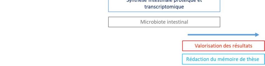
B. Perméabilité intestinale
Le jour du sacrifice (J7), les souris à jeun depuis 12h, seront gavées par une solution de FITC dextran
4 kDa et mises au repos 4h. Le sang sera récupéré lors du sacrifice et la fluorescence mesurée dans
le plasma. Le sang collecté permettra le dosage de la citrulline plasmatique et du GLP-2.
C. Translocation microbienne
La translocation bactérienne sera étudiée dans les tissus périphériques (foie, rate, ganglions
mésentériques) après un challenge par Salmonella enterica serovar typhimurium portant un gène de
résistance à l'ampiciline. Les souches marquées à la GFP de S. typhimurium seront cultivées en
aération modérée pendant 12 h à 37 °C dans un bouillon Luria-Bertani et mis en suspension dans du
PBS à raison de 108 CFU/mL. Quarante huit heure avant le sacrifice, les souris recevront un gavage
des 108 CFU/mL de S typhimurium (100μL en suspension dans du PBS). Les organes cibles seront
prélevés et homogénéisés dans du PBS. Les solutions obtenues seront platées sur gels d’agar
ampicilline-résistant étalées à dilutions successives sur boites de Petri (Ampicilline R).
D. Morphologie intestinale
Après sacrifice, des biopsies des différents segments de l'intestin (jéjunum, iléon et côlon) seront
prélevés. Un score histologique se basant sur la profondeur des cryptes, la taille des villosités,
l’épaisseur de la muqueuse, permettra d’évaluer les dommages tissulaires. L’étude de l’inflammation
sera réalisée par dosage de la myélopéroxydase et par dosage des cytokines TNF-α, IL-1β, IL-6 et KC,
IL-17, IL-23 et IL-10 par ELISA. Le marqueur de prolifération PCNA (proliferating cell nuclear antigen),
les enzymes de la bordure en brosse (intestinal alkaline phosphatase), les jonctions serrées (occludin
et claudin-7) ainsi que l’apoptose (méthode TUNEL) seront étudiés en immunohistochimie afin
d’explorer l’atteinte intestinale.
E. Synthèse intestinale protéique et transcriptomique
Vingt minutes avant le sacrifice, les souris recevront une injection intrapéritonéale de puromycine afin
d’étudier le taux de synthèse protéique (méthode SunSET).
La technique des puces à ADN sera utilisée pour étudier le transcriptome de l’intestin grêle et mettre
en évidence des différences au niveau de l’expression des gènes de la protéosynthèse (voie mTOR),
de la protéolyse (voie du protéasome) et de l’autophagie. La transcriptomique nous permettra
également de mettre en évidence des modifications de l’immunité innée, et en particulier des PRRs
(Pattern Recognition Receptors).
F. Microbiote intestinal
Le dénombrement des grands groupes bactériens sera analysé par qPCR sur la flore adhérente de
l’iléon et sera complété par NGS bactérien.
G. Survie
La survie de ces différents groupes de souris sera étudiée à un mois en poursuivant l’alimentation
qu’elle recevront à J-7.
Après conditionnement par irradiation et allogreffe de moelle, on s’attend à observer chez les souris
recevant l’alimentation hyper-protéique par rapport à celles recevant l’alimentation normo-protéique ou
hypo-protéique : A. une diminution de la sévérité de la GvHDa; B. une moins grande perméabilité
intestinale avec des taux de citrulline et de GLP-2 plus élevés; C. une moindre translocation de S.
typhimurium; D. une diminution des dommages de l’épithélium intestinal avec un taux de prolifération plus
élevé mais un niveau d’apoptose comparable; E. une meilleure synthèse protéique; F. une flore iléale
adhérente plus protectrice; F. une moindre mortalité.
Ces résultats démontreront l’influence de l’apport protéique entéral dans la limitation des dommages de
l’épithélium intestinal provoqué par le conditionnement et dans l’expression de la GvHDa. Ils permettront
également de mieux comprendre par quels mécanismes cette alimentation agit. S’il s’avère que le
renforcement de la barrière intestinale par l’alimentation permet de prévenir l’apparition de la GvHDa, nous
étudierons la meilleure façon d’améliorer cet effet en amont du conditionnement grâce à l’alimentation.
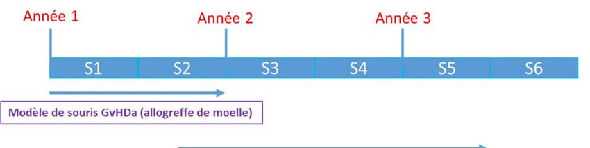
David SEGUY - U995 - Lille – France - 20194) Le programme de travail avec les livrables et l’échéancier prévisionnel
5) Liste de 10 publications portant directement sur le sujet
1.Markey KA, MacDonald KP, Hill GR. The biology of graft‐versus‐host disease: experimental systems instructing
clinical practice. Blood 2014;124:354‐362.
2.Danel Buhl N, Seguy D. Nutritional support in adult patients undergoing allogeneic stem cell transplantation
following myeloablative conditioning. In: al. RRe, ed. Diet and Nutrition in Critical Care. New York: Springer,
2015:593‐605.
3.Hueso T, Coiteux V, Joncquel Chevalier Curt M, Labreuche J, Jouault T, Yakoub‐Agha I, Seguy D. Citrulline and
Monocyte‐Derived Macrophage Reactivity before Conditioning Predict Acute Graft‐versus‐Host Disease. Biol Blood
Marrow Transplant 2017;23:913‐921.
4.Hueso T, Gauthier J, Joncquel Chevalier‐Curt M, Magro L, Coiteux V, Dulery R, Carpentier B, Labreuche J, Damaj
G, Yakoub‐Agha I, Seguy D. Association Between Low Plasma Level of Citrulline Before Allogeneic Hematopoietic
Cell Transplantation and Severe Gastrointestinal Graft vs Host Disease. Clin Gastroenterol Hepatol 2018;16:908‐
917 e2.
5.Seguy D, Berthon C, Micol JB, Darre S, Dalle JH, Neuville S, Bauters F, Jouet JP, Yakoub‐Agha I. Enteral feeding and
early outcomes of patients undergoing allogeneic stem cell transplantation following myeloablative conditioning.
Transplantation 2006;82:835‐839.
6.Seguy D, Duhamel A, Rejeb MB, Gomez E, Buhl ND, Bruno B, Cortot A, Yakoub‐Agha I. Better outcome of patients
undergoing enteral tube feeding after myeloablative conditioning for allogeneic stem cell transplantation.
Transplantation 2012;94:287‐294.
7.Gonzales F, Bruno B, Alarcon Fuentes M, De Berranger E, Guimber D, Behal H, Gandemer V, Spiegel A, Sirvent A,
Yakoub‐Agha I, Nelken B, Duhamel A, Seguy D. Better early outcome with enteral rather than parenteral nutrition
in children undergoing MAC allo‐SCT. Clin Nutr 2018;37:2113‐2121.
8.Azarnoush S, Bruno B, Beghin L, Guimber D, Nelken B, Yakoub‐Agha I, Seguy D. Enteral nutrition: a first option for
nutritional support of children following allo‐SCT? Bone Marrow Transplant 2012;47:1191‐1195.
David SEGUY - U995 - Lille – France - 20199.Gouyer V, Dubuquoy L, Robbe‐Masselot C, Neut C, Singer E, Plet S, Geboes K, Desreumaux P, Gottrand F, Desseyn
JL. Delivery of a mucin domain enriched in cysteine residues strengthens the intestinal mucous barrier. Sci. Rep
2015;5:9577.
10.Desseyn JL, Gouyer V, Gottrand F. Biological modeling of mucus to modulate mucus barriers. Am J Physiol
Gastrointest Liver Physiol 2016;310:G225‐7.
David SEGUY - U995 - Lille – France - 2019English version
Effect of a hyper-protein diet on strengthening the intestinal barrier in acute
graft-versus-host disease
Abstract
Allogeneic hematopoietic stem cell transplantation (allo-SCT) following myeloablative conditioning (MAC)
is the curative treatment for several haematological malignancies. In these patients, acute graft-versus-
host disease (aGvHD), whose target organs are the intestine, liver and skin, is the leading cause of death.
The study of aGvHD in mice has revealed the impact of both intestinal epithelium damage and bacterial
translocation following MAC on the apparition of aGvHD. In humans, we have demonstrated that a low
plasma citrulline level before MAC reflects the presence of subclinical intestinal damage which constitutes
an independent aGvHD risk factor during the post-transplant period. Undernutrition and fasting increase
the risk of aGvHD. In contrast, our team has shown that an early onset of enteral nutrition after allo-SCT
decreases the incidence and severity of aGvHD in both adults and children. This is probably due to enteral
nutrition’s trophic action on the intestine. In humans and mice, a protein deficient diet (or total parenteral
nutrition) induces a decrease in small bowel mass that potentiates bacterial translocation while enteral
amino acid supplementation reverses enterocyte atrophy.
We aim to demonstrate in mice that a high-protein diet limits the occurrence of aGvHD by strengthening
intestinal barrier.
Seven days before transplantation (d-7), female B6D2F1 will be divided into three isoenergetic diet groups:
G1 (high-protein), G2 (medium-protein) and G3 (low-protein). On d0, mice irradiated at d-1 (1100 cGy) will
receive bone marrow allograft from a C57BL/6 donor. On d7, mice will be sacrificed and their tissues
collected.
Compared to groups G2 and G3, we expect to observe in group G1: a decrease in the severity of aGvHD,
intestinal permeability and epithelium damage with higher plasma citrulline and GLP-2 levels; an increase
in intestinal protein synthesis; a reduction of translocation of S. typhimurium with a more protective flora in
the ileum and a better survival.
Research plan
This project has been approved by the Committee of Ethics in Animal Experimentation (N ° # 3546-
2016011210014075). The mice will be housed in an A1 + Exempt Species-Specific Organisms (SPF)
facility.
A. Murine model of allogeneic BMT : female B6D2F1 mice (8-12 weeks old) will receive a non-lethal
irradiation of 1100 cGy. The following day (d0), the mice will be transplanted with 5 x 10⁶ bone marrow
cells and 2 x 10⁶ purified T cells from C57BL/6 donor mice intravenously injected (caudal injection). Injected
lymphocytes will be labeled with CFSE to monitor cell proliferation. Grafted mice will be weighed and
monitored daily.
B. Diets: between d-14 and d-8 before the transplantation, all mice will receive a medium-protein diet
(20% of the total energy intake (TEI) constituted by the proteins). From (D-7), the mice will be divided into
3 groups according to the protein intake contained in the isoenergetic diets which will be given ad libitum:
G1, high-protein (60% of the TEI, n = 10); G2, medium-protein (20% of TEI, n = 10); G3, low-protein (4%
of TEI, n = 10) from Research Diets, New Jersey-USA.
C. Tissue collection: mice will be sacrificed 7 days after graft injection (d7). The blood, liver, spleen,
mesenteric lymph nodes and intestine will be harvested and samples will be frozen. Each experiment will
be carried out in triplicate.
Techniques
A. aGvHD: the clinical score of aGvHD will be daily assessed from d0 to d7. This score reflects the sum
of the following five distinct parameters: percentage of weight loss, posture (hunching), activity, fur texture
and skin integrity. This clinical score will be completed with (i) complete phenotyping of intestinal
intraepithelial lymphocytes (IEL), spleen and mesenteric lymph nodes by FACs analyses (Anti-
David SEGUY - U995 - Lille – France - 2019CD4/CD8/FoxP3 staining) and (ii) cytokine measurements (INF-γ, IL-1β, IL-17 and IL-23) and IHC staining
in these tissues.
B. Intestinal permeability: the day of the sacrifice (d7), 12h fasting mice will receive a gavage with a
solution of FITC dextran 4 kDa. Fluorescence will be measured in plasma derived from blood withdrawn
during the sacrifice, 4 hours after FITC administration. Collected blood will allow the plasma determination
of citrulline and GLP-2 levels.
C. Microbial translocation: Salmonella enterica serovar typhimurium GFP-tagged strains will be grown
for 12 h at 37°c in Luria-Bertani broth, cultured in mild aeration and suspended in PBS (108 CFU/100μL).
Forty-eight hours before sacrifice, the 4h-fasting mice will receive a gavage with 108 CFU of Salmonella
typhimurium (suspended in 100 μL of PBS), then allowed water and food ad libitum. Peripheral tissues
(liver, spleen, mesenteric lymph nodes) will be harvested. The organs will be homogenized in PBS and
plated on ampicillin-resistant agar gels. After dilutions, (10-2 to 10-5) the colonies will be counted and related
back to tissue weight.
D. Intestinal morphology: after sacrifice, jejunum, colon and ileum will be harvested to prepare slides
later stained with Hematoxylin-eosin and AB-PAS. Tissue damage will be evaluated using a histological
score based on crypt depth, size of the villi, mucosa thickness, and goblet cell count. TNF-α, IL-1β, IL-6,
IL-17, IL-23 and IL-10 cytokines will be assessed using ELISA. Proliferating cell nuclear antigen, intestinal
alkaline phosphatase and tight junction proteins (occludin and claudin-7) as well as apoptosis using
TUNEL assay will be assessed by immunohistochemistry (IHC). Plasma citrulline levels will be also
assessed to study the functional enterocyte mass.
E. Intestinal protein synthesis and transcriptomic: twenty minutes before sacrifice, the mice will
receive an intraperitoneal injection of puromycin to study the rate of protein synthesis (SunSET method).
The microarray technique will be used to study the transcriptome of the small intestine and highlight
differences in gene expression of proteosynthesis (mTOR pathway), proteolysis (proteasome pathway)
and autophagy. The transcriptomic will also allow us to highlight changes in innate immunity, and in
particular PRRs (Pattern Recognition Receptors).
F. Intestinal microbiota: The bacterial load of large bacterial groups will be analyzed by qPCR on fresh
feces and sequencing of the bacterial RNAs will be performed.
G. Survival: the survival of these different groups of mice will be studied at one month by continuing the
diet they will receive at d-7.
Data analysis
Comparison of each parameter between the three diet groups will be performed using a Kruskal-Wallis
test and then 2 by 2 using the unpaired Mann-Whitney U test. The one-month overall survival will be
assessed with the Kaplan-Meier method and or each criterion, univariate analysis will be performed using
the log-rank test.
Expected results:
In mice receiving the high-protein diet - compared to those receiving the medium-protein or low-protein
diet - we expect to observe the following: A. a decrease in the severity of aGvHD ; B. a lower intestinal
permeability with higher citrulline and GLP-2 levels; C. less translocation of S. typhimurium; D. a decrease
in the damage to the intestinal epithelium with a higher rate of proliferation but a comparable level of
apoptosis; E. a higher protein synthesis; F. a more protective ileal flora; F. a better survival.
If we confirm that strengthening the intestinal barrier through diet can prevent aGvHD onset, this BMT
murine model will help us to develop specific enteral diets.
In addition to being used with regularity as part of therapeutic treatment for patients receiving allo-SCT,
this approach could also be applied to patients requiring intense and prolonged chemotherapy known to
cause intestinal damage. In such cases, enteral nutrition would be used to limit toxicity and improve the
efficacy of prolonged chemotherapy.
David SEGUY - U995 - Lille – France - 2019Background
Allogeneic hematopoietic stem cell transplantation (allo-SCT) is the curative treatment for several
hematological malignancies. Allo-SCT procedure requires a myeloablative conditioning (MAC)
chemotherapy or total body irradiation preceding the intravenous infusion of stem cells to recover a
functional hematopoietic system. The antitumoral effect of allo-SCT is based on both the cytoreductive
effect of the conditioning regimen and the graft immunological effect related to the recognition and
destruction of tumor cells. However, nearly 50% of patients develop acute graft-versus-host disease
(aGvHD) within 3 months after transplantation. Acute GvHD corresponds to the alloreactivity of the donor
T lymphocytes (LcT) in response to host antigens (recipient), leading to destruction of host tissues
(especially gut, liver and skin). It represents the first cause of morbidity and early mortality (up to 95% of
deaths for grade IV aGvHD)1. The study of murine bone marrow transplantation (BMT) has revealed the
impact of intestinal damage and bacterial translocation following the conditioning regimen, in the initiation
and amplification of aGvHD. Indeed, by crossing intestinal mucosa, microbial products or PAMPs
(Pathogen-Associated Molecular Patterns) such as LPS stimulate the secretion of proinflammatory
cytokines (TNF-α and IL-1) by macrophages and the host antigen presentation by antigen presenting cells
(APC) which enhance the donor LcT alloreactivity2.
In humans, the drop in plasma citrulline levels following MAC suggests the deleterious effect of the latter
on the intestinal epithelium3. Since then, we have demonstrated that a low citrulline level before MAC
reflects the presence of a subclinical impairment of the intestinal barrier which constitutes an independent
risk factor of aGvHD (especially gastrointestinal) and non-relapse mortality in the post transplantation
period4, 5. In these patients, undernutrition and fasting related to anorexia, digestive disorders and dietary
restrictions are factors that increase the frequency and severity of aGvHD6. Conversely, an intestinal
decontamination performed with a combination of broad spectrum antibiotics seems able to reduce the
incidence and the severity of aGvHD7. Our team has also shown that early enteral feeding after allo-SCT,
probably due to its trophic action on the intestine, decrease the incidence and the severity of aGvHD in
both adults8, 9and children10, 11. Indeed, various studies show that enteral feeding, especially thanks to the
amino-acids contained in proteins such as glutamine, plays a determining role in the barrier function of the
intestine12, 13. Two studies suggest that oral intake of glutamine (the preferred food of enterocytes) reduces
the risk of aGvHD in humans (RR = 0.42, 95% CI = 0.21-0.85)14.
Given the difficulty of studying aGvHD in humans, the use of animal models is essential to confirm these
hypotheses. Our team has developed a transgenic murine model that releases a recombinant protein
(rCYSx12) which can increase mucus viscosity in the intestinal lumen. In short, such transgenic mice have
a higher citrulline concentration than their non-mutated sisters (unpublished data). They are more resistant
to chemically induced colitis and bacterial translocation15, 16. Under intense chemotherapy, these murine
models repair their intestinal epithelium faster than their non-mutated sisters (longer villi, deeper crypts,
higher proliferation index and higher citrulline concentration). They also seem less sensitive to Salmonella
typhimurium intestinal translocation. The analysis of their adherent ileal flora by qPCR shows that they
have a protective flora enriched with Clostridium leptum. In mice, feeding is another way to strengthen
intestinal barrier. A protein deficient diet (or a total parenteral nutrition) induces a decrease in small bowel
mass, crypt cell proliferation, crypt and villus heights that potentiated the transmucosal passage of
bacteria17. Free protein diet enriched in glucose involves time-dependent intestinal mucosal atrophy,
whereas amino acids supplementation reverses this atrophy via mTOR complex 1 pathway18. Among the
amino acids that constitute proteins, glutamine is recognized for its positive impact on the intestinal barrier
by reducing permeability and bacterial translocation to physiologic levels and preserving mucosal
integrity19. Consistently, the reintroduction of an ad libitum diet in mice deprived of food reverses the small
bowel atrophy through GLP-2 secretion that is a physiologic regulator of the digestive epithelium in
response to luminal nutrients20.
These data suggest the following:
1. Plasma citrulline concentration reflects the integrity of the intestinal barrier;
2. Intestinal barrier plays a major role in the pathophysiology of aGvHD;
3. Strengthening the intestinal barrier can reduce the frequency and severity of
4. Enteral feeding containing protein is a way to limit conditioning regimen damage and, subsequently,
aGvHD.
We aim to demonstrate that high-protein diet limits the occurrence of aGvHD by strengthening intestinal
barrier in mice.
References
David SEGUY - U995 - Lille – France - 20191.Cahn JY, Klein JP, Lee SJ, Milpied N, Blaise D, Antin JH, Leblond V, Ifrah N, Jouet JP, Loberiza F,
Ringden O, Barrett AJ, Horowitz MM, Socie G. Prospective evaluation of 2 acute graft-versus-host (GVHD)
grading systems: a joint Societe Francaise de Greffe de Moelle et Therapie Cellulaire (SFGM-TC), Dana
Farber Cancer Institute (DFCI), and International Bone Marrow Transplant Registry (IBMTR) prospective
study. Blood 2005;106:1495-1500.
2.Hill GR, Crawford JM, Cooke KR, Brinson YS, Pan L, Ferrara JL. Total body irradiation and acute graft-
versus-host disease: the role of gastrointestinal damage and inflammatory cytokines. Blood 1997;90:3204-
3213.
3.Lutgens LC, Blijlevens NM, Deutz NE, Donnelly JP, Lambin P, De Pauw BE. Monitoring myeloablative
therapy-induced small bowel toxicity by serum citrulline concentration: a comparison with sugar
permeability tests. Cancer 2005;103:191-199.
4.Hueso T, Coiteux V, Joncquel Chevalier Curt M, Labreuche J, Jouault T, Yakoub-Agha I, Seguy D.
Citrulline and Monocyte-Derived Macrophage Reactivity before Conditioning Predict Acute Graft-versus-
Host Disease. Biol Blood Marrow Transplant 2017;23:913-921.
5.Hueso T, Gauthier J, Joncquel Chevalier-Curt M, Magro L, Coiteux V, Dulery R, Carpentier B, Labreuche
J, Damaj G, Yakoub-Agha I, Seguy D. Association Between Low Plasma Level of Citrulline Before
Allogeneic Hematopoietic Cell Transplantation and Severe Gastrointestinal Graft vs Host Disease. Clin
Gastroenterol Hepatol 2018;16:908-917 e2.
6.Mattsson J, Westin S, Edlund S, Remberger M. Poor oral nutrition after allogeneic stem cell
transplantation correlates significantly with severe graft-versus-host disease. Bone Marrow Transplant
2006;38:629-633.
7.Beelen DW, Elmaagacli A, Muller KD, Hirche H, Schaefer UW. Influence of intestinal bacterial
decontamination using metronidazole and ciprofloxacin or ciprofloxacin alone on the development of acute
graft-versus-host disease after marrow transplantation in patients with hematologic malignancies: final
results and long-term follow-up of an open-label prospective randomized trial. Blood 1999;93:3267-3275.
8.Seguy D, Berthon C, Micol JB, Darre S, Dalle JH, Neuville S, Bauters F, Jouet JP, Yakoub-Agha I. Enteral
feeding and early outcomes of patients undergoing allogeneic stem cell transplantation following
myeloablative conditioning. Transplantation 2006;82:835-839.
9.Seguy D, Duhamel A, Rejeb MB, Gomez E, Buhl ND, Bruno B, Cortot A, Yakoub-Agha I. Better outcome
of patients undergoing enteral tube feeding after myeloablative conditioning for allogeneic stem cell
transplantation. Transplantation 2012;94:287-294.
10.Azarnoush S, Bruno B, Beghin L, Guimber D, Nelken B, Yakoub-Agha I, Seguy D. Enteral nutrition: a
first option for nutritional support of children following allo-SCT? Bone Marrow Transplant 2012;47:1191-
1195.
11.Gonzales F, Bruno B, Alarcon Fuentes M, De Berranger E, Guimber D, Behal H, Gandemer V, Spiegel
A, Sirvent A, Yakoub-Agha I, Nelken B, Duhamel A, Seguy D. Better early outcome with enteral rather
than parenteral nutrition in children undergoing MAC allo-SCT. Clin Nutr 2017 (ahead of print).
12.Buchman AL, Moukarzel AA, Bhuta S, Belle M, Ament ME, Eckhert CD, Hollander D, Gornbein J,
Kopple JD, Vijayaroghavan SR. Parenteral nutrition is associated with intestinal morphologic and
functional changes in humans. JPEN J. Parenter. Enteral Nutr 1995;19:453-460.
13.Coeffier M, Claeyssens S, Lecleire S, Leblond J, Coquard A, Bole-Feysot C, Lavoinne A, Ducrotte P,
Dechelotte P. Combined enteral infusion of glutamine, carbohydrates, and antioxidants modulates gut
protein metabolism in humans. Am J Clin Nutr 2008;88:1284-90.
14.Crowther M, Avenell A, Culligan DJ. Systematic review and meta-analyses of studies of glutamine
supplementation in haematopoietic stem cell transplantation. Bone Marrow Transplant 2009;44:413-425.
15.Desseyn JL, Gouyer V, Gottrand F. Biological modeling of mucus to modulate mucus barriers. Am J
Physiol Gastrointest Liver Physiol 2016;310:G225-7.
16.Gouyer V, Dubuquoy L, Robbe-Masselot C, Neut C, Singer E, Plet S, Geboes K, Desreumaux P,
Gottrand F, Desseyn JL. Delivery of a mucin domain enriched in cysteine residues strengthens the
intestinal mucous barrier. Sci. Rep 2015;5:9577.
17.Deitch EA. Bacterial translocation: the influence of dietary variables. Gut 1994;35:S23-7.
David SEGUY - U995 - Lille – France - 201918.Nakamura A, Hara K, Yamamoto K, Yasuda H, Moriyama H, Hirai M, Nagata M, Yokono K. Role of the
mTOR complex 1 pathway in the in vivo maintenance of the intestinal mucosa by oral intake of amino
acids. Geriatr Gerontol Int 2012;12:131-9.
19.Dos Santos R, Viana ML, Generoso SV, Arantes RE, Davisson Correia MI, Cardoso VN. Glutamine
supplementation decreases intestinal permeability and preserves gut mucosa integrity in an experimental
mouse model. JPEN J Parenter Enteral Nutr 2010;34:408-13.
20.Bahrami J, Yusta B, Drucker DJ. ErbB activity links the glucagon-like peptide-2 receptor to refeeding-
induced adaptation in the murine small bowel. Gastroenterology 2010;138:2447-56.
David SEGUY - U995 - Lille – France - 2019Vous pouvez aussi lire