Exploration de l'insuffisance valvulaire aortique en IRM: place par rapport à l'échographie - JM Serfaty, K. Warin Fresse Service d'imagerie ...
←
→
Transcription du contenu de la page
Si votre navigateur ne rend pas la page correctement, lisez s'il vous plaît le contenu de la page ci-dessous
Exploration de l’insuffisance
valvulaire aortique en IRM: place
   par rapport à l’échographie
            JM Serfaty, K. Warin Fresse
     Service d’imagerie cardiaque et vasculaire
                   diagnostique
                   CHU Nantes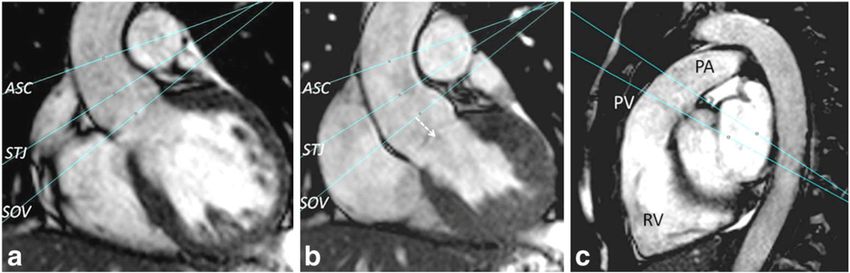
Physiopathologie • Dilatation racine de l’aorte non coaptation des valves – Marfan et appariés – Bicuspidie – Dilatation idiopathique – Dissection – Aortites • Maladie des valves – Calcifications séniles – Bicuspidie (prolapsus valve fusionnée) – Endocardite – RAA +- prolapsus associé des valves
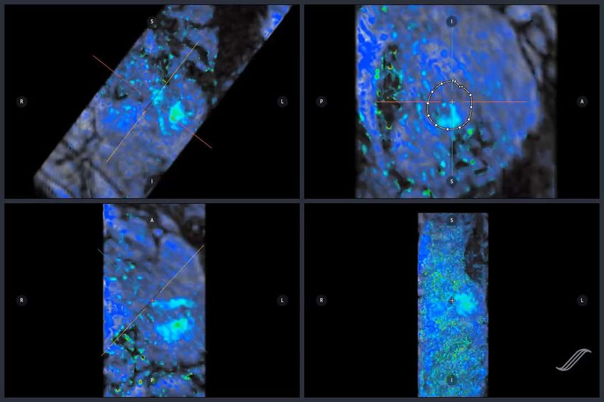
Figure 35.2.1 Definition of aortic root and ascending aortic phenotypes and mechanisms of aortic regurgitation (AR) (echocardiographic
view) and direction of the regurgitant jet (Doppler view).

     Chapter: Aor c regurgita on Author(s): Pilar Tornos Mas and Emmanuel Lansac From: ESC CardioMed (DRAFT)

Downloaded from Oxford Medicine Online. © Oxford University Press, 2018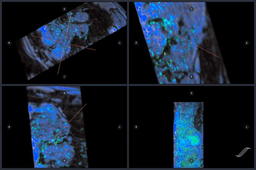
Management of aortic
regurgitation

      Groupe 1

      AORTIQUE

     Chapter: Aor c regurgita on Author(s): Pilar Tornos Mas and Emmanuel Lansac From: ESC CardioMed (DRAFT)

Downloaded from Oxford Medicine Online. © Oxford University Press, 2018
Management of aortic
regurgitation

      Groupe 2
      NON AORTIQUE

      FUITE SEVERE

      symptomatique

     Chapter: Aor c regurgita on Author(s): Pilar Tornos Mas and Emmanuel Lansac From: ESC CardioMed (DRAFT)

Downloaded from Oxford Medicine Online. © Oxford University Press, 2018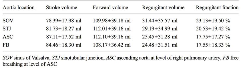
Management of aortic
regurgitation

      Groupe 2
      NON AORTIQUE

      FUITE SEVERE

      Asymptomatique
      FEVG < 50% ou
      Volumes VG augmentés

     Chapter: Aor c regurgita on Author(s): Pilar Tornos Mas and Emmanuel Lansac From: ESC CardioMed (DRAFT)

Downloaded from Oxford Medicine Online. © Oxford University Press, 2018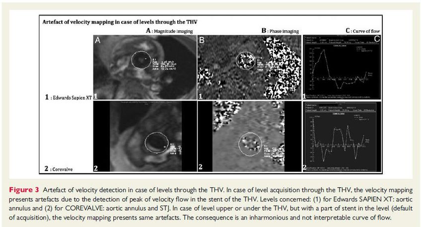
Groupe 1 : AORTIQUE
Marfan sans FDR > 50mm chirurgie INDIQUEE
ESC 2017
Groupe 1 : AORTIQUE
Marfan + FDR, ou mutation TGFBR1-R2 > 45mm
Bicuspidie + FDR ou Coarctation > 50mm chirurgie CONSIDEREE
Tout patient >55mm
FDR: histoire familiale, HTA, >3mm/an
ESC 2017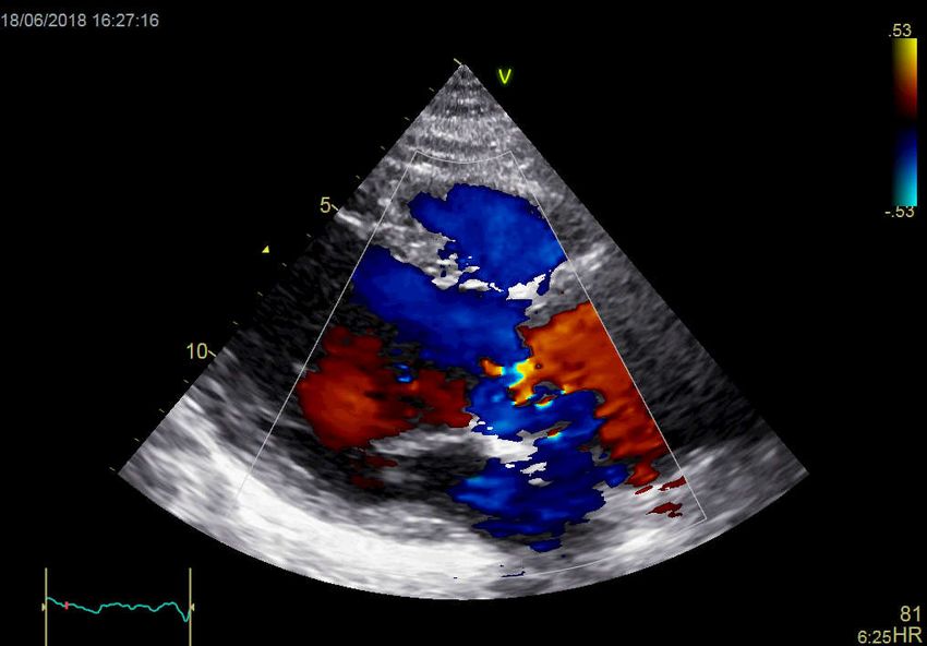
Groupe 1 : AORTIQUE

Réparation valvulaire si : patient jeune + dilatation aortique + valve tricuspide

                                                                     ESC 2017
Groupe 2 : NON AORTIQUE, fuite SEVERE

                                        ESC 2017
Sinus de Valsava
Sinus de Valsava

Prolapsus valve aortique   Prolapsus valve aortique
Figure 35.2.1 Definition of aortic root and ascending aortic phenotypes and mechanisms of aortic regurgitation (AR) (echocardiographic
view) and direction of the regurgitant jet (Doppler view).
Chapter: Aor c regurgita on Author(s): Pilar Tornos Mas and Emmanuel Lansac From: ESC CardioMed (DRAFT)
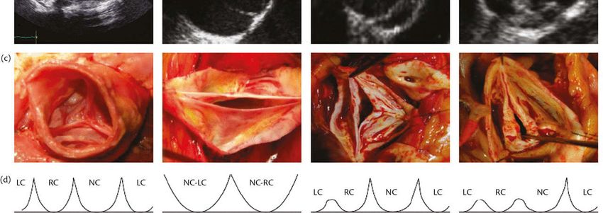
Downloaded from Oxford Medicine Online. © Oxford University Press, 2018Figure 35.2.2 Classification of aortic valve pathology: tricuspid, bicuspid (0 or 1 raphes), and unicuspid. (a) Diagram. (b) Echocardiographic
views. (c) Pathology specimens. (d) LC, left coronary; NC, non-coronary; RC, right coronary.
ETT
Se: 56%
Sp : 86%
Chapter: Aor c regurgita on Author(s): Pilar Tornos Mas and Emmanuel Lansac From: ESC CardioMed (DRAFT)
Downloaded from Oxford Medicine Online. © Oxford University Press, 2018Figure 35.2.4 Definition of coaptation height (cH) and effective height (eH) of aortic valve cusps. STJ,
sinotubular junction.
eH: effective height
Chapter: Aor c regurgita on Author(s): Pilar Tornos Mas and Emmanuel Lansac From: ESC CardioMed (DRAFT)
Downloaded from Oxford Medicine Online. © Oxford University Press, 2018AORTE ASCENDANTE IRM sans injection Scanner injecté synchronisé Bicuspidie type 1
QUANTIFICATION DE LA FUITE (valve NATIVE)

   Utile si discordance entre clinique et mesures écho

   La mesure IRM de la fuite est moins variable que l’ETT et ETO

                                           Précis pour :

                                           Quantification de la fuite

                                           Volume VG
                                           FEVG

                                                           AHA Guidelines 2014
Circ Cardiovasc Imaging. 2013;6:48-57
ECHOGRAPHIE                   IRM

          Circ Cardiovasc Imaging. 2013;6:48-57
Recommendation : Mesure sur la JST Le mieux corrélé à Stroke VG - VD Eur Radiol 2015
Comparaison de
  reflux turbulents à
  des méthodes
  indépendantes de
  ces turbulences

  TE non précisé

  Recommandé :

Stroke Ao – Stroke APulm

    Pediatric Radiol 2014
CIRC 2012
Seuil: Fraction Régurgitation > 33 %
Seuil : Vol VG TD : > 246ml2015
ETT: faible (73%), modérée (10%), sévère (16%)
Un jet régurgitant (33%), 2 jets (60%), 3 jets (7%)
Pas de jet central
7% des patients avaient une fenêtre acoustique limitée

2015 Meilleure mesure : coupe au dessus de la prothèse
Vérifier que le volume éjecté aortique = VE VG, l’absence d’artefact de flux, et
une courbe harmonieuse
2015> 14% Fuite modérée ou sévère
Kappa = 0.33

       Pour les Fuite modérées :
       ETT sous-estime / IRM

                      IRM

ECHO
Mortalité supérieure si fuite post-TAVR 135 patients after TAVR 3 centres Mesure fraction de régurgitation 40 jours après TAVR vs Echo 6 jours post TAVR Suivi 2 ans Endpoint : Mortalité, Rehospitalisation
COPD : BPCO
IRM              Echographie

 Mortalité             Mortalité

Rehospitalisation   Rehospitalisation
• Limites:
   – Pas d’écho 3D
   – Différence de timing entre ETT et IRM

• IRM vs Echo
   – Mesure quantitative vs semi- en écho, dédié à la valve native (fuites
     post-TAVI para-valvulaires excentrées)
   – Mesure reproductible > echo
   – Patients peu échogènes
   – Peu gêné par les jets excentrés > echo
   – Pas de sous-estimation des fuites aortiques > echo
• Indications
– Patients peu échogènes
– ETT et/ou ETO douteuses
– Fuite faible à modérée à l’ETT avec symptômes ou signes
d’insuffisance cardiaque
– Fuite modérée à sévère à l’ETT
• Intérêt du suivi par IRM : quantification de la fuite et
du volume VG
• Décision de gestes complémentaireIRM 3T, 4D flow
Post injection bolus
Arterys
Critères diagnostiques :
•Diamètre jet / Ch chasse VG : 65%
•Longueur jet / longueur VG : 50%
•Jet régurgitant dans l’aorte descendante : non / traces / oui
K=0,88Courtoisie Benjamin Dubourg
Logiciel : Arterys

                     Courtesie : Elie Mousseau
CONCLUSION • Patient avec valve native – Anatomie de l’aorte – Sévérité de la régurgitation – Fraction d’éjection et volume VG • Patient avec valve post-TAVR – Sévérité de la fuite • Technique – 4D flow
Vous pouvez aussi lire