Questions et réponses sur le choix du traitement de la carence en fer et l'utilisation du fer intraveineux dans la pratique clinique courante
←
→
Transcription du contenu de la page
Si votre navigateur ne rend pas la page correctement, lisez s'il vous plaît le contenu de la page ci-dessous
Questions et réponses sur le
choix du traitement de la
carence en fer et l'utilisation
du fer intraveineux dans la
pratique clinique courante
ANNALS OF MEDICINE 2021, VOL. 53, NO. 1, 274–285
Les principaux points marquants de l'article sont les suivants :

   •    La carence en fer est une cause courante de morbidité et peut être la conséquence ou la
        complication de nombreuses maladies.

   •    Le choix du traitement de la carence en fer est basé sur plusieurs facteurs, y compris la présence
        d'une inflammation, le temps disponible pour le réapprovisionnement en fer et le risque anticipé
        d'effets indésirables ou d'intolérance.

   •    Les préparations intraveineuses à base de fer sont indiquées pour le traitement de la carence en
        fer lorsque les préparations orales sont inefficaces ou ne peuvent pas être utilisées et, en
        conséquent, sont applicables dans un large éventail de contextes cliniques, y compris les
        conditions inflammatoires chroniques, les contextes périopératoires et les troubles associés aux
        pertes chroniques de sang.

ANNALS OF MEDICINE 2021, VOL. 53, NO. 1, 274–285
Réplétion de fer avec du fer oral :
• L'objectif du traitement est de réapprovisionner les réserves de fer et, en cas d'anémie,
de normaliser la concentration d'Hb.
• Les suppléments oraux de fer sont absorbés à travers l'épithélium de l'intestin grêle, principalement
dans le duodénum, par l'intermédiaire de transporteurs de fer (DMT1 et ferroportine) sur la surface
apicale et basolatérale des entérocytes.
• En moyenne, seuls ~10% du fer intestinal sont absorbés. Ainsi, sur la dose thérapeutique orale
courante de 60 à 180 mg de fer élémentaire, moins de 20 mg sont absorbés par jour, ce qui signifie
que, théoriquement, 10 à 30 jours de supplémentation continue de fer peuvent être nécessaires pour
obtenir une augmentation de 10 g/l du taux d'Hb, et jusqu'à 6 mois pour normaliser complètement
les taux d'Hb et réapprovisionner les réserves de fer chez les patients anémiques.
• Lorsqu'il est administré par voie orale, le supplément de fer résiduel reste en grande partie non
absorbé dans le tube digestif, ce qui peut endommager les surfaces intestinales et altérer la
composition du microbiome intestinal, entraînant des effets indésirables gastro-intestinaux.
ANNALS OF MEDICINE 2021, VOL. 53, NO. 1, 274–285Réplétion de fer avec du fer intraveineux :
• Les préparations intraveineuses à base de fer contournent l'absorption gastro-intestinale et leurs
complexes fer-glucides sont traités par les macrophages afin de libérer le fer.
Ceci est principalement effectuée par les macrophages dans le foie, la rate et la moelle osseuse,
mais le mécanisme d'absorption et de dégradation ultérieure du complexe est incomplètement
compris.
• Une fois que le fer est libéré dans le cytoplasme, il peut être stocké au sein de la ferritine ou
exporté par la ferroportine dans le plasma.
• En fonction de la préparation intraveineuse spécifique de fer, la quantité totale de fer qui peut être
administrée avec une seule perfusion peut aller de 62,5 mg à >1500 mg. Par conséquent, pour les
préparations délivrant une grande quantité de fer, même une seule dose peut être suffisante pour
combler complètement la carence en fer des patients.
ANNALS OF MEDICINE 2021, VOL. 53, NO. 1, 274–285Traitement à base de fer chez les femmes enceintes :
• Le fer oral peut être utilisé pour la prévention de la carence en fer pendant la grossesse chez les
femmes ayant des réserves réduites en fer (c.-à-d. ferritine 100 g/L), et au cours du premier trimestre lorsque le fer intraveineux est contre-indiqué.
• Le fer intraveineux doit être utilisé à partir du deuxième trimestre en cas d'anémie ferriprive sévère
(c.-à-d. taux d'HbTraitement à base de fer chez les femmes enceintes :
Le fer intraveineux doit être utilisé chez les femmes enceintes dans les cas suivants :
• Les femmes enceintes présentant une anémie modérée (taux d'Hb 95 à 105 g/L) qui ne répondent
pas au fer oral après 4 semaines de prise.
• Femmes enceintes présentant une anémie sévère (taux d’HbTraitement à base de fer chez les femmes ayant des saignements menstruels
abondants :
• Le traitement de la carence en fer avec un traitement oral à base de fer doit être l'approche initiale.
Malheureusement, 20 à 40% des femmes non enceintes qui ingèrent des préparations orales à base
de fer développent des nausées, des vomissements, une constipation et / ou un goût métallique
indésirable, et arrêtent souvent le traitement oral à base de fer. Ces effets indésirables peuvent être
réduits par des changements dans la formulation, l'ajout de laxatifs émollients en cas de
constipation, ou par une administration un jour sur deux.
• En gardant ceci à l’esprit, les femmes doivent être réévaluées et l'échec du traitement (défini par
une augmentation du taux d'HbTraitement à base de fer chez les femmes ayant des saignements menstruels abondants : Le fer intraveineux doit être utilisé chez les filles et les femmes dans les cas suivants : • qui ne tolèrent pas ou ne sont pas aptes à utiliser le fer oral. • qui n'ont pas répondu à une dose appropriée, une formulation et un schéma d'administration de fer oral dans les 30 jours, comme défini par une augmentation du taux d'Hb d'au moins 10 g/l (ou 1 g/l). • qui doivent subir une chirurgie gynécologique élective et qui, de l'avis du chirurgien, sont peu susceptibles d'obtenir un taux d'Hb d'au moins 110 g/l (11 g/dl) le jour de l'intervention en utilisant des formulations orales. ANNALS OF MEDICINE 2021, VOL. 53, NO. 1, 274–285
Utilisation du traitement à base de fer dans l’Insuffisance rénale chronique :
   • Le fer intraveineux est utilisé systématiquement chez presque tous les patients nécessitant une
     hémodialyse (HD), et il est requis de manière variable chez les patients présentant une
     Insuffisance rénale chronique qui ne dépend pas de la dialyse, ceux sous dialyse péritonéale
     (DP) et les receveurs de transplantation rénale.

   • Pour les Insuffisance rénale chronique moins avancées (stade 1 à 3), la supplémentation orale
     en fer est acceptable comme option de première intention. Cependant, le fer intraveineux peut
     s'avérer nécessaire pour les patients qui y répondent mal ou qui développent des effets
     indésirables gastro-intestinaux gênants.

ANNALS OF MEDICINE 2021, VOL. 53, NO. 1, 274–285
ANNALS OF MEDICINE
2021, VOL. 53, NO. 1, 274–285
https://doi.org/10.1080/07853890.2020.1867323
REVIEW ARTICLE
Questions and answers on iron deficiency treatment selection and the use
of intravenous iron in routine clinical practice
Toby Richardsa , Christian Breymannb, Matthew J. Brookesc,d, Stefan Lindgrene, Iain C. Macdougallf,
Lawrence P. McMahong, Malcolm G. Munroh,i, Elizabeta Nemethj, Giuseppe M. C. Rosanok, Ingolf Schiefkel
€nter Weissm,n
and Gu
a
Faculty of Health and Medical Sciences, University of Western Australia, Perth, Australia; bObstetric Research-Feto Maternal
Haematology Unit, University Hospital Zurich, Zurich, Switzerland; cGastroenterology Unit, Royal Wolverhampton NHS Trust,
Wolverhampton, UK; dResearch Institute in Healthcare Science (RIHS), University of Wolverhampton, Wolverhampton, UK; eDepartment
of Gastroenterology and Hepatology, Lund University, Skåne University Hospital, Malm€o, Sweden; fDepartment of Renal Medicine, King’s
College Hospital, London, UK; gDepartments of Renal Medicine and Obstetric Medicine, Eastern Health Clinical School, Monash
University, Melbourne, Australia; hDepartment of Obstetrics and Gynecology, David Geffen School of Medicine, University of California,
Los Angeles, CA, USA; iDepartment of Obstetrics and Gynecology, Kaiser-Permanente, Los Angeles Medical Center, Los Angeles, CA, USA;
j
Center for Iron Disorders, David Geffen School of Medicine, University of California, Los Angeles, CA, USA; kDepartment of Medical
Sciences, IRCCS San Raffaele, Roma, Italy; lDepartment of Gastroenterology, Hepatology, Diabetology and Endocrinology, Klinikum St.
Georg, Leipzig, Germany; mDepartment of Internal Medicine II, Medical University Innsbruck, Innsbruck, Austria; nChristian Doppler
Laboratory for Iron Metabolism and Anemia Research, University of Innsbruck, Innsbruck, Austria
ABSTRACT ARTICLE HISTORY
Iron deficiency is a common cause of morbidity and can arise as a consequence or complication Received 24 September 2020
from many diseases. The use of intravenous iron has increased significantly in the last decade, Revised 3 December 2020
but concerns remain about indications and administration. Modern intravenous iron prepara- Accepted 15 December 2020
tions can facilitate rapid iron repletion in one or two doses, both for absolute iron deficiency
KEYWORDS
and, in the presence of inflammation, functional iron deficiency, where oral iron therapy is inef- Anaemia; iron-deficiency;
fective or has not worked. A multidisciplinary team of experts experienced in iron deficiency cardiovascular diseases;
undertook a consensus review to support healthcare professionals with practical advice on man- erythrocyte transfusion;
aging iron deficiency in gastrointestinal, renal and cardiac disease, as well as; pregnancy, heavy inflammatory bowel
menstrual bleeding, and surgery. We explain how intravenous iron may work where oral iron diseases; infusions;
has not. We provide context on how and when intravenous iron should be administered, and intravenous; iron;
informed opinion on potential benefits balanced with potential side-effects. We propose how menorrhagia; renal
intravenous iron side-effects can be anticipated in terms of what they may be and when they insufficiency; chronic;
may occur. The aim of this consensus is to provide a practical basis for educating and preparing pregnancy complications
staff and patients on when and how iron infusions can be administered safely and efficiently.
KEY MESSAGES
Iron deficiency treatment selection is driven by several factors, including the presence of
inflammation, the time available for iron replenishment, and the anticipated risk of side-
effects or intolerance.
Intravenous iron preparations are indicated for the treatment of iron deficiency when oral
preparations are ineffective or cannot be used, and therefore have applicability in a wide
range of clinical contexts, including chronic inflammatory conditions, perioperative settings,
and disorders associated with chronic blood loss.
Adverse events occurring with intravenous iron can be anticipated according to when they
typically occur, which provides a basis for educating and preparing staff and patients on how
iron infusions can be administered safely and efficiently.
Introduction impact of iron deficiency on clinical outcomes and
The use of intravenous iron has grown substantially in quality of life. Current intravenous iron preparations
the past decade due to heightened awareness of the are indicated for the treatment of iron deficiency
CONTACT Toby Richards toby.richards@uwa.edu.au Faculty of Health and Medical Sciences, The University of Western Australia (M581), 35
Stirling Highway, Perth, 6009, Australia
This article has been republished with minor changes. These changes do not impact the academic content of the article.
ß 2021 The Author(s). Published by Informa UK Limited, trading as Taylor & Francis Group
This is an Open Access article distributed under the terms of the Creative Commons Attribution License (http://creativecommons.org/licenses/by/4.0/), which permits
unrestricted use, distribution, and reproduction in any medium, provided the original work is properly cited.ANNALS OF MEDICINE 275
when oral preparations are ineffective or cannot be however, occur only when iron storage has been
used [1]. They have a wide range of applicability in largely depleted.
clinical contexts including; nutritional deficiency, mal- Functional iron deficiency describes a state of
absorption, disorders associated with chronic blood impaired iron mobilization that arises in conditions
loss, and chronic inflammatory conditions [2–5]. The associated with inflammation, infection or chronic dis-
availability of modern preparations allows rapid iron ease, where the normal pathways of iron transport
repletion in just one or two doses facilitating ease of and iron metabolism are disrupted [11,12]. In these
treatment [6,7]. settings, cytokines, particularly IL6, upregulate the
Intravenous iron preparations are approved with master regulator of iron homeostasis, hepcidin.
broad labels and the clinical trials testing efficacy span Increased hepcidin (normally seen in response to iron
many indications and disease groups, this has led to a loading) leads to reduced dietary iron absorption and
proliferation of guidelines and consensus statements. failure of cellular iron export into plasma, with reten-
However, there remains a need for more practical tion of iron within macrophages. Consequently, this
“how to” guidance on intravenous iron administration reduces circulating levels of iron and its availability for
as patients and staff may express concerns over erythropoiesis despite the presence of adequate
potential side-effects, adverse events, reactions and iron stores.
complications, or contra-indications, which may be
confused or misperceived. What is the difference between iron deficiency
Here, we aim to support healthcare professionals anaemia and anaemia of chronic disease?
by summarizing the current recommendations for
Definitions of iron deficiency are frequently inconsist-
intravenous iron, alongside practical advice on their
ent because absolute and functional iron deficiency
administration and informed opinion on the antici-
are often used interchangeably. Absolute iron defi-
pated risks versus expected benefits of treatment.
ciency is commonly defined by reduced ferritin levels
(276 T. RICHARDS ET AL.
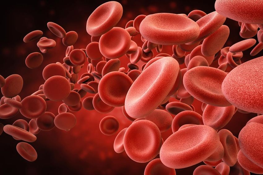
Oral iron supplement Intravenous iron infusion
Lumen Iron
complex
Macrophage
DMT1 Iron
complex
Ferritin
Duodenal
enterocytes Fe Fe
Ferritin
FPN FPN
Blood Blood
Hepcidin Hepcidin
Fe Fe
Inflammation,
iron loading
Figure 1. The mechanism of oral and intravenous iron treatments. Iron administered via an oral iron supplement is absorbed by
duodenal enterocytes. Divalent metal transporter 1 (DMT1) imports iron across the apical surface of enterocytes, whereas ferropor-
tin exports iron across the basolateral surface. The hormone hepcidin, which is increased by iron loading or inflammation, impairs
cellular iron export into blood by causing ferroportin degradation. Intravenous iron preparations are administered by infusion, and
the iron–carbohydrate complex is taken up and processed by macrophages in the liver, spleen and marrow. Once iron is released
into the cytoplasm, it is either stored in ferritin or exported from macrophages through ferroportin (FPN). Intravenous iron can
overcome the hepcidin-mediated block of iron absorption from the gut.
heart failure, obesity or chronic obstructive pulmonary supplements are absorbed across the small intestinal
disease [12]. In addition, patients with chronic kidney epithelium, mainly in the duodenum, via iron trans-
disease (CKD) also have features of functional iron porters (DMT1 and ferroportin) on the apical and
deficiency with increased circulating hepcidin levels basolateral surface of enterocytes (Figure 1). Only
due to subtle inflammation and often impaired urinary 10% of intestinal iron is absorbed on average [18].
hepcidin excretion [14] (Figure 1). Thus, of the common therapeutic oral dose of
Current guidelines typically suggest that a serum 60–180 mg elemental iron, less than 20 mg is absorbed
ferritin level below 100 mg/L is considered indicative of per day, meaning that theoretically 10–30 days of con-
absolute iron deficiency in the setting of an inflamma- tinuous iron supplementation may be required to
tory disease. Alternative laboratory determinates achieve a 10 g/L increase in Hb, and as long as
include; soluble transferrin receptor (sTfR) levels, sTfR/ 6 months to fully normalize Hb levels and replenish
log ferritin ratio, percentage of hypochromic red blood iron stores in anaemic patients. When given orally,
cells, reticulocyte Hb content, hepcidin, and red cell residual iron supplement remains largely unabsorbed
indices may be helpful to identify absolute iron defi- in the digestive tract, which can injure intestinal surfa-
ciency in patients with ferritin levels >100 mg/L, but ces and alter the composition of the gut microbiome,
these are mostly used in academic settings [15,16]. leading to gastrointestinal side-effects.
The use of hepcidin levels for diagnostic purposes or Intravenous iron preparations bypass gastrointes-
to guide efficacy of therapeutic iron supplementation tinal absorption and their iron–carbohydrate com-
is not currently validated in clinical practice [17]. plexes are processed by macrophages in order to
release iron [19]. This is primarily performed by macro-
phages in the liver, spleen and bone marrow, but the
How does iron repletion differ with oral
mechanism of the uptake and subsequent degradation
versus intravenous iron?
of the complex is incompletely understood. Once iron
The treatment goal is to refill iron stores and in cases is released into the cytoplasm, it can be stored within
of anaemia, normalize Hb concentration. Oral iron ferritin or exported by ferroportin into plasmaANNALS OF MEDICINE 277
(Figure 1). Depending on the specific intravenous iron marrow depression). After treatment, follow-up is man-
preparation, the total amount of iron that can be datory to avoid recurrence of iron deficiency, particu-
administered through a single infusion may range larly in active IBD and patients may require in excess
from 62.5 mg to >1500 mg. Therefore, for those prepa- of 3000 mg annually.
rations delivering a large amount of iron, even a single In summary, we recommend that intravenous iron
dose can be sufficient for the complete replacement should be used first line in the following IBD patient
of iron deficit in patients. groups: (1) those with active inflammation; (2) those
Ferroportin, the transporter required for iron export with moderate to severe anaemia (Hb278 T. RICHARDS ET AL.
intravenous iron and its relationship to the dose of acute HF; reporting a reduction in the risk of subse-
ESA administered in this group is uncertain. Many quent HF hospitalizations but with no apparent effect
physicians will manage anaemia in this population in on the risk of cardiovascular death [39].
a similar manner to other non-dialysis-dependent Oral iron supplementation failed to improve exer-
CKD patients. cise capacity in HF patients with iron deficiency [40].
In summary, we recommend that intravenous iron Ongoing randomized controlled clinical trials are
should be used as follows: expected to provide further evidence regarding the
effects of intravenous iron supplementation on sur-
First-line in haemodialysis patients in order to opti- vival and long-term safety in patients. Based on cur-
mize iron stores, especially in the context of con- rent data, intravenous iron should be considered in
current ESA use. iron deficiency defined as serum ferritinANNALS OF MEDICINE 279 In postpartum anaemia (i.e. Hb
280      T. RICHARDS ET AL.

                                                                                        Flu-like symptoms
                Brain fog                            Flushing

                                 Staff anxiety                         Anaphylaxis                          Low phosphate

                                                                                                            Ð PO4

                  -24
ANNALS OF MEDICINE 281
placement before the infusion starts. The can- the response. In most cases, hypersensitivity reac-
nula should be well secured and checked dur- tions are limited with flushing, itching and urtica-
ing the infusion. At completion, disconnect the rial rash.
iron infusion and undertake a further10 mL N/ Management in mild cases is to stop the infu-
saline flush to ensure there is no residual iron sion and monitor the patient as symptoms
in the cannula that could leak out and stain pass and settle in about 10 minutes, after
the vein or skin on removal. which time the infusion can be recommenced
Stains may resolve over time, but this can take at a reduced rate and finished (providing no
up to 2 years and is not guaranteed, therefore further symptoms occur) [74,75].
referral to dermatology for laser therapy may In moderate cases, a steroid injection can be
be considered [69]. considered (e.g. hydrocortisone 200 mg), with
3. Flushing and Fishbane reaction: An initial slower or without a 500 mL fluid bolus according to
rate of infusion is recommended over the first the patient’s observations. Patients who experi-
couple of minutes of an intravenous iron adminis- ence symptoms should be monitored for fur-
tration as some patients may experience flushing ther progression.
with associated light-headedness, dizziness or Serious hypersensitivity reactions (anaphylaxis)
nausea. This is believed to be caused by unbound are rare and typically characterized by a sud-
labile iron interacting with the endothelium lead- den onset of symptoms or progressive worsen-
ing to the release of nitric oxide, referred to as a ing in some cases. Extensive guidance on risk
“Fishbane reaction” [70]. These acute effects are minimization and protocols for management of
self-limiting, typically lasting a couple of minutes. hypersensitivity reactions were published by
Management is to stop the infusion for several other consensus groups [76,77]. Staff should
minutes, reassure the patient, and provide a be familiar with these protocols and trained in
glass of water. The iron infusion can then be the management of severe hypersensitivity
restarted slowly and usually completed. reactions including access to resuscita-
Patients should made aware of the possibility tion facilities.
of flushing reactions prior to administration 5. Post-infusion flu: Patients often report flu-like
and advised to arrive well hydrated. symptoms 2–5 days after receiving an iron infu-
Intravenous irons should also be prepared in sion. These include; myalgia, aching, bone pain
the prescribed volume of saline (100–250 mL) and, in some cases, increased temperature [78,79].
and not over diluted as this may increase the These types of symptoms may be more common
free or labile iron during infusion. than most institutions document, affecting up to
4. Hypersensitivity reactions: The rate of hypersen- one-third of all patients [80]. Symptoms are self-
sitivity reactions with intravenous iron is less than limiting and typically last 24–48 hours; however,
0.1% [71]; the risk is enhanced for patients with this can be alarming to affected patients. The con-
known allergies, a history of severe asthma, dition should not be confused with an “allergic
eczema or other atopic allergy, and patients with reaction” or hypersensitivity, which is rare once an
immune or inflammatory conditions. In this latter infusion is completed.
high-risk group, premedication with a steroid Patients should be reminded of the possibility
injection can be considered (e.g. hydrocortisone of symptoms before leaving the institution and
200 mg), we would not suggest intravenous advised to stay well hydrated and take ibupro-
Piriton (chlorphenamine maleate) as the side- fen if needed.
effects of the medication are often mistaken or 6. Hypophosphataemia: One potential consequence
worse than any effects of the iron infusion [72,73]. of intravenous iron recognized recently is a fall in
From a clinical perspective, reactions may start serum phosphate with several formulations, par-
within seconds of commencing the infusion and ticularly ferric carboxymaltose [81]. Concentrations
often be confused with a flushing reaction. Some fall below the normal range in up to 40% of cases
hypersensitivity reactions can occur in the30- and relate to release of a hormone, fibroblast
minute period of observation after completion of growth factor-23 (FGF-23), from osteocytes
the infusion (so it is advisable to leave the can- [81–83]. This prompts receptor-mediated renal
nula in situ). Diagnosis and treatment depends on phosphate excretion, a reduction in circulating
the relevant clinical picture and the severity of parathyroid hormone levels, and indirectly reduces282 T. RICHARDS ET AL.
gut phosphate absorption through an FGF-23- and chronic disease on iron deficiency. Intravenous
mediated reduction in 1,25-di-OH vitamin D [83]. iron has a significant role to play in patients’ health
The effect is usually asymptomatic and reaches a but it is important to be well informed of potential
nadir between 1 and 2 weeks [81]. In most cases, adverse events and timing thereof to help staff and
phosphate concentrations have returned to base- patients ensure treatment is administered safely and
line by 12 weeks [82,84]. Isolated case reports, efficiently.
often in the setting of other metabolic disorders
and mostly in patients receiving multiple infu-
Acknowledgements
sions, have shown a potential association between
longer periods of hypophosphataemia and osteo- The rationale and concept for this manuscript was proposed
by the authors at an advisory board meeting sponsored by
malacia and fractures [83,85]. These events are
Vifor Pharma in September 2019. Ben Holtom, DPhil
extremely rare and there are currently no intra- (Elements Communications Ltd, Westerham, Kent) provided
venous iron-related clinical trial data that report medical editorial assistance, which was financially supported
an association between clinical adverse events by Vifor Pharma. Vifor Pharma provided a medical accuracy
and low phosphate levels [82,86]. review of the final draft. The authors were not compensated
Patient management should be based on an and retained full editorial control over the content of
the paper.
assessment of risk.
There is a strong consensus that patients with
IBD should be checked for micronutrient defi- Disclosure statement
ciencies on a regular basis (regardless of iron Outside of this submitted work, TR reports grants, personal
administration), with specific deficits appropri- fees and non-financial support from Pharmocosmos and
ately corrected [3,87]. Vifor Pharma, grants and personal fees from Acelity, and per-
Monitoring of serum phosphate levels may be sonal fees from Amgen, Medtronic, and TIASH. TR is a dir-
indicated in patients at risk for low serum ector of The Iron Clinic Ltd and Veincare London Ltd, and
vascular lead for 18 Week Support Ltd. CB reports personal
phosphate who require a repeat course of
fees from Pharmacosmos and Vifor Pharma. MJB reports
treatment [88,89]. grants and personal fees from Vifor Pharma and Tillotts
Pharma. SL reports personal fees from Pharmacosmos and
In summary, clarification over the sequence of Vifor Pharma. ICM reports personal fees from Vifor Pharma.
events and likely risks following an iron infusion is LPM reports non-financial support from Vifor Pharma. MGM
important for both staff and patients to ensure that reports personal fees from Vifor Pharma, Daiichi Sankyo, and
intravenous iron is administered in the safest manner American Regent. EN reports stock ownership of Intrinsic
LifeSciences and Silarus Therapeutics, and consulting for
for patient benefit. Given that iron deficiency and the
Vifor Pharma, Ionis Pharmaceuticals, and Protagonist
acute but non-life-threatening side-effects of intraven- Therapeutics. GMCR has no disclosures to report. IS reports
ous iron are both associated with an array of general- personal fees from Vifor Pharma. GW reports personal fees
ized symptoms, it is possible that concerns over from Vifor Pharma. No Data Availability Statement for
hypersensitivity and hypophosphataemia may be mis- the manuscript.
construed with more common symptomatic events,
such as anxiety, Fishbane reactions, and post-infusion ORCID
flu-like symptoms. Reassurance and knowledge of the
Toby Richards http://orcid.org/0000-0001-9872-4653
clinical trial data are important, where adverse events
are well-documented and independently adjudicated,
to inform staff and patients of the benefits and poten- References
tial risk of intravenous iron. [1] Camaschella C. Iron-deficiency anemia. N Engl J Med.
2015;372:1832–1843.
[2] Cheong Y, Cameron IT, Critchley HOD. Abnormal uter-
Conclusion
ine bleeding. Br Med Bull. 2019;131:119.
Intravenous iron repletion is a transformative therapy [3] Forbes A, Escher J, Hebuterne X, et al. ESPEN guide-
that can have a major bearing on clinically relevant line: clinical nutrition in inflammatory bowel disease.
Clin Nutr. 2017;36:321–347.
outcomes and overall well-being. Treating physicians
[4] Kidney Disease: Improving Global Outcomes (KDIGO)
should always aim to establish the underlying cause Anemia Work Group. KDIGO Clinical Practice
of bleeding, address it with appropriate therapy when Guideline for Anemia in Chronic Kidney Disease.
possible, and understand the impact of inflammation Kidney Int (Suppl). 2012;2:279–335.ANNALS OF MEDICINE 283
[5] Ponikowski P, Voors AA, Anker SD, Bueno H, et al., management of iron deficiency and anaemia in
ESC Scientific Document Group. 2016 ESC Guidelines inflammatory bowel diseases. J Crohns Colitis. 2015;9:
for the diagnosis and treatment of acute and chronic 211–222.
heart failure: The Task Force for the diagnosis and [24] Stoffel NU, Zeder C, Brittenham GM, et al. Iron
treatment of acute and chronic heart failure of the absorption from supplements is greater with alternate
European Society of Cardiology (ESC)Developed with day than with consecutive day dosing in iron-defi-
the special contribution of the Heart Failure cient anemic women. Haematologica. 2020;105:
Association (HFA) of the ESC. Eur Heart J. 2016;37: 1232–1239.
2129–2200. [25] Macdougall IC, Bock AH, Carrera F, on behalf of the
[6] Cappellini MD, Musallam KM, Taher AT. Iron defi- FIND-CKD Study Investigators, et al. FIND-CKD: a
ciency anaemia revisited. J Intern Med. 2020;287: randomized trial of intravenous ferric carboxymaltose
153–170. versus oral iron in patients with chronic kidney dis-
[7] Auerbach M, Gafter-Gvili A, Macdougall IC. ease and iron deficiency anaemia. Nephrol Dial
Intravenous iron: a framework for changing the man- Transplant. 2014;29:2075–2084.
agement of iron deficiency. Lancet Haematol. 2020;7: [26] Richardson D, Bartlett C, Jolly H, et al. Intravenous
e342–e50. iron for CAPD populations: proactive or reactive strat-
[8] Muckenthaler MU, Rivella S, Hentze MW, et al. A red egies? Nephrol Dial Transplant. 2001;16:115–119.
carpet for iron metabolism. Cell. 2017;168:344–361. [27] Roger SD, Tio M, Park HC, et al. Intravenous iron and
[9] Oexle H, Gnaiger E, Weiss G. Iron-dependent changes erythropoiesis-stimulating agents in haemodialysis: a
in cellular energy metabolism: influence on citric acid systematic review and meta-analysis. Nephrology
cycle and oxidative phosphorylation. Biochim Biophys (Carlton). 2017;22:969–976.
Acta. 1999;1413:99–107. [28] Macdougall IC, White C, Anker SD, et al. Intravenous
[10] Beard JL. Iron biology in immune function, muscle iron in patients undergoing maintenance hemodialy-
metabolism and neuronal functioning. J Nutr. 2001; sis. N Engl J Med. 2019;380:447–458.
131:568S–579S. discussion 80S. [29] Ganz T, Aronoff GR, Gaillard CAJM, et al. Iron adminis-
[11] Camaschella C. Iron deficiency. Blood. 2019;133:30–39. tration, infection, and anemia management in CKD:
[12] Weiss G, Ganz T, Goodnough LT. Anemia of inflamma- Untangling the effects of intravenous iron therapy on
tion. Blood. 2019;133:40–50. immunity and infection risk. Kidney Med. 2020;2:
[13] Goddard AF, James MW, McIntyre AS, et al., British 341–353.
Society of Gastroenterology. Guidelines for the man- [30] Wish JB, Aronoff GR, Bacon BR, et al. Positive iron bal-
agement of iron deficiency anaemia. Gut. 2011;60: ance in chronic kidney disease: How much is too
1309–1316. much and how to tell? Am J Nephrol. 2018;47:72–83.
[14] Ganz T, Nemeth E. Iron balance and the role of hepci- [31] Jankowska EA, Rozentryt P, Witkowska A, et al. Iron
din in chronic kidney disease. Semin Nephrol. 2016; deficiency: an ominous sign in patients with systolic
36:87–93. chronic heart failure. Eur Heart J. 2010;31:1872–1880.
[15] Goodnough LT, Nemeth E, Ganz T. Detection, evalu- [32] Rocha BML, Cunha GJL, Menezes Falc~ao LF. The bur-
ation, and management of iron-restricted erythropoi- den of iron deficiency in Heart Failure: Therapeutic
esis. Blood. 2010;116:4754–4761. Approach. J Am Coll Cardiol. 2018;71:782–793.
[16] Weiss G. Anemia of chronic disorders: new diagnostic [33] Anker SD, Comin Colet J, Filippatos G, et al. Ferric car-
tools and new treatment strategies. Semin Hematol. boxymaltose in patients with heart failure and iron
2015;52:313–320. deficiency. N Engl J Med. 2009;361:2436–2448.
[17] Girelli D, Nemeth E, Swinkels DW. Hepcidin in the [34] Anker SD, Kirwan BA, van Veldhuisen DJ, et al. Effects
diagnosis of iron disorders. Blood. 2016;127: of ferric carboxymaltose on hospitalisations and mor-
2809–2813. tality rates in iron-deficient heart failure patients: an
[18] Cook JD, Reddy MB. Efficacy of weekly compared individual patient data meta-analysis. Eur J Heart Fail.
with daily iron supplementation. Am J Clin Nutr. 2018;20:125–133.
1995;62:117–120. [35] Jankowska EA, Tkaczyszyn M, Suchocki T, et al. Effects
[19] Bhandari S, Pereira DIA, Chappell HF, et al. of intravenous iron therapy in iron-deficient patients
Intravenous irons: from basic science to clinical prac- with systolic heart failure: a meta-analysis of random-
tice. Pharmaceuticals (Basel. ). 2018;11:82. ized controlled trials. Eur J Heart Fail. 2016;18:
[20] Sangkhae V, Nemeth E. Regulation of the iron homeo- 786–795.
static hormone hepcidin. Adv Nutr. 2017;8:126–136. [36] Kapoor M, Schleinitz MD, Gemignani A, et al.
[21] Camaschella C, Pagani A. Advances in understanding Outcomes of patients with chronic heart failure and
iron metabolism and its crosstalk with erythropoiesis. iron deficiency treated with intravenous iron: a meta-
Br J Haematol. 2018;182:481–494. analysis. Cardiovasc Hematol Disord Drug Targets.
[22] Dibb M, Subramanian S. Anaemia in inflammatory 2013;13:35–44.
bowel disease. Frontline Gastroenterol. 2014;5: [37] Ponikowski P, van Veldhuisen DJ, Comin-Colet J, for
190–196. the CONFIRM-HF Investigators, et al. Beneficial effects
[23] Dignass AU, Gasche C, Bettenworth D, European of long-term intravenous iron therapy with ferric car-
Crohn’s and Colitis Organisation [ECCO], et al. boxymaltose in patients with symptomatic heart fail-
European consensus on the diagnosis and ure and iron deficiency. Eur Heart J. 2015;36:657–668.284 T. RICHARDS ET AL.
[38] Filippatos G, Farmakis D, Colet JC, et al. Intravenous [53] Hallberg L, Hogdahl AM, Nilsson L, et al. Menstrual
ferric carboxymaltose in iron-deficient chronic heart blood loss-a population study. Variation at different
failure patients with and without anaemia: a subanal- ages and attempts to define normality. Acta Obstet
ysis of the FAIR-HF trial. Eur J Heart Fail. 2013;15: Gynecol Scand. 1966;45:320–351.
1267–1276. [54] Fraser IS, McCarron G, Markham R. A preliminary
[39] Ponikowski P, Kirwan BA, Anker SD, et al. Ferric car- study of factors influencing perception of menstrual
boxymaltose for iron deficiency at discharge after blood loss volume. Am J Obstet Gynecol. 1984;149:
acute heart failure: a multicentre, double-blind, rando- 788–793.
mised, controlled trial. Lancet. 2020;396:1895–1904. [55] Liu Z, Doan QV, Blumenthal P, et al. A systematic
[40] Lewis GD, Malhotra R, Hernandez AF, et al., for the review evaluating health-related quality of life, work
NHLBI Heart Failure Clinical Research Network. Effect impairment, and health-care costs and utilization in
of oral iron repletion on exercise capacity in patients abnormal uterine bleeding. Value Health. 2007;10:
with heart failure with reduced ejection fraction and 183–194.
iron deficiency: The IRONOUT HF randomized clinical [56] Schoep ME, Nieboer TE, van der Zanden M, et al. The
trial. JAMA. 2017;317:1958–1966. impact of menstrual symptoms on everyday life: a
[41] Kotecha D, Ngo K, Walters JA, et al. Erythropoietin as survey among 42,879 women. Am J Obstet Gynecol.
a treatment of anemia in heart failure: systematic 2019;220:569 e1–e7.
review of randomized trials. Am Heart J. 2011;161: [57] Fraser IS, Mansour D, Breymann C, et al. Prevalence of
822–831.e2. heavy menstrual bleeding and experiences of affected
[42] Swedberg K, Young JB, Anand IS, et al., RED-HF women in a European patient survey. Int J Gynaecol
Investigators. Treatment of anemia with darbepoetin Obstet. 2015;128:196–200.
alfa in systolic heart failure. N Engl J Med. 2013;368: [58] Henry C, Ekeroma A, Filoche S. Barriers to seeking
1210–1219. consultation for abnormal uterine bleeding: system-
[43] World Health Organisation. Iron deficiency anaemia: atic review of qualitative research. BMC Womens
Health. 2020;20:123.
assessment, prevention, and control; 2001. WHO/
[59] Munro MG, Critchley HO, Broder MS, et al., FIGO
NHD/01.3.
Working Group on Menstrual Disorders. FIGO classifi-
[44] Crispin P, Stephens B, McArthur E, et al. First trimester
cation system (PALM-COEIN) for causes of abnormal
ferritin screening for pre-delivery anaemia as a patient
uterine bleeding in nongravid women of reproductive
blood management strategy. Transfus Apher Sci.
age. Int J Gynaecol Obstet. 2011;113:3–13.
2019;58:50–57.
[60] Munro MG, Critchley HOD, Fraser IS, Committee FMD.
[45] Daru J, Zamora J, Fernandez-Felix BM, et al. Risk of
The two FIGO systems for normal and abnormal uter-
maternal mortality in women with severe anaemia
ine bleeding symptoms and classification of causes of
during pregnancy and post partum: a multilevel ana-
abnormal uterine bleeding in the reproductive years:
lysis. Lancet Glob Health. 2018;6:e548–e554.
2018 revisions. Int J Gynaecol Obstet. 2018;143:
[46] Bergmann RL, Richter R, Bergmann KE, et al.
393–408.
Prevalence and risk factors for early postpartum
[61] Cancelo-Hidalgo MJ, Castelo-Branco C, Palacios S,
anemia. Eur J Obstet Gynecol Reprod Biol. 2010;150: et al. Tolerability of different oral iron supplements: a
126–131. systematic review. Curr Med Res Opin. 2013;29:
[47] Breymann C, Honegger C, Hosli I, et al. Diagnosis and 291–303.
treatment of iron-deficiency anaemia in pregnancy [62] Shand AW, Bell J, Henry A, et al. Rapid increase in
and postpartum. Arch Gynecol Obstet. 2017;296: intravenous iron therapy for women of reproductive
1229–1234. age in Australia. Med J Aust. 2020;213:85–86.
[48] Lewkowitz AK, Gupta A, Simon L, et al. Intravenous [63] Chertow GM, Mason PD, Vaage-Nilsen O, et al.
compared with oral iron for the treatment of iron- Update on adverse drug events associated with par-
deficiency anemia in pregnancy: a systematic review enteral iron. Nephrol Dial Transplant. 2006;21:
and meta-analysis. J Perinatol. 2019;39:519–532. 378–382.
[49] Sultan P, Bampoe S, Shah R, et al. Oral vs intravenous [64] Wang C, Graham DJ, Kane RC, et al. Comparative risk
iron therapy for postpartum anemia: a systematic of anaphylactic reactions associated with intravenous
review and meta-analysis. Am J Obstet Gynecol. 2019; iron products. JAMA. 2015;314:2062–2068.
221:19–29.e3. [65] Patton K, Borshoff DC. Adverse drug reactions.
[50] Holm C, Thomsen LL, Norgaard A, et al. Single-dose Anaesthesia. 2018;73:76–84.
intravenous iron infusion or oral iron for treatment of [66] Avni T, Bieber A, Grossman A, et al. The safety of
fatigue after postpartum haemorrhage: a randomized intravenous iron preparations: systematic review and
controlled trial. Vox Sang. 2017;112:219–228. meta-analysis. Mayo Clin Proc. 2015;90:12–23.
[51] NICE, editor. Heavy menstrual bleeding. London (UK): [67] Morales Mateluna CA, Scherer Hofmeier K, Bircher AJ.
National Institute for Health and Clinical Excellence; Approach to hypersensitivity reactions from intraven-
2007. ous iron preparations. Allergy. 2017;72:827–830.
[52] National Institute for Health and Care Excellence. [68] Canning M, Grannell L. A stain on iron therapy. Aust
Heavy menstrual bleeding: assessment and manage- Prescr. 2020;43:160–163.
ment. NICE guideline (NG88). London; 2018 March 14, [69] Eggenschwiler CDC, Dummer R, Imhof L. Use of lasers
2018. for iron-induced accidental tattoos: experience at aANNALS OF MEDICINE 285
tertiary referral center. Dermatol Surg. 2020;46: [81] Wolf M, Koch TA, Bregman DB. Effects of iron defi-
1176–1182. ciency anemia and its treatment on fibroblast growth
[70] Fishbane S, Ungureanu VD, Maesaka JK, et al. The factor 23 and phosphate homeostasis in women. J
safety of intravenous iron dextran in hemodialysis Bone Miner Res. 2013;28:1793–1803.
patients. Am J Kidney Dis. 1996;28:529–534. [82] Rosano G, Schiefke I, Go €hring UM, et al. A pooled
[71] Szebeni J, Fishbane S, Hedenus M, et al. analysis of serum phosphate measurements and
Hypersensitivity to intravenous iron: classification, ter- potential hypophosphataemia events in 45 interven-
minology, mechanisms and management. Br J tional trials with ferric carboxymaltose. J Clin Med.
Pharmacol. 2015;172:5025–5036. 2020;9:3587.
[72] Auerbach M, Adamson J, Bircher A, et al. On the [83] Zoller H, Schaefer B, Glodny B. Iron-induced hypo-
safety of intravenous iron, evidence trumps conjec- phosphatemia: an emerging complication. Curr Opin
ture. Haematologica. 2015;100:e214–e215. Nephrol Hypertens. 2017;26:266–275.
[73] Barton JC, Barton EH, Bertoli LF, et al. Intravenous [84] Vifor Pharma UK Ltd. Ferinject (ferric carboxymaltose)
iron dextran therapy in patients with iron deficiency summary of product characteristics. 2020 Oct.
and normal renal function who failed to respond to Available from: https://www.medicines.org.uk/emc/
or did not tolerate oral iron supplementation. Am J product/5910/smpc
Med. 2000;109:27–32. [85] Glaspy JA, Lim-Watson MZ, Libre MA, et al.
[74] DeLoughery TG. Safety of Oral and Intravenous Iron.
Hypophosphatemia associated with intravenous iron
Acta Haematol. 2019;142:8–12.
therapies for iron deficiency anemia: a systematic lit-
[75] Gomez-Ramırez S, Shander A, Spahn DR, et al.
erature review. Ther Clin Risk Manag. 2020;16:
Prevention and management of acute reactions to
245–259.
intravenous iron in surgical patients. Blood Transfus.
[86] Wolf M, Rubin J, Achebe M, et al. Effects of iron iso-
2019;17:137–145.
maltoside vs ferric carboxymaltose on hypophospha-
[76] Lim W, Afif W, Knowles S, et al. Canadian expert con-
sensus: management of hypersensitivity reactions to temia in iron-deficiency anemia: two randomized
intravenous iron in adults. Vox Sang. 2019;114: clinical trials. JAMA. 2020;323:432–443.
363–373. [87] Lamb CA, Kennedy NA, Raine T, IBD guidelines
[77] Rampton D, Folkersen J, Fishbane S, et al. eDelphi consensus group, et al. British Society of
Hypersensitivity reactions to intravenous iron: guid- Gastroenterology consensus guidelines on the man-
ance for risk minimization and management. agement of inflammatory bowel disease in adults.
Haematologica. 2014;99:1671–1676. Gut. 2019;68:s1–s106.
[78] Auerbach M, Ballard H, Glaspy J. Clinical update: intra- [88] Huang LL, Lee D, Troster SM, Kent AB, et al. A con-
venous iron for anaemia. Lancet. 2007;369:1502–1504. trolled study of the effects of ferric carboxymaltose
[79] Bircher AJ, Auerbach M. Hypersensitivity from intra- on bone and haematinic biomarkers in chronic kidney
venous iron products. Immunol Allergy Clin North disease and pregnancy. Nephrol Dial Transplant. 2018;
Am. 2014;34:707–23. x–xi. 33:1628–1635.
[80] Drexler C, Macher S, Lindenau I, et al. High-dose intra- [89] Sto€hr R, Sandstede L, Heine GH, et al. High-dose ferric
venous versus oral iron in blood donors with iron carboxymaltose in patients with HFrEF induces signifi-
deficiency: the IronWoMan randomized, controlled cant hypophosphatemia. J Am Coll Cardiol. 2018;71:
clinical trial. Clin Nutr. 2020;39:737–745. 2270–2271.Vous pouvez aussi lire