The History of Electro-Encephalography and Epileptology in Geneva
←
→
Transcription du contenu de la page
Si votre navigateur ne rend pas la page correctement, lisez s'il vous plaît le contenu de la page ci-dessous
The History of Electro-Encephalography and Epileptology in Geneva

                                                                          Serge Vulliémoz, Anne-Chantal Héritier Barras and
                                                                          Margitta Seeck
                                                                          Unité d‘EEG et d‘Exploration des Epilepsies, University
                                                                          Hospital and Faculty of Medicine of Geneva

    Acknowledgements                                                      technique moderne d‘imagerie tridimensionnelle.
                                                                          Notre centre a également réalisé des travaux pion-
        The authors thank Mme Annette Beaumanoir, Prof.                   niers concernant la combinaison simultanée d‘EEG et
    Michel Magistris, Prof. Pierre Jallon and Dr Stephen Per-             IRM fonctionnelle. L‘article passe finalement en revue
    rig for interesting discussions and supporting docu-                  les activités actuelles et les perspectives d‘avenir.
    ments and references.
                                                                          Mots clés : Genève, EEG, épilepsie, histoire

    Summary
                                                                          Die Geschichte des EEGs und der Epileptologie in
        This article describes the history of EEG and epilep-             Genf
    tology in Geneva, characterised first by the early semio-
    logical work of Herpin in the XIXth century and, later,                   Dieser Artikel beschreibt die Geschichte des EEG
    by the installation of one of the first 3 EEG systems                 und der Epileptologie in Genf, zuerst charakterisiert
    in Switzerland in the beginning of the 1940‘. The role                durch das frühe semiologische Werk von Herpin im 19.
    of the principal actors in the field is described with a              Jahrhundert, später durch die Einrichtung eines der er-
    particular focus on the important increase in the epi-                sten drei EEG-Systeme in der Schweiz am Anfang der
    leptological and electrophysiological activity in Geneva              1940er Jahre. Die Rolle der Hauptakteure auf diesem
    in the past 20 years. The epilepsy surgery program, in                Gebiet wird beschrieben mit besonderem Fokus auf der
    strong collaboration with Lausanne, and the combined                  wichtigen Zunahme der epileptologischen und elektro-
    work of clinicians and neuroscientists have trans-                    physiologischen Aktivität in Genf in den vergangenen
    formed EEG into a modern tridimensional neuroimag-                    20 Jahren. Das Epilepsiechirurgie-Programm in enger
    ing technique. Geneva has also pioneered the combina-                 Zusammenarbeit mit Lausanne und die kombinierten
    tion of 3D EEG with functional MRI. The article finishes              Anstrengungen von Klinikern und Neurowissenschaft-
    by browsing current activities and future perspectives.               lern haben das EEG in eine moderne dreidimensionale
                                                                          Bildgebungstechnik verwandelt. Genf spielte auch eine
                               Epileptologie 2015; 32: 4 – 10             Pionierrolle in der Kombination von 3D EEG mit funkti-
                                                                          onellem MRI. Am Schluss werden noch laufende Aktivi-
    Key words: Geneva, EEG, epilepsy, history                             täten und Zukunftsperspektiven angesprochen.

                                                                          Schlüsselwörter: Genf, EEG, Epilepsie, Geschichte
    L‘histoire de l‘EEG et de l‘épileptologie à Genève

        Cet article décrit l‘histoire de l‘EEG et de l‘épilepto-          The early years
    logie à Genève, caractérisée tout d‘abord par les tra-
    vaux sémiologiques précoces de Herpin au XIX siècle                      The early history of epileptology in Geneva is per-
    puis par l‘installation de l‘un des 3 premiers systèmes               sonalised by Théodore Joseph Dieudonné Herpin (1799-
    EEG en Suisse au début des années 1940. Le rôle des                   1865). Born in Lyon, France, he studied in Geneva and
    acteurs principaux dans le domaine est décrit avec un                 Paris before returning to Geneva where he practised
    intérêt particulier pour l‘augmentation importante                    as a medical doctor and surgeon in Geneva and the
    de l‘activité en épileptologie et électrophysiologie à                nearby city of Carouge. Besides his clinical activities,
    Genève dans les 20 dernières années. Le programme                     Herpin exerted several mandates for the local govern-
    de chirurgie de l‘épilepsie, en collaboration intense                 ment as a deputy and vice-president of the “Conseil
    avec Lausanne et les travaux combinés de cliniciens                   de santé” (Health Board). On top of being the founder
    et de neuroscientifiques ont transformé l‘EEG en une                  and director of the Medical Society of Geneva (1823),

4    Epileptologie 2015; 32          The History of Electro-Encephalography and Epileptology in Geneva | S. Vulliémoz, A.-C. Héritier Barras, M. Seeck
Herpin shall be remembered essentially for two of his bornée à la partie supérieure du corps ; bientôt elle est
publications that contain pioneering observations and devenue générale ; si l’enfant est debout ou marche,
interpretations on patients suffering from epilepsy, no- il peut tomber, mais cela est rare, s’il y a chute ; il se
tably with respect to juvenile myoclonic epilepsy and to relève à l’instant même. Il lâche ou lance ce qu’il tient à
the semiology of focal onset seizures. la main, surtout à la main droite.
In the first of these works, “Du traitement et du Le patient dit qu’il perd la vue au moment de la
pronostic des épilepsies”, he describes 38 patients commotion, mais il la recouvre immédiatement après ;
treated with monotherapies of valeriane or zinc oxyde la mère donne à ces accidents le nom de tremblement,
but “never both simultaneously” (sic!). Important ob- le père les appelle secousses.... L’attaque est précédée de
servations still valid today were reported, namely that deux ou plusieurs commotions (on en a observé jusqu’à
seizures were controlled in half of the patients and that neuf consécutives) dont la dernière, au moins, avec cri.
26% of patients did not respond to the available treat- Les mains se projettent en avant et la chute aurait tou-
ments. Thus the concept of pharmaco-resistance was jours lieu si on ne retenait pas le patient ; yeux renversés
born, still valid today. en haut, rotation de la tête, rigidité générale, prompte-
Herpin also noticed that early treatment seemed to ment accompagnée de convulsions cloniques, signes
improve seizure outcome. Herpin’s greatest contribu- d’asphyxie, lèvres violettes plus tard pâleur, émission
tion to epileptology can be found in his work “des accès salivaire, râle guttural.” (At the onset of the attack, the
incomplets d’épilepsie” (Figure 1) where he provides an commotion or jerk was limited to the upper part of the
exquisite description of a patient with juvenile myo- body; soon it became generalised; if the child was stand-
clonic epilepsy, using the work “commotion” to refer to ing or walking, fall could follow but this is rare; then the
myoclonic jerks: child would get up again right away, dropping or throw-
“A l’origine du mal, la commotion ou secousse était ing whatever is in the hand, especially the right hand.The
patient says that he loses eyesight during the commo-
tion but recovers it immediately after ; the mother gives
to these accidents the name of shaking, the father calls
them jerks... The fit is preceded by two or more commo-
tions (up to nine in a row were observed) and the last,
at least with a scream. The hands are projected forward
and the fall would always follow if the patient was not
refrained; eyes turned upwards, rotation of the head,
general rigidity, quickly accompanied by clonic convul-
sions, signs of asphyxia, purple lips, later pallor, salivary
emission, guttural moan) (translated by the authors) [1].
In his work, Herpin precisely describes the semio-
logical features “peripheral motor seizures” (Jacksonian),
“visceral” (epigastric aura) “encephalic” (sensory sei-
zures, “déjà-vu”) and “concussions” (myoclonic jerks).
Herpin also introduced the concept of localisation
and propagation, usually attributed to John Hughlings
Jackson whose description was done later than Herpin’s:
« Chez la même personne, toutes les crises commencent
toujours de la même manière, bien que le développe-
ment d’une crise peut par la suite varier d’une crise à une
autre ». (In the same person, all seizures always start in
the same way, although the development of a seizure
can thereafter vary from one seizure to another) (trans-
lated by the authors) [1].
Indeed, Jackson, more often remembered in the epi-
lepsy textbooks than Herpin, insisted on the heritage of
Herpin:
« I take this opportunity of advising the younger
medical neurologists to study carefully Herpin’s writing
on epilepsy. I have long known his valuable work “Du
pronostic et du traitement curatif de l’épilepsie” (1852)
but I have only recently heard of his still more valuable
work “Des accès incomplets d’épilepsie” (1867) » (John
Figure 1: Théodore Joseph Dieudonné Herpin: “Des accès in- Hughlings Jackson , 1899 [2]).
complets d’épilepsie”, 1867
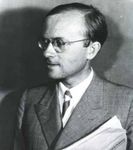
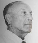
The History of Electro-Encephalography and Epileptology in Geneva | S. Vulliémoz, A.-C. Héritier Barras, M. Seeck Epileptologie 2015; 32 5Figure 2: Marcel Monnier, (1907-1996), director of the „Labo- Figure 3: Ferdinand Morel (1888-1957), who introduced the
ratoire d‘EEG clinique neurologique“ and the „Laboratoire de EEG in the psychiatric hospital of Bel-Air in 1957
recherche en neurophysiologie appliquée“.
Electroencephalography (EEG) and modern epi- Monnier already existed before 1950. The use of EEG in
leptology in Geneva psychiatry was later strongly supported by Prof. Julian
de Ajuriaguerra in the 1960’ notably by the creation
In 1941, the neurophysiologist PD Dr Marcel Mon- of the “Laboratoire d’EEG et de Neurophysiologie ap-
nier (Figure 2) and the engineer Marc Marchand pro- pliquée” in 1964, which included beds, equipped with
vided the first description of the installation of an EEG EEG, for the study of sleep. This Lab was initially direct-
equipment in Geneva, in the Institute of Physiology. ed by René Tissot, a French neurologist trained in Paris
The system was based on an electrocardiogram cou- notably under Alajouanine and Lhermitte [3].
pled to a preamplifier and a powerful amplifier allow- In 1966 Dr Annette Beaumanoir, a French neurolo-
ing the recording of electrical activity in a bipolar or gist, took the lead of the EEG laboratory in the hopital
monopolar montage, as proposed by Berger and Tön- cantonal of Geneva (Figure 4). A. Beaumanoir had been
nies. Monnier and Marchand initially presented the trained in the famous epileptology school of Marseille
localisation of head trauma using EEG. In 1952, Mon- under Henri Gastaut. She contributed to the char-
nier became director of the newly created “Laboratoire acterisation of paroxystic occipital discharges of idi-
d’EEG clinique neurologique” and the “Laboratoire de opathic focal occipital epilepsies, today referred to as
recherche en neurophysiologie appliquée”[3]. “Gastaut syndrome” and “Panayiotopoulos syndrome”.
In 1958, Prof. François Martin took over from M. The arrival of A. Beaumanoir in Geneva is strongly re-
Monnier. Under his direction, Geneva partnered the lated to her political activities. Member of the French
University Hospital of Zurich in EEG teaching: the Resistance at age 18, she was a member of the Com-
Swiss School of Electroencephalography and Epileptol- munist Party and later supported the war for Algeria’s
ogy. Between 1970-72 Martin also served as president independence (“Front National de Libération”). She be-
of the Swiss Society for Clinical Neurology, founded in came a member of the Algerian cabinet before being
1948 (as the “Schweizerische Arbeitsgemeinschaft für forced to leave the country after a coup in 1965, and
Elektroencephalographie”) [4]. found refuge in Geneva where she directed the EEG
EEG also became an important instrument in Swiss Unit until her retirement. She continued working with
psychiatric institutions, in the early 1950’. In Geneva, Gastaut, notably in descriptions of the Lennox-Gastaut
an EEG Laboratory was installed in 1957 by Prof. Fer- syndrome with an interest in pediatric EEG, installed
dinand Morel (Figure 3), a theologian, medical doctor the first EEG-telemetry with videoscopic recordings in
and director of the psychiatric hospital of Bel-Air out- Switzerland (and one of the first in Europe) and also
side Geneva. Actually, close collaborations between reported important observations on non-convulsive
psychiatrists Serge Mutrux, Claude Horneffer and M. status epilepticus, “état de mal à expression confu-
6 Epileptologie 2015; 32 The History of Electro-Encephalography and Epileptology in Geneva | S. Vulliémoz, A.-C. Héritier Barras, M. Seeckfailure [7] as well as work investigating the origin of
periodic lateralised discharges [8]. Jallon was also dedi-
cated to educational aspects and destigmatisation of
epileptic patients, as reflected by his activity in “l’Ecole
de l’Epilepsie” and two mountaineering expeditions to
4000m summits uniting patients and epileptologists
(“Sport et Epilepsie” at the Bishorn 4153 m, Valais and
the Gran Paradiso 4061 m, Piedmont, Figure 5). Jallon
witnessed the transition from ink and paper EEG to
digital recordings at the very end of the XXth century.
This radical evolution offered not only an easier offline
clinical interpretation, but opened new avenues for EEG
research.
Epilepsy surgery and EEG-based neuroimaging
In 1995 the Unit for Presurgical Epilepsy Evaluation
was founded by Prof. Theodor Landis, head of Neurol-
ogy. Its direction was given to Prof Margitta Seeck, a
German epileptologist trained in Germany and Bos-
Figure 4: Annette Beaumanoir (1923*) and her „Salle Annette ton, USA. Under her leadership, in close collaboration
Beaumanoir“, still perpetuating her memory in the University with Prof. Paul-André Despland, head of the EEG labo-
Hospital of Geneva. ratory in the Centre Hospitalier Universitaire Vaudois
(CHUV), the presurgical evaluation unit (Figure 6) and
the epilepsy surgery program was quickly established.
sionnelle”, together with her colleague PD Dr Josette Le A sophisticated multimodal electrophysiological and
Floch-Rohr. imaging platform flourished with intense collabora-
In 1989, she was followed by Prof. Pierre Jallon, an- tions with clinical experts in the Neuroradiology, Nu-
other French epileptologist, trained in Bordeaux and clear Medicine, Neuropsychology and Psychiatry as well
former director of the EEG unit of the French military as neuroscientists for the development and applica-
hospital of Val de Grâce in Paris. Interested in epide- tion of new imaging techniques. The surgical program
miology, Jallon conducted several prevalence and in- is placed in the context of an intense and fruitful col-
cidence studies in Switzerland, notably and on the in- laboration with the CHUV and the Institution of Lavi-
cidence of status epilepticus (EPISTAR) [5], new-onset gny (see the Lausanne contribution in this issue) defin-
epilepsy (EPIGEN) [6] as well as similar studies in French ing a Geneva-Vaud epilepsy surgery program with the
overseas territories (EPIREUN, EPIMART). From an EEG partnership of the surgical teams of Prof. JG Villemure,
perspective, he reported toxic-metabolic encepha- Prof. N. de Tribolet and Dr C. Pollo, followed by Prof. K.
lopathies related to the accumulation of the antibiotic Schaller and Prof. R. Daniels, PD Dr Momjian.
agent cefepime in patients with concomitant renal The program, celebrating its 20th anniversary in
2015, currently offers all aspects of epilepsy surgery
including invasive peroperatory and extra-operatory
EEG recordings using subdural electrodes or stereotac-
tically placed depth electrodes tailored to the clinical
need. Resective potentially curative surgery as well as
palliative surgery such as corpus callosotomy can be
proposed. Functional surgical approaches using vagal
nerve stimulation and deep brain stimulation, in the
amygdalo-hippocampal structures or the nucleus an-
terior of the thalamus are also part of the therapeu-
tic armamentarium. Cognitive studies involving some
patients of our centre with intracranially implanted
electrodes have led to some important scientific con-
tributions for the understanding of brain functions.
Probably the most striking is the localisation of feelings
of “out-of-body experiences” to the temporo-parietal
Figure 5: Epileptic patients and epileptologists, (Pr Jallon and junction [9, 10], giving an unambiguous organic sub-
colleagues) after the ascension of Gran Paradiso, 2005. strate to these symptoms often perceived as having a
The History of Electro-Encephalography and Epileptology in Geneva | S. Vulliémoz, A.-C. Héritier Barras, M. Seeck Epileptologie 2015; 32 7Seeck who continued to expand both the clinical and
scientific activity in EEG and epileptology. Margitta
Seeck also served as president of the Swiss Society for
Clinical Neurophysiology in 2011-15. The clinical re-
search axis led to enhanced validation of ESI as a pre-
surgical localising technique in larger patient groups
thanks to invasive validation in patients with subse-
quent intracranial EEG or post-surgical follow-up [14,
15]. Clinical studies in simultaneous EEG-fMRI also fol-
lowed the earlier first validation of this new technique
with invasive EEG [16].
The focus on epilepsy imaging was further devel-
oped by PD Dr Serge Vulliémoz, an epileptologist and
physicist trained in Geneva and in Queen Square, Lon-
don, who further developed EEG-fMRI recordings and
its combination with ESI, contributing to its increasing
consideration as a clinically relevant imaging tool. A
seminal study showed that even in the absence of vis-
ible interictal epileptic spikes, epileptogenic activity can
be identified and localized [17]. The current brain imag-
ing research focuses on the mapping of functional and
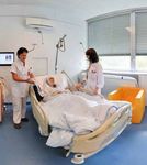
Figure 6: Today‘s view of the presurgical evaluation unit structural relationships between brain regions involved
in the occurrence of epileptic activity (Figure 7) [18].
Nowadays, ESI and EEG-fMRI are routinely performed in
purely psychiatric origin. the presurgical work-up of patients with focal epilepsy
Approximately at the same time as the Presurgi- and their results integrated in the clinical case discus-
cal Evaluation Unit was founded, Professor Landis also sion.
founded the Functional Brain Mapping laboratory The pediatric aspect of epileptology and electroen-
(1994), led by Prof. Christoph M. Michel, a neuroscien- cephalography and epilepsy surgery has also increased
tist and EEG expert, trained by Prof. Dietrich Lehmann in recent years, corresponding to about a third of the
in Zurich. C. Michel became a renowned international presurgical evaluations. Geneva has the largest Swiss
figure in the analysis of scalp voltage topography and pediatric presurgical evaluation program, performed on
Electric Source Imaging, consisting in the estimation of children referred from most parts of the country under
the brain electric generators of scalp EEG. This approach the joint supervision of Prof. Seeck’s team and neuro-
largely benefitted from the development of high densi- pediatricians. Initially, the pediatric part of the program
ty EEG systems with up to 256 scalp electrodes, initially was developed together with Prof. Eliane Roulet-Perez
used only for non-clinical human research. Dr L. Spinelli, in Lausanne who has special interest in the role of epi-
physicist, developed a head model called SMAC (Spheri- lepsy in the development of the child and potential of
cal Model with Anatomical Constraints) describing the children to catch up developmental retardation if the
propagation of electromagnetic fields across the head operation is carried out early and successfully [19].
and allowing the localisation of electrical sources tak- The pediatric epilepsy surgery team was later joined by
ing into account the individual brain anatomy [11]. This PD Dr Christian Korff, expert in pediatric epileptology
model has proved very reliable in clinical studies and trained in Geneva and later in Chicago and Dr Joël Fluss,
not inferior than more sophisticated later models [12]. expert in cognitive aspects trained in Paris and Toronto.
More broadly, the presurgical evaluation unit and PD Dr Fabienne Picard, a French neurologist trained
the functional brain mapping lab established a very in Strasbourg, is particularly interested in specific ge-
strong collaboration that led to pioneering methodo- netic forms of epilepsies, notably Autosomal Dominant
logical advances and clinical applications, in the field Nocturnal Frontal Lobe Epilepsy syndrome that she has
of functional and structural brain imaging in patients contributed to characterise in several large families [20]
with epilepsy, while pursuing stringent validation us- and which is related to a mutation in the nicotinic re-
ing concordance with invasive EEG or resection area in ceptor. These studies led her to further investigate the
patients with post-operative seizure-freedom. Electrical functional aspects of the insula and the distribution of
Source Imaging (ESI) has also been applied to the lo- nicotinic receptors in the brain, using nuclear imaging.
calisation of eloquent brain regions such as the soma- Psycho-social and education aspects have been
tosensory cortex [13]. further developed by Dr Anne-Chantal Héritier Barras,
At the retirement of Prof. Jallon, in 2007, the routine a Swiss epileptologist trained in Geneva and expert in
EEG and epilepsy clinic was fused with the presurgical therapeutic education of patients with chronic medical
evaluation unit under the unified direction of Prof. M. conditions. She introduced modern methodology in pa-
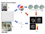
8 Epileptologie 2015; 32 The History of Electro-Encephalography and Epileptology in Geneva | S. Vulliémoz, A.-C. Héritier Barras, M. SeeckFigure 7: In the past 20 years, collaboration between the EEG and Epilepsy Unit and the Functional Brain Mapping Lab has
contributed to the transformation towards a neuro-imaging technique. Electrical Source Imaging: (a) high resolution EEG re-
cordings and (b) topographic representation of the scalp voltage together with sophisticated mathematical algorithms allow
to estimate the 3D localisation of the cortical generators of interictal epileptic activity (spikes) (c) . (d) EEG combined to simul-
taneous fMRI and the estimation of the neurovascular coupling (e) allow to map the hemodynamic changes correlated to the
occurence of interictal epileptic activity (spikes) (f).
tient’s interviews [21] and management to offer them cal research team with internationally recognised EEG
better strategies to understand and accept their con- expertise ensures that new development in EEG-based
dition and improve their care for themselves and their neuroimaging are rigorously validated and then rapidly
quality of life. integrated into the clinical work-up for an increased
Other more recent developments in epileptology in- quality of care.
clude a first seizure clinic aiming to rapid extensive di- The very recent large reorganisation of the neu-
agnostic evaluation and follow-up of patients with first rosciences in Geneva in the Biotech Campus in 2014,
seizure of probable epileptic origin. joining engineering, neuroscientific and clinical teams
Beyond their joint epilepsy program, the centres of from the University of Geneva and the Ecole Polytech-
Geneva and Lausanne have also strongly collaborated nique Fédérale de Lausanne will enhance multidiscipli-
since 2006 by organising the “Université d’Eté d’EEG”, a nary collaborations with a special focus on new imag-
yearly training program for Neurologists and EEG tech- ing techniques and neuromodulation. New implant-
nicians. able diagnostic and therapeutic devices using electro-
physiological recordings and electrical stimulation, as
well as neuroprosthetic tools developed in the Geneva-
Conclusion and perspectives Lausanne area will hopefully be part of epileptological
care in the not so far future.
The complementarity of the medical, nursing and
technical team as well as the strong local, regional,
nationwide and international clinical and scientific col- References
laborations allow our centre to continue evolving to
offer patients the best diagnostic and therapeutic ap- 1. Herpin. T. Des Accès Incomplets d’Epilepsie. Paris : JB Baillère et Fils, Li-
proaches, from diagnostic clarification to presurgical braires de l’académie impériale de médecine, 1867
evaluation and to comprehensive care of the patients’ 2. Taylor J (ed): Selected Writings of John Hughlings Jackson. Vol. 1. London:
condition. A great number of clinical and non-clinical Staples Press, 1958
collaborators worked towards this goal over the years. 3. Pidoux V. Expérimentation et clinique électroencéphalographiques en-
They could not be explicitely named in this article but tre physiologie, neurologie et psychiatrie (Suisse, 1935-1965). Revue
their contribution should be strongly acknowledged. d’Histoire des Sciences 2010; 63: 439-472
The proximity of a strong methodological and clini- 4. Hess CW. 50 Jahre Schweizerische Ges für Klinische Neurophysiologie.
The History of Electro-Encephalography and Epileptology in Geneva | S. Vulliémoz, A.-C. Héritier Barras, M. Seeck Epileptologie 2015; 32 9Schweiz Arch Neurol Psychiatr 1998; 149: 257 - 260 Address for correspondence:
5. Coeytaux A, Jallon P, Galobardes B, Morabia A. Incidence of status epi- Dr Serge Vulliémoz
lepticus in French-speaking Switzerland: (EPISTAR). Neurology 2000; 55: Unité d’EEG et d‘Exploration des Epilepsies
693-697 Service de Neurologie
6. Jallon P, Goumaz M, Hänggeli C, Morabia A. Incidence of first epileptic University Hospital and Faculty of Medicine
seizures in the canton of Geneva, Switzerland. Epilepsia 1997; 38: 547- Rue Gabrielle-Perret-Gentil 4
552 CH 1211 Geneva 14
7. Jallon P, Fankhauser L, Du Pasquier R et al. Severe but reversible encepha- Phone 0041 22 3728352
lopathy associated with cefepime. Neurophysiol Clin 2000; 30: 383-386 Fax 0041 22 3728340
8. Assal F, Papazyan JP, Slosman DO et al. SPECT in periodic lateralized epi- serge.vulliemoz@hcuge.ch
leptiform discharges (PLEDs): a form of partial status epilepticus? Seizure
2001; 10: 260-265
9. Arzy S, Seeck M, Ortigue S et al. Induction of an illusory shadow person.
Nature 2006; 443: 287
10. Blanke O, Ortigue S, Landis T, Seeck M. Stimulating illusory own-body
perceptions. Nature 2002; 419: 269-270
11. Spinelli L, Andino SG, Lantz G et al. Electromagnetic inverse solutions in
anatomically constrained spherical head models. Brain Topogr 2000; 13:
115-125
12. Birot G, Spinelli L, Vulliémoz S et al. Head model and electrical source
imaging: a study of 38 epileptic patients. Neuroimage Clin 2014; 5: 77-
83
13. Lascano AM, Brodbeck V, Lalive PH et al. Increasing the diagnostic value
of evoked potentials in multiple sclerosis by quantitative topographic
analysis of multichannel recordings. J Clin Neurophysiol 2009; 26: 316-
325
14. Brodbeck V, Spinelli L, Lascano AM et al. Electroencephalographic source
imaging: a prospective study of 152 operated epileptic patients. Brain
2011 ; 134 : 2887-2897. doi : 10.1093/brain/awr243
15. Mégevand P, Spinelli L, Genetti M et al. Electric source imaging of
interictal activity accurately localises the seizure onset zone. J Neu-
rol Neurosurg Psychiatr 2014 ; 85 : 38-43. Doi : 10.1136/jnnp-2013-
305515. Epub 2013 Jul 30
16. Seeck M, Lazeyras F, Michel CM et al. Non-invasive epileptic focus locali-
zation using EEG-triggered functional MRI and electromagnetic tomo-
graphy. Electroencephalogr Clin Neurophysiol 1998; 106: 508-512
17. Grouiller F, Thornton RC, Groening K et al. With or without spikes: lo-
calization of focal epileptic activity by simultaneous electroencephalo-
graphy and functional magnetic resonance imaging. Brain 2011; 134:
2867-2886
18. Pittau F, Mégevand P, Sheybani L et al. Mapping epileptic activity: sour-
ces or networks for the clinicians? Front Neurol 2014; 5: 218
19. Roulet-Perez E, Davidoff V, Mayor-Dubois C et al. Impact of severe epi-
lepsy on development: recovery potential after successful early epilepsy
surgery. Epilepsia 2010; 51: 1266-1276
20. Picard F, Bertrand S, Steinlein OK et al. Mutated nicotinic receptors re-
sponsible for autosomal dominant nocturnal frontal lobe epilepsy are
more sensitive to carbamazepine. Epilepsia 1999; 40: 1198-1209
21. Héritier Barras AC. Patients et soignants face à l’épilepsie. Enquête quali-
tative de besoins. Editions Universitaires Européennes, 2014
10 Epileptologie 2015; 32 The History of Electro-Encephalography and Epileptology in Geneva | S. Vulliémoz, A.-C. Héritier Barras, M. SeeckVous pouvez aussi lire
